G E N E R A L R E V I E W UDC: 616.65-07-037 DOI: 10.2298/VSP1505447P
Benign prostatic hyperplasia and prostate–specific antigen
Benigna hiperplazija prostate i prostata specifi
č
ni antigen
Tomislav Pejčić*, Miodrag Aćimović*, Zoran Džamić*, Milan Radovanović*, Jovan Hadži-Djokić†
*Clinic of Urology, Clinical Center of Serbia, Belgrade, Serbia; †Serbian Academy of Sciences and Arts, Belgrade, Serbia
Key words:
prostatic hyperplasia; prostate-specific antigen; prognosis.
Ključne reči:
prostata, hiperplazija; prostata, specifični antigen; prognoza.
Androgens and prostate function
Testosterone (T) and 5α-dihidrotestosterone (DHT) play a crucial role in the fetal prostate development. These androgens stimulate mesenchyme, while mesenchyme in-duces the proliferation of the epithelial buds from the uro-genital sinus. This process, called “mesenchyme-epithelial interaction”, starts in the 10th gestational week and continues in the adult age 1, 2. Testosterone induces the development of the seminal vesicles and Wolffian ducts, while DHT induces the development of the prostate, penis and scrotum.
Prostatic tissue is composed of stroma and epithelium. Prostatic stroma is composed of stromal cells (fibroblasts, endothelial capillary cells, lymph vessels and smooth muscle cells), neuroendocrine (NE) cells, neural cell axons, intercel-lular liquid and collagen fibers3. Prostatic epithelium is com-posed of secretory, basal, intermediary and NE cells. Secre-tory cells synthesize and secrete various proteins, like pros-tate specific antigen (PSA), prostatic acid phosphatase (PAP), androgen receptor (AR) and make the greatest part of the prostatic epithelium. It is believed that NE cells induce growth, differentiation, and secretory functions of the pro-static epithelium4.
Numerous factors regulate prostatic growth: endocrine, neuroendocrine, paracrine, or growth factors (GF), autocrine and intracrine factors. However, the action of the endocrine factors is the best known. Testosterone is the most important serum androgen in the male, with the average serum concen-tration of 611 ± 186 ng/dL, while the average DHT serum concentration is 56 ± 20 ng/dL. However, the average con-centration of active, free T is only 12.1 ± 3.7 ng/dL, while the rest is bound to the globulins and albumins. The major androgen in the prostatic tissue is DHT, with the average
tis-sue concentration of 2.4–5.1 ng/g. The average tistis-sue con-centration of T is 3–5 times lower and measures 0.9 ng/g 5–7.
Only free T molecules can enter the prostatic cell, by diffusion. In the cytosol, one part of T molecules transforms into DHT. Both T and DHT bind to AR and form androgen-AR complexes. Subsequently, those complexes make pairs, entering the nucleus and bind to androgen-responsive ele-ments (ARE) on DNA. After the information was tran-scripted from DNA to mRNA, mRNA leaves the nucleus and comes on ribosomes, where the information is translated into protein. Enzyme 5-alpha reductase (5αR) performs the con-version of T to DHT. There are two isoforms of 5αR: type 5αR-2 is dominant in the prostatic stroma and accessory genital tissues. Type 5αR-1 is present in the skin and prostate epithelium.
Benign prostate hyperplasia
much higher: it is 10% for men in IV decade, 20% for men in V decade, 50–60% in men in VII decade and 80–90% in men in VIII and IX decade 11.
Etiology of benign prostatic hyperplasia
The most common factors that induce the development of BPH are steroid hormones, growth factors, interactions between stroma and epithelium, and the regulation of apop-tosis (Figure 1). The gradual decrease of T concentration is characteristic for an aging man; however, DHT concentration increases in prostatic tissue, or remains unchanged. The role of estrogens and estrogen receptors (ER) in the etiology of BPH is very possible. It is proved that dog prostate contains large amounts of ER and that estrogen administration in-duces stromal growth in dogs. In humans, epithelial prolif-eration is stimulated by fibroblast growth factors (FGF) and inhibited by transforming growth factors (TGF). The concen-tration of TGF-β is decreased in BPH 12. In brief, the devel-opment of BPH requires androgenic influence in young age and long-term androgen stimulation in adult age. In old age, characteristic events are the decrease of T concentration and the increase of DHT and estrogen concentration, increased FGF activity, decreased TGF-β activity and the decreased apoptosis.
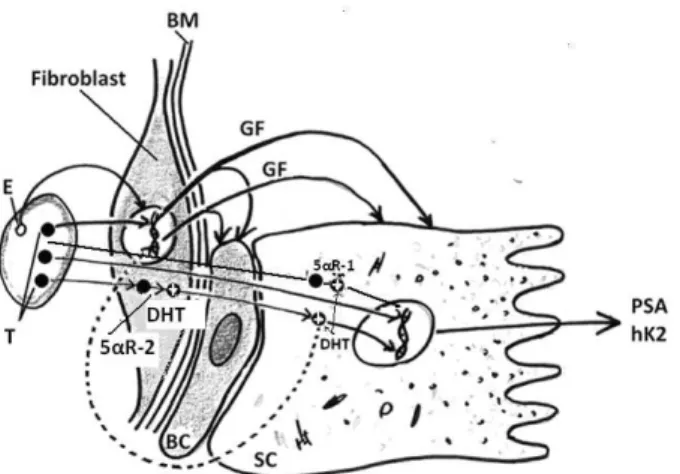
Fig. 1 – Stromal-epitelial interaction (Autor T. Pejčić) E – estrogen; T – testosterone; DHT – dihydrotestosterone; 5αR-1 – 5-alfa reductase type-1; 5αR-2 – 5-alfa reductase type-2; BM – besement membrane; GF – growth factor; BC – basal cell; SC – secretory cell; PSA – prostate-specific antigen; hK2 –
human kalikrein 2.
Benign prostatic hyperplasia is common among mem-bers of Western civilization; however, there is an increasing incidence of BPH in Asian countries, with traditionally low prevalence of BPH 13. It is believed that the increase in inci-dence is associated with the lifestyle of the modern man. In fact, today's man is fed differently than his ancestors, live longer and retains sexual activity long after the generative period. Humans have drastically changed diet about 15,000 years ago, when domesticated animals and, of obligate her-bivores, became carnivorous 14. Excessive intake of meat, fried and baked foods and obesity, lead to hormonal distur-bances and oxidative DNA damage 15. In developed
coun-tries, the average human life span is today over 80 years, while the length of human life in the Neolithic period was only 20 years 16, 17. In addition, the man retains sexual activ-ity for a long period: 20–35% of men aged 60–69 years have one intercourse per week 18. Unlike most of primates, man's sexual activity does not have seasonal variations. Therefore, long-term hormonal stimulation of the prostate, oxidative stress and increased incidence of genetic changes associated with aging are all important combined etiological factors for the development of BPH.
Pathology and pathophysiology of benign prostatic hyperplasia
Prostatic hyperplasia increases urethral resistance, which leads to a compensatory increase in detrusor pressure and the reduction in bladder capacity. It is believed that the capsule of the prostate plays a very important role in the de-velopment of lower urinary treat symptoms (LUTS), because it transmits the pressure of the hyperplastic tissue on the ure-thra.
The important characteristic of BPH is the increase of the total number of cells, not only an increase in cell size. McNeal has shown that early periurethral nodules have a stromal structure, while the early nodules in the transition zone (TZ) represent the proliferation of the glandular tissue. Glandular nodules rise from the newly formed small ducts, arising as buds on existing ducts; these ducts grow and branch out, creating an entirely new ductal system within the nodule. During the first 20 years, the development of BPH is characterized by an increased number of slowly growing nodules. Thereafter, in the second stage, major nodules show significant growth 18.
Clinical characteristics of benign prostatic hyperplasia
Common characteristics of BHP are progressive en-largement of the prostate, voiding symptoms and increased PSA. Total prostate volume (TPV) increases from 25 mL in men aged 30–35 years, to 45 mL in men over 70, while the transition zone (TZ) volume increases from 15 mL to 25 mL. Transrectal ultrasound (TRUS) provides the most accurate measurement of TPV. The Olmsted study revealed that TPV grows 0.4 mL per year in men aged 40–59, and 1.2 mL per
year in men aged 60–79. The overall TPV growth was 0.6 mL, or 1.9% per year.
from 0–35. However, the obstruction can be assessed objec-tively by the measurement of the urinary flow, or Uroflow. It is accepted that the maximum urine flow, Qmax < 10 mL/sec, carries a high probability for the presence of the ob-struction, while Qmax > 15 mL/sec carries a low probability. Some authors tried to express urine flow through the single number, Qi. Index Qi is the result of multiplying Qmax and average flow, Qave: Qi = Qmax × Qave. Pejčić et al. 20 found that 71% of men with IPSS > 7 had Qi < 100, while 75% healthy men with IPSS < 7 had Qi > 100.
Prostate – specific antigen
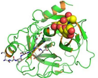
The main secretory proteins of the prostate gland are prostate-specific antigen (PSA), human glandular kalikrein (hK2), prostatic acid phosphatase (PAP) and prostate-specific protein (PSP-94). Molecular weight of PSA is 34 kDa; PSA molecule consists of one chain with 240 amino-acids and four carbohydrate lateral chains (Figure 2).
Fig. 2 – Human prostate-specific antigen (PSA/KLK3) with bound substrate from complex with antibody 21.
Prostate-specific antigen was isolated in seminal plasma in 1966; for a long time, PSA has been used in forensic evi-dence of rape 22. It has been estimated that the PSA concen-tration in seminal plasma was very high (1.5 mg/mL) and lower in urine (250 ng/mL). However, Wang et al 23 were the first to predict the possible use of PSA determination in the blood in the diagnosis of the prostate diseases. The average PSA concentration in the prostatic tissue ranges from 10,000 ng/mg of tissue to 76,000 ng/mg of tissue 24, 25. In other words, prostate gland weighing 20 mL contains 0.2–1.5 mg PSA. The expression of PSA is high in benign epithelial cells, low in malignant cells and progressively decreases with the degree of anaplasia 26–31.
Synthesis and secretion of prostate-specific antigen
The intensity of PSA synthesis largely depends on the concentration of DHT in the prostatic tissue 32. DHT mole-cules bind to AR and form DHT-AR complexes. Those com-plexes enter the nucleus and bind to ARE on the DNA. The
following is a transcription to mRNA; after that, mRNA leaves the nucleus and goes to the ribosomes, where the translation and PSA synthesis take place 33, 34. Alternative PSA synthesis pathway was demonstrated in the tissue cul-ture. This pathway goes via the membrane steroid receptor; it is considerably faster than the genomic process and lasts 1– 30 minutes 35.
The first product of the synthesis is the preproPSA molecule, with a leading sequence of 17 amino acids. Pre-proPSA molecules are placed in a number of prostate secre-tary granules (PSGs), which migrate towards the apical part of the cell. During the following biochemical process, lead-ing sequence of 17 amino acids is separated from the pre-proPSA molecule. This product is now referred to as proPSA, and it is ejected as an inactive proenzyme from a cell ie secreted into the lumen of the acinus. In the lumen of the acini, hK2 separates another seven amino acids from the proPSA molecule, which results in the creation of active en-zyme PSA. The last reaction which happens in the acini is very important: acinar enzymes change the conformal struc-ture of the PSA molecule, after which it becomes inactive as an enzyme. In normal acini, 25–30% of the PSA molecules are inactivated and thereafter diffuse into the systemic circu-lation. The remaining PSA molecules enter blood as active enzymes, where they rapidly form complexes with heavy plasma proteins. One fraction of PSA molecules does not en-ter blood, but leaks down the acini and ducts, as a part of prostatic secretion. Secretion is moving towards prostatic urethra due to the difference in hydrostatic pressure and stromal smooth muscle tone.
Prostate-specific antigen in serum
Under normal conditions, 25–30% of the inactive PSA molecules and 70–75% of active PSA molecules enter blood stream. As soon as the active PSA molecules enter blood, they are immediately bound by protease inhibitors, such as antichymotrypsin (ACT) and alpha 2-macroglobulin (alpha-2M). This process is fast and efficient because the molar concentration of the inhibitors exceeds molar PSA concen-tration over 100,000 times. PSA-ACT complex is the most common PSA form in serum, and is formed in the 1 : 1 molar ratio, in the irreversible reaction. These molecules can be de-tected in blood tests and they are named “complexed PSA”. On the other hand, inactive PSA molecules can be detected as the fraction of free molecules and they are named “free PSA”. In the presence of prostatic acinar lesion, smaller frac-tion of active PSA molecules completes the transformafrac-tion to an inactive PSA; that is the explanation why free PSA frac-tion is lower in patients with prostate cancer (PCa).
It is not yet exactly known how PSA molecules enter blood. However, it is known that PSA molecules have to cross the so-called “prostatic blood-barrier”, consisting of prostatic basal cells, basement membrane of the duct, the ex-tracellular space, the capillary basement membrane and the layer of capillary endothelial cells. All processes which lead to damage of the prostatic blood-barrier enable massive PSA transfer in blood 42, 43.
Determination of PSA in serum in the diagnosis of BPH
Clinical application and research related to PSA over the last 25 years have been so extensive, that this period of urology is called the "PSA era". What is even more impor-tant, the occurrence of PSA strongly influenced the tremen-dous changes in the diagnostics and treatment of prostate cancer (PCa) and the development of new strategies and technologies for the treatment of this disease. However, the phenomenon of elevated PSA in BPH patients has always been regarded as the "artifact", which complicates the diag-nosis of localized PCa.
It has long been thought that normal PSA level is below 4.0 ng/mL. However, subsequent studies have shown two confusing facts. First, it became clear that a significant num-ber of patients with localized PCa had PSA < 4.0 ng/mL; soon, the new PSA cut-off value of 2.5 ng/mL was estab-lished 44, 45. Second, it has been proven that 70–80% of peo-ple without PCA after biopsy, had PSA within the "gray zone” i.e., 4.0–10.0 ng/mL. It became clear that the PSA level in each subject depends on many factors. In the first place, the synthesis of PSA depends on the concentration of DHT, as well as the presence of BPH. Then, the level of PSA depends on the hormonal status, obesity, body weight, total blood volume and so on. In patients with localized PCa, the size and location of the tumor also influence the PSA level. Finally, serum PSA level depends on the concentration of PSA in prostatic tissue surrounding the growing tumor 46.
The so-called "PSA derivatives" were introduced in or-der to distinguish the patients with BPH and PCA, with the
PSA in gray zone. PSA density (PSAD) was introduced with the aim to reduce the impact of prostate size on the interpre-tation of the PSA. For the threshold is taken PSAD = 0.1, i.e., patients with PSAD > 0.1 are more likely to have PCa 47. Similarly, subjects with PSA velocity (PSAV) > 0.8 ng/mL
per year, have greater risk for the presence of PCa. Those with PSAV ≤ 0.8 ng/mL per year are more likely to have BPH. A derivative called “PSA doubling time” (PSADt) ex-presses the increase of PSA in time (t) more precisely; it is calculated using the formula: PSADt = log 2t / log final PSA – log initial PSA. Prostate cancer has shorter PSADt than BPH; in addition, the more aggressive the tumor is, PSADt is shorter 48, 49. Free/total PSA ratio (f/t PSA) is frequently used to distinguish the persons with BPH and PCa having PSA in the gray zone and normal digital rectal examination (DRE). Normal values of f/t PSA are 0.18 to 0.22 50; however, the patients with f/t PSA < 0.1 have 56% chance to have PCa 51.
Today, it becomes quite clear that BPH is the main rea-son for the PSA values from 4.0–10 ng/mL, or 2.5–10 ng/mL. The American Urological Association (AUA) states that a very high risk for the presence of PCa, about 90%, is present in PSA > 20 ng/mL. It is not difficult to conclude that the increase in PSA, caused by the presence of BPH, was the main reason for the unnecessary diagnosis of a large number of clinically insignificant PCa. This is one of the rea-sons why the AUA reduced the range of PSA screening for men aged 55 to 69 years in 2013.
On the other hand, in the field of BPH, the situation is far less complicated and PSA is a precise parameter of dis-ease progression. Several large multicenter studies have de-fined the precise parameters for monitoring the growth and progression of BPH. The most well-known studies are: Pro-scar Long-Term Efficacy and Safety Study (PLESS), Medi-cal Therapy of Prostatic Symptoms (MTOPS), "Olmsted County Study of Urinary Symptoms and Health Status Among Men" and The Combination of Avodart and Tamsu-losin (CombAT).
MTOPS study included 3,047 patients, who were fol-lowed for 4.5 years. Factors that indicated the progression of BPH were TPV ≥ 31 mL, PSA ≥ 1.6 ng/mL, Qmax < 10.6 ml/s, residual urine, RU ≥ 39 mL and the age ≥ 62 years 52. In patients who were taking finasteride, the average reduc-tion in TPV for 4.5 years was 19%. However, men with TPV > 40 mL had an average reduction in TPV by 25% 53, 54. CombAT study included 4,844 men aged over 50 years with a clinical diagnosis of BPH, IPSS > 12, TPV > 30 mL, PSA in the range of 1.5–10 ng/mL and at least two urinations with Qmax of 5–15 mL/s 55.
Determination of PSA in the urine in the diagnosis of BPH
Determination of PSA concentration in urine (uPSA) has never been used in the diagnosis of BPH and monitoring of BPH progression. It is interesting that even in 1987, Tremblay et al. 59 found that the average uPSA concentration was 216 ng /mL and that people with BPH had higher uPSA values than young men. However, over the following years, researches have focused mainly on the ability to distinguish BPH and PCa and to detect early relapse after radical pros-tatectomy (RP). From 1994 to 2000, few works on this topic concluded that uPSA was higher in BPH than in PCa, but that it cannot help in differentiating those two diseases 60–63. The hope that uPSA will become a marker of the early recur-rence after RP, was closed when Iwakiri et al. 64 demon-strated that PSA was normally present in urine in all patients after RP and that it originated from the urethral glands 64. In some studies, it has been found that men with alopecia had higher values of urethral PSA after RP 65.
All researchers agree that uPSA is highly androgen-dependent marker for monitoring of the hormonal treatment, in both men and women 66, 67. Also, uPSA can be used as an early noninvasive marker of the appearance of puberty in boys 68, 69. In most primates, the seasonal uPSA increase in-dicates the beginning of the breeding season 70. Except the determination in fresh urine, uPSA can be determined in the dried urine, on filter paper, where it remains stable over a long period of time 71. However, most researchers agree that the methodology of PSA determination in urine is still incon-sistent 72.
In recent years, several papers that trigger the clinical use of uPSA were published. In a group of patients with PSA of 2.5–10.0 ng/mL, Bolduc et al. 73 found a significant dif-ference in mean uPSA in BPH (123.2 ng/mL) and PCa (52.6 ng/mL). With the uPSA threshold > 150 ng/mL, the sensitiv-ity of the test was 92.5%. The authors believe that subjects with PSA of 2.5–10.0 ng/mL and uPSA > 150 ng/mL, could be exempted from prostate biopsy, in the absence of suspi-cious lesions on DRE and TRUS. In some studies, it has been found that larger tumors had lower uPSA than smaller tumors, probably due to the obstruction of the drainage of secretions 74–76.
However, only one paper described the methodology of uPSA usage as a prognostic marker of BPH 77. In a group of 265 patients without PCa, uPSA, PSA, TPV and patients` age were determined. According to MTOPS criteria, TPV ≥ 31 mL, PSA ≥ 1.6 ng/mL and age ≥ 62 years were used as cutoff values of BPH progression. Persons with TPV < 31
mL had significantly lower uPSA, than patients with TPV ≥ 31 mL (119.3 ± 124.5 and 255.5 ± 204.9 ng/mL, respec-tively; p < 0.0001). In addition, persons in the so-called “non-progressive BPH” group (TPV < 31 mL, PSA < 1.6 ng/mL, age < 62 yrs) had significantly lower uPSA than pa-tients from the “progressive BPH” group (86.8 ± 82.4 ng/mL and 274.9 ± 208.3 ng/mL, respectively; p < 0.0001). Urinary PSA correlated significantly with TPV (r = 0.32, p < 0.0001).
The urinary PSA cutoff level of 150 ng/mL discrimi-nated the patients with non-progressive BPH and progressive BPH with specificity of 0.83 and sensitivity of 0.67. In that issue, Pejčić et al. 77 conclude that uPSA reflects prostatic hormonal activity and correlates with TPV, PSA and age. Therefore, uPSA level ≥ 150 ng/mL can be used as an addi-tional predictive parameter of BPH progression.
Conclusion
Testosterone and 5α-dihidrotestosterone play a crucial role in the prostate fetal development, growth and function. Testos-terone is the most important serum androgen in the male, but the major androgen in the prostatic tissue is 5α-dehidrotestosterone.
Benign prostatic hyperplasia is the fourth most common disease and affects 6% of general population. Biochemical char-acteristics of benign prostatic hyperplasia are decreased testos-terone, increased 5α-dehidrotestosterone and estrogen concen-tration, increased fibroblast growth factor and decreased trans-forming growth factor-beta activity.
Prostate-specific antigen is the main secretory product of the prostate gland; its synthesis largely depends on the 5α -dehidrotestosterone concentration in the prostatic tissue. Normal healthy prostate gland secretes 0.01–0.02 mg prostate-specific antigen per day, while hyperplastic prostate secretes ten times larger amounts of specific antigen. Secreted prostate-specific antigen is washed out from the urethra during voiding and can be detected in the urine.
However, the phenomenon of elevated prostate-specific antigen in benign prostatic hyperplasia patients has always been regarded as the "artifact", which complicates the diagno-sis of localized prostate cancer. Nevertheless, recent studies precisely established that serum prostate-specific antigen ≥ 1.6 ng/mL is suggestive for benign prostatic hyperplasia progres-sion in men with prostate volume ≥ 31 mL and age ≥ 62 years. In addition, urinary prostate-specific antigen concentration is significantly higher in subjects with benign prostatic hyperpla-sia; urinary prostatic antigen level ≥ 150 ng/mL can be used as additional predictive parameter of benign prostatic hyperplasia progression.
R E F E R E N C E S
1. Berman D, Rodriguez R, Veltri RW. Development, Molecular Biology, and Physiology of the Prostate. In: Wein AJ, Kavoussi LR, Partin AW, Craig PA, Novick AC, editors. Campbell-Walsh Urology. 10th ed. Philadelphia: Saunders; 2012. p. 2533−69.
2. Cunha GR. Role of mesenchymal-epithelial interactions in normal and abnormal development of the mammary gland and prostate. Cancer 1994; 74(Suppl 3): 1030−44.
4. Vashchenko N, Abrahamsson P. Neuroendocrine differentiation in prostate cancer: implications for new treatment modalities. Eur Urol 2005; 47(2): 147−55.
5. Wurzel R, Ray P, Major-Walker K, Shannon J, Rittmaster R. The effect of dutasteride on intraprostatic dihydrotestosterone concentra-tions in men with benign prostatic hyperplasia. Prostate Cancer Prostatic Dis 2006; 10(2): 149−54.
6. Mohler JL, Gregory CW, Ford HO, Kim D, Weaver CM, Petrusz P, et al. The androgen axis in recurrent prostate cancer. Clin Cancer Res 2004; 10(2): 440−8.
7. Titus MA, Schell MJ, Lih FB, Tomer KB, Mohler JL. Testosterone and dihydrotestosterone tissue levels in recurrent prostate cancer. Clin Cancer Res 2005; 11(13): 4653−7.
8. Naslund MJ, Issa MM, Grogg AL, Eaddy MT, Black L. Clinical and economic outcomes in patients treated for enlarged prostate. Am J Manag Care 2006; 12(Suppl 4): 111−6.
9. Vos T, Flaxman AD, Naghavi M, Lozano R, Michaud C, Ezzati M, et al. Years lived with disability (YLDs) for 1160 sequelae of 289 dis-eases and injuries 1990-2010: a systematic analysis for the Global Burden of Disease Study 2010. Lancet 2012; 380(9859): 2163−96. 10. Verhamme KM, Dieleman JP, Bleumink GS, van der Lei J, Sturkenboom MC, Artibani W, et al. Incidence and prevalence of lower urinary tract symptoms suggestive of benign prostatic hyperplasia in pri-mary care-the Triumph project. Eur Urol 2002; 42(4): 323−8. 11. Roehrborn C, Mcconnell J. Etiology, pathophysiology, epidemiology
and natural history of benign prostatic hyperplasia. In: Walsh P, Retik A, Vaughan E, WeinA, editors. Campbell’s Urology. 8th ed. Philadelphia: Saunders; 2002. p. 1297−36.
12. Cohen P, Nunn SE, Peehl DM. Transforming growth factor-beta in-duces growth inhibition and IGF-binding protein-3 production in prostatic stromal cells: abnormalities in cells cultured from benign prostatic hyperplasia tissues. J Endocrinol 2000; 164(2): 215−23. 13. Gu F. Epidemiological survey of benign prostatic hyperplasia and
prostatic cancer in China. Chin Med J 2000; 113(4): 299−302. 14. Coffey DS. Similarities of prostate and breast cancer: Evolution,
diet, and estrogens. Urology 2001; 57(4 Suppl 1): 31−8.
15. Bethel CR, Chaudhary J, Anway MD, Brown TR. Gene expression changes are age-dependent and lobe-specific in the brown Norway rat model of prostatic hyperplasia. Prostate 2009; 69(8): 838−50. 16. Life expectancy. Available from:
http://en.wikipedia.org/wiki/Life_expectancy
17. Oded G, Omer M. The Neolithic Revolution and Contemporary Variations in Life Expectancy. 2007. [cited 2010 September 12]. Available from:
http://www.kinseyinstitute.org/resources/FAQ.html
18. The Kinsey institute. Frequently asked sexuality questions to the Kinsey Institute. Available from:
http://www.kinseyinstitute.org/resources/FAQ.html
19. Roehrborn CG. Benign Prostatic Hyperplasia: Etiology, Pathophysi-ology, EpidemiPathophysi-ology, and Natural History. In: Wein AJ, Kavoussi LR, Partin AW, Peters CA, Novick AC, editors. Campbell-Walsh Urology. 10th ed. Philadelphia: Saunders; 2012. p. 2570−610. 20. Pejcic T, Argirovic DJ, Crnomarkovic D. Uroflow Index (pF x mF):
More Precise Interpretation of the Results. Eur Urol Meeting 2006; 1(2): 2.
21. 21 EAS. Humahn prostate specific antigen (PSA/KLK3) with bound substrate from complex with antibody (PDB id: 2ZCK) [cited 2011 September 21]. Available from:
commons.wikimedia.ortg/.../File:PSA_KLK3_PD
22. Graves HC, Sensabaugh GF, Blake ET. Postcoital detection of a male-specific semen protein. Application to the investigation of rape. N Engl J Med 1985; 312(6): 338−43.
23. Wang MC, Papsidero LD, Kuriyama M, Valenzuela LA, Murphy GP, Chu TM. Prostate antigen: a new potential marker for prostatic cancer. Prostate 1981; 2(1): 89−96.
24. Vesey SG, Goble M, Ferro MA, Stower MJ, Hammonds JC, Smith PJ. Quantification of prostatic cancer metastatic disease using pros-tate-specific antigen. Urology 1990; 35(6): 483−6.
25. Erickson DR, Hlavinka TC, Rockwood AP, Metter JD, Novicki DE, Fried MG. Prostatic acid phosphatase, beta-glucuronidase and prostate specific antigen assays in fine needle aspirates from be-nign and malignant prostates. J Urol 1991; 146(5): 1402−7. 26. Denmeade SR, Sokoll LJ, Chan DW, Khan SR, Isaacs JT.
Concentra-tion of enzymatically active prostate-specific antigen (PSA) in the extracellular fluid of primary human prostate cancers and human prostate cancer xenograft models. Prostate 2001; 48(1): 1−6. 27. Jung K, Brux B, Lein M, Rudolph B, Kristiansen G, Hauptmann S, et al.
Molecular forms of prostate-specific antigen in malignant and be-nign prostatic tissue: biochemical and diagnostic implications. Clin Chem 2000; 46(1): 47−54.
28. Ersev A, Ersev D, Turkery L, Ilker Y, Simsek F, Kullu S, et al. The re-lation of prostatic acid phosphatase and prostate specific antigen with tumor grade in prostatic adenocarcinoma: an immunohisto-chemical study. Prog Clin Biol Res 1990; 357: 129−34.
29. Bostwick DG. Prostate specific antigen and pathology of the pros-tate. Eur Urol 1995; 27(Suppl 2): 5.
30. Pretlow TG, Pretlow TP, Yang B, Kaetzel CS, Delmoro CM, Kamis SM, et al. Tissue concentrations of prostate-specific antigen in prostatic carcinoma and benign prostatic hyperplasia. Int J Cancer 1991; 49(5): 645−9.
31. Weir EG, Partin AW, Epstein JI. Correlation of serum prostate spe-cific antigen and quantitative immunohistochemistry. J Urol 2000; 163(6): 1739−42.
32. Zhu Y, Cai L, You X, Cordero JJ, Huang Y, Imperato-McGinley J. An-drogen-induced prostate-specific antigen gene expression is medi-ated via dihydrotestosterone in LNCaP cells. J Androl 2003; 24(5): 681−7.
33. Balk SP, Ko Y, Bubley GJ. Biology of prostate-specific antigen. J Clin Oncol 2003; 21(2): 383−91.
34. Zhu Y, Sun G. 5α-Reductase Isozymes in the Prostate. J Med Sci 2005; 25(1): 1−12.
35. Kampa M, Papakonstanti EA, Hatzoglou A, Stathopoulos EN, Stourna-ras C, Castanas E. The human prostate cancer cell line LNCaP bears functional membrane testosterone receptors that increase PSA secretion and modify actin cytoskeleton. FASEB J 2002; 16(11): 1429−31.
36. Kabalin JN, Hornberger JC. Prostate specific antigen is not excreted by human kidney or eliminated by routine hemodialysis. Urology 1991; 37(4): 308−10.
37. Shibata K, Kajihara J, Kato K, Hirano K. Purification and characteri-zation of prostate specific antigen from human urine. Biochim Biophys Acta 1997; 1336(3): 425−33.
38. Schieferstein G. Prostate-specific antigen (PSA) in human seminal plasma. Arch Androl 1999; 42(3): 193−7.
39. Lilja H. Role of hK2, free PSA, and complexed PSA measure-ments in the very early detection of prostate cancer. Eur Urol 2001; 39(Suppl 4): 47−8.
40. Lilja H. Free and total PSA: background information and rationale for use. In: Tindal DJ, editor. Recent advances in prostate cancer: basic science discoveries and clinical. New York: Parthenon Pub-lishing Group; 1997. p. 195−7.
41. Robert M, Gagnon C. Semenogelin I: a coagulum forming, multi-functional seminal vesicle protein. Cell Mol Life Sci 1999; 55(6−7): 944−60.
42. Ellis WJ, Brawer MK. PSA in benign prostatic hyperplasia and pro-static intraepithelial neoplasia. Urol Clin North Am 1993; 20(4): 62115.
44. Catalona WJ, Ramos CG, Carvalhal GF, Yan Y. Lowering PSA cut-offs to enhance detection of curable prostate cancer. Urology 2000; 55(6): 791−5.
45. Gilbert SM, Cavallo CB, Kahane H, Lowe FC. Evidence suggesting PSA cutpoint of 2. 5 ng/mL for prompting prostate biopsy: re-view of 36, 316 biopsies. Urology 2005; 65(3): 549−53.
46. Pejcić T, Hadzi-Djokić J, Topuzović C, Basić D, Marjanović A, Djurasic L. The analysis of some factors that influence on serum PSA level in localized prostate cancer patients: mathematical model. Acta Chir Iugosl 2011; 58(1): 81−7.
47. Catalona WJ, Southwick PC, Slawin KM, Partin AW, Brawer MK, Flanigan RC, et al. Comparison of percent free PSA, PSA density, and age-specific PSA cutoffs for prostate cancer detection and staging. Urology 2000; 56(2): 255−60.
48. Schmid HP, Prikler L, Sturgeon CM, Semjonow A. Diagnosis of pros-tate cancer-the clinical use of prospros-tate-specific antigen. EAU Up-date Series 2003; 1: 3−8.
49. Kakehi Y, Kamoto T, Shiraishi T, Kato T, Tobisu K, Akakura K, et al. Correlation of initial PSA level and biopsy features with PSA-doubling time in early stage prostate cancers in Japanese men. Eur Urol 2002; 41(1): 47−53.
50. Horninger W, Reissigl A, Rogatsch H, Volgger H, Studen M, Klocker H, et al. Prostate cancer screening in the Tyrol, Austria: experience and results. Eur J Cancer 2000; 36(10): 1322−35.
51. Heidenreich A, Bellmunt J, Bolla M, Joniau S, Mason M, Matveev V, et al. EAU guidelines on prostate cancer. Part 1: screening, diagnosis, and treatment of clinically localised disease. Eur Urol 2011; 59(1): 61−71.
52. Crawford E, Wilson SS, McConnell JD, Slawin KM, Lieber MC, Smith JA, et al. Baseline factors as predictors of clinical progression of benign prostatic hyperplasia in men treated with placebo. J Urol 2006; 175(4): 1422−6.
53. de la Taille A. Contribution of the PCPT trial to finasteride treat-ment of micturition disorders due to benign prostatic hyperplasia. Prog Urol 2008; 18 Suppl 3: S53−7. (French)
54. Kaplan SA, Roehrborn CG, Mcconnell JD, Meehan AG, Surynawanshi S, Lee JY, et al. Long-term treatment with finasteride results in a clinically significant reduction in total prostate volume compared to placebo over the full range of baseline prostate sizes in men en-rolled in the MTOPS trial. J Urol 2008; 180(3): 1030−2.
55. Roehrborn CG, Siami P, Barkin J, Damião R, Becher E, Miñana B, et al. The Influence of Baseline Parameters on Changes in International Prostate Symptom Score with Dutasteride, Tamsulosin, and Combination Therapy among Men with Symptomatic Benign Prostatic Hyperplasia and an Enlarged Prostate: 2-Year Data from the CombAT Study. Eur Urol 2009; 55(2): 461−71.
56. Chung BH, Roehrborn CG, Siami P, Major-Walker K, Morrill BB, Wil-son TH, et al. Efficacy and safety of dutasteride, tamsulosin and their combination in a subpopulation of the CombAT study: 2-year results in Asian men with moderate-to-severe BPH. Prostate Cancer Prostatic Dis 2009; 12(2): 152–9.
57. Roehrborn CG. BPH progression: concept and key learning from MTOPS, ALTESS, COMBAT, and ALF-ONE. BJU Int 2008; 101(Suppl 3): 17−21.
58. Siami P, Roehrborn CG, Barkin J, Damiao R, Wyczolkowski M, Duggan A, et al. Combination therapy with dutasteride and tamsulosin in men with moderate-to-severe benign prostatic hyperplasia and prostate enlargement: the CombAT (Combination of Avodart and Tamsulosin) trial rationale and study design. Contemp Clin Trials 2007; 28(6): 770−9. .
59. Tremblay J, Frenette G, Tremblay RR, Dupont A, Thabet M, Dube JY. Excretion of three major prostatic secretory proteins in the urine of normal men and patients with benign prostatic hypertrophy or prostate cancer. Prostate 1987; 10(3): 235−43.
60. Breul J, Pickl U, Hartung R. Prostate-specific antigen in urine. Eur Urol 1994; 26(1): 18−21.
61. Hillenbrand M, Bastian M, Steiner M, Zingler C, Muller M, Wolff JM, et al. Serum-to-urinary prostate-specific antigen ratio in patients with benign prostatic hyperplasia and prostate cancer. Anticancer Res 2000; 20(6D): 4995−6.
62. Irani J, Millet C, Levillain P, Doré B, Begon F, Aubert J. Serum-to-urinary prostate specific antigen ratio: its impact in distinguishing prostate cancer when serum prostate specific antigen level is 4 to 10 ng./ml. J Urol 1997; 157(1): 185−8.
63. Pannek J, Rittenhouse HG, Evans CL, Finlay JA, Bruzek DJ, Cox JL, et al. Molecular forms of prostate-specific antigen and human kal-likrein 2 (hK2) in urine are not clinically useful for early detection and staging of prostate cancer. Urology 1997; 50(5): 715−21. 64. Iwakiri J, Granbois K, Wehner N, Graves HC, Stamey T. An analysis of
urinary prostate specific antigen before and after radical prostatec-tomy: evidence for secretion of prostate specific antigen by the periurethral glands. J Urol 1993; 149(4): 783−6.
65. Pejcić T, Hadzi-Djokić J, Marković B, Lalić N, Glisić B. What are the possible reasons for urethral PSA varieties after radical prostatec-tomy. Acta Chir Iugosl 2010; 57(2): 31−5.
66. Pejcić T, Dimitrijević V, Hadzi-Djokić J. Urinary PSA in monitoring of patients with prostate cancer. Acta Chir Iugosl 2012; 59(1): 57−60.
67. Zaviacic M, Ruzicková M, Jakubovský J, Danihel L, Babál P, Blazeková J. The significance of prostate markers in the orthology of the fe-male prostate. Bratisl Lek Listy 1994; 95(11): 491−7. (Slovak) 68. Obiezu CV, Giltay EJ, Magklara A, Scorilas A, Gooren LJG, Yu H, et
al. Serum and urinary prostate-specific antigen and urinary human glandular kallikrein concentrations are significantly increased after testosterone administration in female-to-male transsexuals. Clin Chem 2000; 46(6 Pt 1): 859−62.
69. Sato I, Yoshikawa A, Shimizu K, Ishiwari A, Mukai T, Iwamoto T. Uri-nary prostate-specific antigen is a noninvasive indicator of sexual development in male children. J Androl 2007; 28(1): 155−7. 70. Sato I, Yoshikawa A, Ishiwari A, Shimizu K. Seasonal Changes in
Urinary Prostate-Specific Antigenic Activity in Male Japanese Ma-caques (Macaca fuscaa fuscata). J Androl 2007; 28(6): 821−6. 71. Sağlam HS, Köse O, Ozdemir F, Adsan O. Do the values of prostate
specific antigen obtained from fresh and dried urine reflect the se-rum measurements. Urol Ann 2013; 5(2): 99−102.
72. Hekal IA. Urinary prostate specific antigen, usefulness is still a matter of controversy. Urol Ann 2013; 5(2): 102.
73. Bolduc S, Lacombe L, Naud A, Gregoire M, Fradet Y, Tremblay RR. Urinary PSA: a potential useful marker when serum PSA is be-tween 2. 5 ng/mL and 10 ng/mL. Can Urol Assoc J 2007; 1(4): 377−81.
74. Pejcić T, Hadzi-Djokić J, Marković B, Dragićević D, Glisić B, Lalić N, et al. Urinary PSA level and relative tumor volume after prostate bi-opsy. Acta Chir Iugosl 2009; 56(2): 17−21.
75. Pejcic T, Hadzi-Djokic J, Acimovic M, Topuzovic C, Milkovic B, Janjic A. Urinary prostate specific antigen: is the clinical use likely? Acta Chir Iugosl 2005; 52(4): 69−74.
76. Pejčić TP. Prostata specifični antigen u urinu. Beograd: Zadužbina Andrejević; 2005. (Serbian)
77. Pejcic TP, Tulic CD, Lalic NV, Glisic BD, Ignjatovic SD, Markovic BB, et al. Urinary prostate-specific antigen: predictor of benign pro-static hyperplasia progression. Can J Urol 2013; 20(2): 6707−13.