Artic le Numb e r: E4D4F4346171
ISSN 1996-0816
C o p yrig ht © 2014
Autho r(s) re ta in the c o p yrig ht o f this a rtic le
http :/ / www.a c a d e mic jo urna ls.o rg / AJPP
African Journal of Pharmacy and
Pharmacology
Review
Potential involvement of oxidative stress in induction of
neurodegenerative diseases: Actions, mechanisms and
neurotherapeutic potential of natural antioxidants
George Laylson da Silva Oliveira
1, Francisco Rodrigo de Asevedo Mendes de Oliveira
1and
Rivelilson Mendes de Freitas
1*
Pharmaceutical Sciences, Federal University of Piaui, Teresina, Piaui, 64049-550, Brazil.
Received 1 December, 2013; Accepted 2 July, 2014Several studies have shown that oxidative damage to various organs of a given organism is involved in
causing various diseases, and in this context, the brain is highly vulnerable to oxidative stress because
of its high metabolic rate, low capacity of cell regeneration, small amounts of enzymatic and
non-enzymatic antioxidants. Oxidative stress is caused by an imbalance in cell redox state, in which there is
excessive production of reactive oxygen species and/or failure of antioxidant defense systems.
Consequently, oxidative stress in brain has shown direct implications in the pathogenesis of several
neuro-degenerative diseases such as Alzheimer's, Parkinson's, Huntington's, Schizophrenia and
amyotrophic lateral sclerosis. In this review, the critical role of oxidative stress in neuro-degenerative
diseases as well as some aspects of antioxidant compounds (phenolic compounds and vitamins)
regarding prevention and/or treatment of Alzheimer's, Parkinson's, Huntington's, Schizophrenia and
amyotrophic lateral sclerosis were evaluated.
Key words:
Natural antioxidants, brain, neurodegenerative diseases, oxidative stress.
INTRODUCTION
Oxidative stress causes damage to various cellular
processes in a deleterious manner, and consequently
plays important roles in the development of many
diseases, including those that affect the central nervous
system such as neuro-degenerative diseases. Thus,
many studies have aimed at researching new treatment
strategies using antioxidants as a therapy for
neuro-degenerative diseases (Choi et al., 2012; Johri and Beal,
2012; Jin et al., 2013a; Li et al., 2014; Deslauriers et al.,
2014). This review reports a general view on oxidative
stress in pathophysiology of neuro-degenerative diseases
and also discussed the use of antioxidants of natural
origin as neurotherapy in diseases such as Alzheimer's,
Parkinson's,
Huntington's,
Schizophrenia
and
amyotrophic lateral sclerosis.
This review was made based on a literature search
using Science Direct (http://www.sciencedirect.com),
Scopus
(http://www.scopus.com),
Pub
Med
(http://www.ncbi.nlm.nih.gov/pubmed), Web of Science
(http://wokinfo.com)
and
SciFinder
(http://cas.org/products/scifindr/index.html). The search
was conducted until May 2014 using the following terms:
*C o rre sp o nd ing a utho r. E-ma il: rive lilso n@ p q .c np q .b r. Te l: +55 86 3215 5870. Fa x: +55 86 3215 5870.
686 Afr. J. Pharm. Pharmacol.
oxidative stress, reactive oxygen species, reactive
nitrogen species, brain, antioxidants, neurodegenerative
diseases, Alzheimer's disease, Parkinson's disease,
amyotrophic
lateral
sclerosis,
Schizophrenia,
Huntington's disease, phenolic compounds and vitamins.
These words were also used in various combinations,
being considered valid original research article, full text
and that provided the search terms in the title and/or
abstract. The duplicate articles in scientific databases
were excluded and the remaining ones were evaluated
by their eligibility regarding the inclusion and exclusion
criteria, by reading and analyzing title and abstract.
Articles in English published up to 2014 were analyzed.
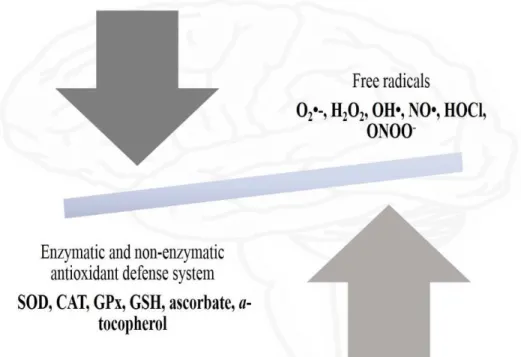
OXIDATIVE STRESS IN BRAIN
The chemical substances that have one or more unpaired
electrons are considered free radicals, which have as
main characteristic that facility to donate their electrons to
other molecules causing chain reactions and oxidative
damage (Liochev, 2013). Free radicals and related
molecules are classified as reactive oxygen species
(ROS) and reactive nitrogen species (RNS) and a wide
variety of these radicals are produced during normal
metabolism in biological systems, which are counter
balanced by cellular antioxidant mechanisms. However,
the imbalance by excessive ROS and RNS and
decreased antioxidant defense systems at cellular level
cause oxidative stress (Figure 1), which can be induced
to damage by peroxidation of cellular structures, protein
oxidation, DNA damage and inhibition of electron
transport chain in mitochondria (Dasuri et al., 2013).
ROS are represented by a group of chemicals that
include the superoxide anion (O
2•-), hydroxyl (HO
•),
peroxyl (RO
2•), hydroperoxyl (HO
2•), lipid hydroperoxide
(LOOH), hypochlorous acid (HClO), singlet oxygen (O
2)
and hydrogen peroxide (H
2O
2). RNS produced in cells
are represented by nitric oxide (NO) and peroxynitrite
(ONOO
•) (Roberts et al., 2010; Koskenkorva-Frank et al.,
2013). The superoxide anion, hydroxyl and hydrogen
peroxide are the main ROS. Among these, the
superoxide anion is formed due to reduction of molecular
oxygen by various enzymatic systems (enzymes in the
electron transport chain in mitochondria, xanthine
oxidase, cyclooxygenase and NADPH-oxidase). In turn,
the enzyme superoxide dismutase (SOD) converts the
superoxide anion into molecules of hydrogen peroxide.
Hydrogen peroxide in other reactions, in the presence of
transition metals (iron and copper) is converted into a
very reactive ROS, the hydroxyl radical (Milenkovic et al.,
2013; Koskenkorva-Frank et al., 2013). One of the main
mechanisms responsible by production of ROS in cells of
the brain tissue is illustrated in Figure 2.
Several studies have considered that oxidative damage
to various organs of a given organism is involved in the
onset of various diseases. In this context, the brain is an
organ highly vulnerable to oxidative stress due to its high
metabolic rate, which accounts for 20% of oxygen
consumption (Sultana et al., 2013). In addition, the brain
has low capacity for cellular regeneration, small amount
of antioxidant enzymes (catalase, superoxide dismutase,
glutathione peroxidase and glutathione reductase) and
antioxidant non-enzymatic (reduced glutathione,
-tocopherol and ascorbic acid) and as in any other tissue,
oxidative stress in brain can damage the neurons by
peroxidation of poly-unsaturated fatty acids (arachidonic
acid and docosahexaenoic acid) in cell membrane,
oxidative damage to DNA, RNA, proteins and induction of
apoptosis (Figure 3) (Nunomura et al., 2012; Smith et al.,
2013).
Peroxidation of the cell membrane has as consequence
the increase of stiffness, decreased activity of membrane
bound enzymes, deficiency of membrane receptors and
alters the permeability. ROS and RNS can also attack
directly the proteins of the lipid membrane and cause
fragmentation of proteins, cross-links between proteins
and the formation of carbonyl, which contributes to the
integrity of the cell membrane and interferes with various
cellular processes. ROS and RNS can still attack the
nucleic acids causing the crosslinking of the protein-DNA,
modification of DNA bases, breakage and mutations in
the DNA chain (Sultana et al., 2013).
OXIDATIVE STRESS AND NEURODEGENERATIVE
DISEASES
Oxidative stress and Alzheimer's diseases
Alzheimer's disease (AD) is clinically characterized by
progressive memory loss and severe cognitive function
decline. The accumulation of extracellular amyloid protein
deposited senile plaques and intracellular neurofibrillary
tangles made of abnormal and hyper-phosphorylated tau
protein, regionalized neuronal death, loss of synaptic
connections in selective brain regions, proliferation of
astrocytes and microglia activation, and define
neuro-pathologically the AD. In addition, the oxidative stress
and mitochondrial dysfunction are related to the
development of AD (Gubandru et al., 2013).
Mitochondria are responsible for ATP production,
oxidative phosphorylation, cell cycle control, cell growth
and apoptosis (Chaturvedi and Beal, 2013). Thus, a
problem in energy metabolism contributes to reduced
production of ATP, beyond the generation of excessive
ROS, such as superoxide, hydroxyl radicals and
hydrogen peroxide. The literature describes the
mitochondrial damage induced by ROS and vascular
hyperfusion as key initiators for the development of AD
(Butterfield et al., 2006; Aliev et al., 2010; Massaad et al.,
2009; Parihar and Brewer, 2007; Kovacic and
Somanathan, 2012; Schrag et al., 2013).
Figure 1. Representation of oxidative stress.
688 Afr. J. Pharm. Pharmacol.
Figure 3. Oxidative damage caused by ROS and RNS to various biomolecules.
are stable and used as markers of ROS. Moreover,
exogenous antioxidants and antioxidant enzymes can
indirectly measure the levels of ROS. In that sense, in
lipid peroxidation, significant levels of reactive aldehydes
may
be
evidenced,
including
4-hydroxynonal,
malondialdehyde (MDA), and 2-propenal (acrolein) and
are chemically and metabolically stable iso-prostanoids
including F2-isoprostanes and F4-neuroprostanes (Wang
et al., 2013a; Gubandru et al., 2013; Sultana et al., 2013).
Several investigations show that in the oxidation of
proteins, the most investigated markers are carbonyls
and 3-nitrotyrosine, final product of the interaction of
peroxy nitrite with tyrosine residues (Aksenov et al.,
2001; Castegna et al., 2002; Good et al., 1996; Tohgi et
al., 1999; Butterfield et al., 2001; Castegna et al., 2003;
Reed et al., 2009).
In the oxidation of the genetic material (DNA/RNA), the
hydroxyl
deoxyguanosine
8-(8-OHdG)
and
is growing demand for a treatment capable of protecting
neurons and inhibiting oxidative damage, may be an
effective alternative evidenced in natural antioxidants
(Bonda et al., 2010).
Oxidative stress and Parkinson's diseases
Parkinson's disease (PD) is a chronic neurological
progressive disease and associated with a loss of
dopaminergic neurons in the compact part of the
substantia nigra (SN), clinically characterized by cardinal
symptoms, resting tremor, rigidity, brady kinesia and
postural instability. In addition to dopamine depletion, PD
is neuropathologically defined by the deposition of Lewis
bodies and Lewis neurites in vulnerable neurons. The
etio-pathogenesis of PD is still not fully understood. In
most cases the disease is eventual: a multifactorial
idiopathic disease, seems to arise from a combination of
genetic susceptibility and environmental exposures
(Perfeito et al., 2013; Dickson et al., 2009; Halliday et al.,
2011; Hauser and Hastings, 2013).
Some environmental toxins are involved in the process
of oxidative stress, including paraquat, rotetone, maneb
and
MPTP
(1-methyl-4-phenyl-1,2,3,6-tetrahydropyridine). The MPTP, for example, crosses the
blood-brain barrier due to their lipophilicity and is oxidized
to
a
toxic
substance,
MPP+
(1-methyl-4-phenylpyridinium) by monoamine oxidase in glial cells.
MPP+ is accumulated in mitochondria of dopaminergic
neurons. The MPP+ inhibits mitochondrial complex I,
causing break in electron transport chain and as a result
the reduction in ATP production and increased production
of ROS (Subramaniam and Chesselet, 2013; Chen and
Le, 2006).
Moreover, evidence shows an association between
oxidative stress and mitochondrial DNA (mtDNA)
mutations with the onset of PD. Associated with constant
exposure to ROS, the lack of protection by histones, and
limited DNA repair mechanisms, make mtDNA
particu-larly vulnerable to oxidative damage, which can lead to
harmful mutations. As a result, a positive feedback is
generated by the accumulation of these mutations, which
can gradually cause a reduction in the efficiency of the
electron transport chain (ETC) stimulating decreased
ATP production and increased production of ROS,
followed by eventual cell death (Yan et al., 2013; Sanders
and Greenamyre, 2013).
In addition, ROS can irreversibly damage protein, for
example, amino groups in side chains of specific amino
acids can be modified to carbonyl groups (Greenamyre
and Sanders, 2013). It recently shows an association
be-tween elevated levels of protein carbonyls and increased
oxidative stress (Hyun et al., 2002). Another study
showed damage of mitochondrial complex I subunit in PD
brains related to high levels of protein carbonyl (Keeney
et al., 2006).
Similarly, the oxidized lipids can exercise deleterious
effects on neuronal function and lead to PD. In general,
lipids involved in membrane fluidity and permeability, can
store energy that participate in inflammatory processes
and in apoptosis signaling. The poly-unsaturated fatty
acids are more prone to lipid peroxidation, particularly in
the brain there is a large amount of two of these, the
arachidonic acid and docosahexaenoic acid. After the
adipose tissue, the organ with the highest lipid content is
the brain and thus, it is very prone to lipid peroxidation
(Ruipérez et al., 2010; Bochkov et al., 2002; Roberts II
and Fessel, 2004; Chen et al., 2008). Evidence supports
an association between lipid peroxidation and PD (Hyun
et al., 2002; Hoepken et al., 2007; Lee et al., 2001).
Oxidative stress and Huntington's diseases
Huntington disease (HD) is a progressive
neuro-degenerative disorder characterized by presence of
emotional and movement disorders and dementia
(Ayala-Peña, 2013). HD has autosomal dominant transmission
with the mutant gene termed IT15, responsible for the
disease located on chromosome 4. The mutation
respon-sible by this disease consists in an excessive number of
abnormal tri-nucleotide cytosine-adenine-guanosine
(CAG). The mutant gene encodes a protein called
huntingtin, which interacts with several proteins that are
involved in transcription, cell signaling and intracellular
transport. As a result of repeated CAG, may be
polyglutamine expansion located near the N-terminus of
protein molecule. The expanded poly-glutamine chains
lead to fragmentation of protein, which tends to
accumu-late inside the neuron. The aggregation of protein
fragments causes changes in neuronal functioning and
possibly plays a role in neuronal death process (Goldberg
et al., 1994; DiMauro and Schon, 2008; Harjes and
Wanker, 2003; Cattaneo et al., 2005; Ha et al., 2012).
It is known that oxidative stress is mediated by
increased ROS, including superoxide, hydrogen peroxide
and hydroxyl radical. These ROS impair cell function by
degrading proteins, lipids and nucleic acids and
consequently, there is a vicious cycle of mitochondrial
oxidative damage in Huntington's diseases. In context of
mutant Huntington expression, the ROS overproduction
causes accumulation and exhaustion of mtDNA damage
and
reduced
mitochondrial
bioenergetics.
690 Afr. J. Pharm. Pharmacol.
Johri and Beal, 2012; Ayala-Peña, 2013). Furthermore,
oxidative modification of proteins (protein carbonyl) and
lipids (malondialdehyde and 4-hydroxynonenal) are also
increased in HD in the brain (Beal and Browne, 2006).
Relevant characteristics that support the relationship
between oxidative stress and HD, can be evidenced by
accumulation of several markers, such as lipofuscin
derived from peroxidation of unsaturated fatty acids,
3-nitrotyrosine acids (nitrated protein) as well as
8-hydroxy-2-deoxyguanosine (OH8dG) in mtDNA (Dhillon and
Fenech, 2013).
Oxidative stress and Schizophrenia
Schizophrenia is a chronic debilitating
neuro-degenerative disorder and is characterized by several
positive symptoms such as delusions, hallucinations,
disorganized speech and negative symptoms, including
deficits in cognitive and social capacity and blunted affect
(Yao and Keshavan, 2011). The initial pathophysiology of
schizophrenia collaborates to increase the generation of
reactive oxygen species in the brain (Bitanihirwe and
Woo, 2011). In addition, genetic evidence demonstrates
that decreased capacity of synthesizing antioxidant
enzymes, such as glutathione (GSH) under conditions of
oxidative stress favor the appearance of schizophrenia
(Gysin et al., 2007; From et al., 2009). As previously
discussed, excessive ROS cause damage to important
macromolecules such as DNA, proteins and lipids. It can
affect the genetic material by modifying gene expression,
protein oxidation, making them non-functional and
peroxidative damage in lipids, which results in damage to
cell membrane and cell organelles (Wu et al., 2013).
Additionally, dopamine, an auto-oxidizable
neuro-transmitter, has a dihydroquinone structure that can be
oxidized by molecular oxygen to form hydrogen peroxide
and
o
-quinone. Thus, the catecholamines oxidation
combined to deficient antioxidant system in the brain,
produces an excess of free radicals resulting in oxidative
stress (Bošković et al., 2011).
Oxidative stress and amyotrophic lateral sclerosis
Amyotrophic lateral sclerosis (ALS) is one of the major
neurodegenerative
disorders
regarding
the
neuromuscular junction. The disease is characterized by
selective degeneration of upper motor neurons
(corticospinal) and lower (spinal and bulbar), which
results in pathological mis-communication between nerve
and muscle (Pansarasa et al., 2013). The majority of ALS
cases are of unknown etiology (sporadic). However, the
identification of mutant genes that predispose to these
disorders has provided the means to better understand
its pathogenesis. It is known that this condition occurs in
a change mutant gene that encodes Cu/Zn superoxide
dismutase type 1 (SOD1), enzyme that removes free
radicals and protects cells (motor neurons) from oxidative
stress (Halter et al., 2010; Rosen et al., 1993). Studies
have shown that SOD1 in its mutant version can cause
mitochondrial dysfunction. The accumulation of SOD1
aggregates in membrane of mitochondria can cause
blockage of proteins import, excessive ROS production
and eventual cell death by apoptosis (Vijayvergiya et al.,
2005; Mattiazzi et al., 2002; Takeuchi et al., 2002).
NATURAL
ANTIOXIDANTS
WITH
NEUROTHERAPEUTIC POTENTIAL
Recently, natural antioxidants have gained great
importance in treatment or prevention of diseases such
as cancer, diabetes, cardiovascular and mainly
neuro-degenerative disorders. Scientific studies have already
developed and discussed in preceding paragraphs
demonstrate that the generation of ROS and RNS have
direct implications in neurodegenerative diseases such
as Alzheimer's, Parkinson's, amyotrophic lateral sclerosis
and Huntington's disease. Thus, there is great interest in
studies aiming to investigate the therapeutic potential of
natural antioxidants for preventing or controlling oxidative
stress in neurodegenerative diseases, since it is known
that brain tissue is vulnerable to oxidative damage by
having abundant lipid content and relative scarcity of
antioxidant enzymes when compared with other tissues
(Figure 4).
Antioxidants such as vitamins, phenolic compounds
and flavonoids have been extensively investigated as
potential therapeutic agents
in vitro
and
in vivo
for
prevention of neurodegenerative diseases (Ebrahimi and
Schluesener, 2012; Choi et al., 2012). Most of these
antioxidant compounds can be found in fruits, vegetables,
plants or synthesized, and the antioxidant mechanisms
can act by induction of gene expression of endogenous
antioxidant defense systems (activation of nuclear factor
kappa B, NF-kB), regulation of ROS and RNS, interaction
with oxidative pathways and the capacity to chelate metal
ions responsible for the formation of free radicals (Dajas,
2012) (Figure 5).
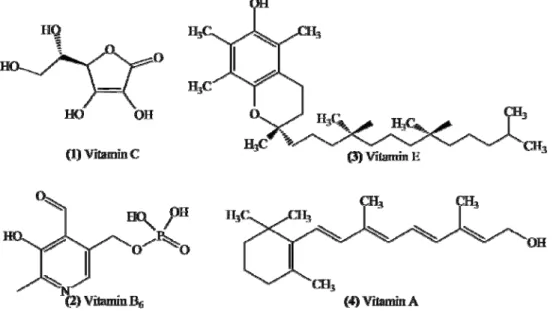
VITAMINS
Vitamins C, A, E and B
6shown in Figure 6, are
substances with high capacity to remove ROS and RNS
in vitro
and
in vivo
studies, and therefore can act in
Figure 4. Number of publications addressing antioxidants and neurodegenerative diseases, published between 2003 and 2013. Source: Pubmed and Science Direct.
Figure 5. Main antioxidant mechanisms against ROS/RNS.SOD (superoxide dismutase), CAT (catalase), GPx (glutathione peroxidase).
Vitamins C and E are natural compounds well known
for their high antioxidant capacity in
in vitro
and
in vivo
experimental models. Once there is an involvement of
ROS and RNS in neurodegenerative diseases, vitamins
C and E have been considered as potential therapeutic
agents for neurodegenerative diseases such as
Alzheimer's, Parkinson's, Huntington's and amyotrophic
lateral sclerosis (Heo et al., 2013). For example, Miyake
et al. (2011) investigated the relationship between intake
692 Afr. J. Pharm. Pharmacol.
Figure 6. Chemical structures of vitamins C, B6, E and A.
disease.
Even considering the pharmacological potential of
Vitamin C, there are conflicting scientific studies on its
potential in treatment of various neurodegenerative
diseases, including Alzheimer's disease. For example, in
the study of Arlt et al. (2012) involving the 12 patients
with Alzheimer's disease, was observed that
supplemen-tation with vitamin C did not have significant effect on the
prevention of Alzheimer's disease over 1 year. In another
study involving 57 patients with Alzheimer's disease and
treated with vitamin E, it has been observed that the
oxidative stress was not inhibited when it was considering
the oxidized glutathione levels (Lloret et al., 2009).
Vitamins addressed in Figure 6 are part of human diet
and are considered substances with high antioxidant
capacity. Thus, these antioxidant properties have
attrac-ted great attention for the treatment of neurodegenerative
diseases, but studies to date are contradictory regarding
the therapeutic potential for neurodegenerative diseases.
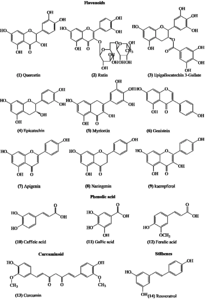
PHENOLIC COMPOUNDS
Phenolic compounds have one or more hydroxyl groups
bonded to a benzene ring, being chemical constituents
found in a wide variety of plants and which are
represented by a variety of classes of compounds that
can be divided according to their chemical structures.
Among these classes, there are phenolic acids
(hydroxybenzoic acids (C
6-C
1) and hydroxycinnamic
acids (C
6-C
3), flavonoids (C
6-C
3-C
6, which includes
antho-cyanins, flavonols, flavones, flavanones and isoflavones),
stilbenes (C
6-C
2-C
6), lignans (C
6-C
3-C
6-C
3) and
curcuminoids (C
3-C
6-C
3-C
1-C
6)) (Figure 7) (Cheynier et al.,
2013).
Antioxidant capacity of phenolic compounds occurs
primarily by elimination of free radicals, chelation capacity
and modulation of enzymes, as well as their effects on
cell signaling pathways and gene expression, being these
mechanisms dependent on the chemical characteristics
of the compounds (De Mello and Fasolo, 2014). Animal
studies, clinical and epidemiological, support the critical
role of phenolic compounds in the prevention and
treatment of various diseases such as neurodegenerative
disorders (Table 2).
Among the studies of phenolic compounds approached
in Figure 7, quercetin (flavonoid) has been shown to be
promising in treating various neurodegenerative diseases
(Jazvinšćak et al., 2012; Pandey et al., 2012; Denny
Joseph and Muralidhara, 2013). In a model of induced
Huntington's disease in rats, treatment with quercetin at a
dose of 25 mg/kg was able to reverse the inhibition of
mitochondrial electron transport chain, restore ATP
levels, prevent the mitochondrial dysfunction by inhibiting
oxidative stress and increase the SOD and CAT activities
(Sandhir and Mehrotra, 2013). In other
in vitro
and
in vivo
studies, quercetin has demonstrated a role in therapeutic
strategies to treatment of neurodegenerative diseases in
clinical settings to provide neuroprotection related to
suppression of oxidative stress, improvement in
behavioral function, reduction in infarct volume, cerebral
edema and cell injury. In contrast, some studies
contra-dict the neuroprotective potential of quercetin and other
flavonoids (Ossola et al., 2009; Huebbe et al., 2010).
694 Afr. J. Pharm. Pharmacol.
Table 1. Main vitamins with therapeutic potential for the treatment of neurodegenerative diseases.
Vitamin Neurodegenerative diseases Mechanisms involved References
1
Alzheimer's disease Parkinson's disease Huntington's disease Amyotrophic lateral sclerosis Schizophrenia
Elimination of ROS Chelating activity
Lipid peroxidation inhibition
Inhibition of amyloid-β (Aβ) aggregation
Ballaz et al. (2013), Heo et al. (2013), Engelhart et al. (2002), Kontush et al. (2001), Li et al. (2012), Quinn et al. (2003), Montilla-López et al. (2002), De Oliveira et al. (2012), Morales et al. (1989), Rebec et al. (2006), Heiser et al. (2010), Sivrioglu et al. (2007), Dadheech et al. (2006), Dakhale et al. (2005), and Arvindakshan et al. (2003)
2 Alzheimer's disease Elimination of ROS Chelating activity Hashim et al. (2011) and Nassiri-asl et al. (2012);
3
Alzheimer's disease Parkinson's disease Huntington's disease Schizophrenia
Elimination of ROS
Regulation of antioxidant enzymes Lipid peroxidation inhibition
Morrissey and Hill (2011), Ricciarelli et al. (2007), Ahmed (2012), Morris et al. (2005), Viña et al. (2004), Guan et al. (2012), Kontush and Schekatolina (2004); Kalonia et al. (2010), Peyser et al. (1995), Rao et al. (2003), Dadheech et al. (2006), Sivrioglu et al. (2007) and Arvindakshan et al. (2003)
4 Alzheimer's disease Parkinson's disease
Elimination of ROS/RNS Chelating activity
Inhibition of amyloid-β (Aβ) aggregation
Lee et al. (2009), Ono et al. (2004), Ono and Yamada (2012), Gackowski et al. (2008), Sutachan et al. (2012), King et al. (1992) and Rao et al. (2003)
Table 2. Phenolic compounds with therapeutic potential for the treatment of neurodegenerative diseases.
Phenolic compounds Neurodegenerative diseases Mechanisms involved References
1 Alzheimer's, Parkinson's, Huntington's and Schizophrenia
Elimination of ROS, chelating activity; Regulation of antioxidant enzymes; Lipid peroxidation inhibition; Inhibition of amyloid-β (Aβ) aggregation; Attenuation of deficits in motor coordination.
Liu et al. (2013), Sandhir and Mehrotra (2013), Zhu et al. (2013), Huebbe et al. (2010), Ansari et al. (2009), Lavoie et al. (2009), and Chakrabortyet al. (2013)
2 Alzheimer's and Parkinson's Elimination of ROS; Lipid peroxidation inhibition; Inhibition of amyloid-β (Aβ) aggregation Wang et al. (2012), Javed et al. (2012), Moshahid et al. (2012), and Islam et al. (2012)
3 Alzheimer's, Parkinson's, Huntington's and Amyotrophic lateral sclerosis
Elimination of ROS, Inhibition of amyloid-β (Aβ); Modulation of cell signaling pathways
Zhang et al. (2013), Lee et al. (2013), He et al. (2012), Levites et al. (2001), Kim et al. (2010b), Avramovich-Tirosh et al. (2007), Ehrnhoefer et al. (2006), Xu et al. (2006), and Koh et al. (2006)
4 Alzheimer's, Parkinson's, and Schizophrenia Elimination of ROS; Inhibition of amyloid-β (Aβ) aggregation; Inhibition of the aggregation of tau proteins; Lipid peroxidation inhibition.
George et al. (2013), Ejaz Ahmed et al. (2013), Teixeira et al. (2013), Dietrich-Muszalska et al. (2012), and Lim et al. (2013)
Table 2. Contd.
6 Alzheimer's, Parkinson's, and Amyotrophic lateral sclerosis Elimination of ROS; Inhibition of amyloid-β (Aβ) aggregation Bagheri et al. (2011), Baluchne et al. (2009), Liu et al. (2008), Trieu and Uckun (1999)
7 Alzheimer's Elimination of ROS, chelating activity; Regulation of antioxidant enzymes; Inhibition of amyloid-β (Aβ)
aggregation Zhao et al. (2013a, b)
8 Alzheimer's and Parkinson's Elimination of ROS; Lipid peroxidation inhibition; Inhibition of amyloid-β (Aβ) aggregation Heo et al. (2004), Khan et al. (2012), Ma et al. (2013), Zbarsky et al. (2005), and Sonia et al. (2013)
9 Alzheimer's, Parkinson's and Huntington's Elimination of ROS; Inhibition of amyloid-β (Aβ) aggregation Kim et al. (2010a), Sharoar et al. (2012), Filomeni et al., (2012), Li and Pu (2011), and Lagoa et al. (2009)
10 Alzheimer's, Parkinson's and Amyotrophic lateral sclerosis Elimination of ROS, chelating activity; Lipid peroxidation inhibition; Inhibition of amyloid-β (Aβ) aggregation Barros et al. (2013), Huang et al. (2013a), and Fontanilla et al. (2011)
11 Alzheimer's Elimination of ROS, chelating activity; Regulation of antioxidant enzymes; Inhibition of amyloid-β (Aβ)
aggregation Zhao et al. (2013a, b)
12 Alzheimer's Elimination of ROS; Inhibition of amyloid-β (Aβ) aggregation Mori et al. (2013) and Pi et al. (2012)
13 Alzheimer's, Parkinson's, Huntington's and Schizophrenia Elimination of ROS; chelating activity; Inhibition of amyloid-β (Aβ) aggregation
Wang et al. (2013b), Huang et al. (2013b), Li et al. (2013), Yu et al. (2013), Agrawal et al. (2012), Du et al. (2012), Mansouri et al. (2012), Sandhir et al. (2013), Hickey et al. (2012), Bishnoi et al. (2008) and Lavoie et al. (2009)
14 Alzheimer's, Parkinson's, Huntington's and Amyotrophic lateral sclerosis
Elimination of ROS; Inhibition of amyloid-β (Aβ)
aggregation; Regulation of antioxidant enzymes; Chelating activity, regulating of expression of SIRT1
Khan et al. (2010), Jin et al. (2008b), Feng et al. (2013), Porquet et al. (2013), Ho et al. (2010), Wang et al. (2011), and Kim et al. (2007)
by streptozotocin (STZ) (model used to study the
Alzheimer's disease) in rats and the results
showed that the gallic acid attenuated the
cognitive deficits, increased levels of enzymatic
antioxidants (GPx, CAT and SOD) and inhibited
lipid peroxidation by the reduction of the MDA
levels in brain tissue.
Another substance with great therapeutic
potential for treatment of neurodegenerative
diseases is resveratrol (Figure 7), a phenolic
non-flavonoid compound found in grapes and red
wine. This antioxidant compound has been
investigated in different
in vitro
and
in vivo
studies
to treatment of Parkinson's disease, Alzheimer's,
Huntington's and amyotrophic lateral sclerosis. In
a model of Parkinson's disease induced in rats, it
was observed that resveratrol (20 mg/kg) in
dopaminergic neurons showed neuroprotective
effect by regulating glutathione, glutathione
peroxidase, glutathione reductase, catalase,
superoxide dismutase and decreasing levels of
thiobarbituric acid reactive substances (TBARS).
Furthermore, it was observed decreased carbonyl
protein levels, phospholipase A2 activity and
improvement in content of dopamine (Khan et al.,
2010). Resveratrol also showed neuroprotective
effects in a cellular model of amyotrophic lateral
sclerosis and in a model of Alzheimer's disease in
mice, being observed in the hippocampus a
significant increase of neuronal survival (Kim et
al., de 2007).
696 Afr. J. Pharm. Pharmacol.
prevention of neurodegenerative diseases, the results of
Bournival et al. (2009) have clearly demonstrated that
resveratrol and quercetin are compounds that effectively
protect neurons against apoptotic cascade induced by
oxidative stress.
Most of the works discussed in this article are mainly
supported by data obtained in animal models and thus
more studies regarding the therapeutic potential for
human health are needed to unravel the role of these
antioxidant compounds, which are becoming increasingly
important as alternative or complementary therapies to
neurodegenerative diseases (Figure 4).
CONCLUSION
Based on the literature, there is good evidence that
oxidative stress plays an important role in the
pathogenesis of neurodegenerative diseases such as
Alzheimer's, Parkinson's, Huntington's, Schizophrenia
and amyotrophic lateral sclerosis. In this context, natural
antioxidant compounds with different chemical structures
that neutralize the damaging effects of ROS have been
investigated in preventing neurodegenerative diseases.
Most of the items discussed in this review demonstrated
the efficacy of antioxidant compounds in treatment of
neurodegenerative disorders using animal models or in
small clinical studies. However, some studies showed
contradictory results regarding the therapeutic potential of
antioxidants of natural origin. Thus, new studies
approa-ching clinical applications of antioxidants in the treatment
or prevention of neurodegenerative diseases are needed.
Conflict of interest
The author(s) declare(s) that they have no conflicts of
interest to disclose.
REFERENCES
Agrawal SS, Gullaiya S, Dubey V, Singh V, Kumar A, Nagar A, Tiwari P (2012). Neurodegenerative Shielding by Curcumin and Its Derivatives on Brain Lesions Induced by 6-OHDA Model of Parkinson's Disease in Albino Wistar Rats. Cardiovasc. Psychiatry. Neurol. 1-8.
Ahmed HH (2012). Modulatory effects of vitamin E, acetyl-l-carnitine and α-lipoic acid on new potential biomarkers for Alzheimer's disease in rat model. Exp. Toxicol. Pathol. 64:549-556.
Aksenov MY, Aksenova MV, Butterfield DA, Geddes JW, Markesbery WR (2001). Protein oxidation in the brain in Alzheimer's disease. Neuroscience 103:373-383.
Aliev G, Palacios HH, Gasimov E, Obrenovich ME, Morales L, Leszak J, Bragin V, Herrera AS, Gokhman D (2010). Oxidative stress induced mitochondrial failure and vascular hyperfusion as a key initiator for the development of Alzheimer’s disease. Pharmaceuticals 3:158-187. Ansari MA, Abdul HM, Joshi G, Opii WO, Butterfield DA (2009).
Protective effect of quercetin in primary neurons against Aβ(1-42): relevance to Alzheimer's disease. J. Nutr. Biochem. 20:269-275. Arlt S, Müller-Thomsen T, Beisiegel U, Kontush A (2012). Effect of
One-Year Vitamin C- and E-Supplementation on Cerebrospinal Fluid Oxidation Parameters and Clinical Course in Alzheimer’s Disease.
Neurochem. Res. 37:2706-2714.
Arvindakshan M, Ghate M, Ranjekar PK, Evans DR, Mahadik SP (2003). Supplementation with a combination of ω-3 fatty acids and antioxidants (vitamins E and C) improves the outcome of schizophrenia. Schizophr. Res. 62:195-204.
Ayala-Peña S (2013). Role of oxidative DNA damage in mitochondrial dysfunction and Huntington’s disease pathogenesis. Free. Rad. Biol. Med. 62:102-110.
Bagheri M, Joghataei MT, Mohseni S, Roghani M (2011). Genistein ameliorates learning and memory deficits in amyloid β(1-40) rat model of Alzheimer’s disease. Neurobiol. Learn. Mem. 95:270-276. Ballaz S, Morales I, Rodríguez M, Obeso JA (2013). Ascorbate prevents
cell death from prolonged exposure to glutamate in an in vitro model
of human dopaminergic neurons. J. Neurosci. Res. 91:1609-1617. Barros Silva R, Santos NAG, Martins NM, Ferreira DAS, Barbosa Jr F,
Oliveira Souza VC, Kinoshita Â, Baffa O, Del-Bel E, Santos AC (2013). Caffeic acid phenethyl ester protects against the dopaminergic neuronal loss induced by 6-hydroxydopamine in rats. Neuroscience 233:86-94.
Bishnoi M, Chopra K, Kulkarni SK (2008). Protective effect of Curcumin,
the active principle of turmeric (Curcuma longa) in
haloperidol-induced orofacial dyskinesia and associated behavioural, biochemical and neurochemical changes in rat brain. Pharmacol. Biochem. Behav. 88:511-522.
Bitanihirwe BKY, Woo TUW (2011). Oxidative stress in schizophrenia: An integrated approach. Neurosci. Biobehav. Rev. 35:878-893. Bochkov VN, Kadl A, Huber J, Gruber F, Binder BR, Leitinger N (2002).
Protective role of phospholipid oxidation products in endotoxin-induced tissue damage. Nature 419:77-81.
Bonda DJ, Wang X, Perry G, Nunomura A, Tabaton M, Zhu X, Smith MA (2010). Oxidative stress in Alzheimer disease: A possibility for prevention. Neuropharmacology 59:290-294.
Bošković M, Vovk T, Plesničar BK, Grabnar I (2011). Oxidative stress in schizophrenia. Curr. Neuropharmacol. 9:301-312.
Bournival J, Quessy P, Martinoli MG (2009). Protective Effects of Resveratrol and Quercetin Against MPP+ -Induced Oxidative Stress Act by Modulating Markers of Apoptotic Death in Dopaminergic Neurons. Cell. Mol. Neurobiol. 29:1169-1180.
Browne SE, Beal MF (2006). Oxidative damage in Huntington’s disease pathogenesis. Antioxid. Redox Signal 8:2061-2073.
Butterfield DA, Kanski J (2001). Brain protein oxidation in age-related neurodegenerative disorders that are associated with aggregated proteins. Mech. Ageing Dev. 122:945-962.
Butterfield DA, Perluigi M, Sultana R (2006). Oxidative stress in Alzheimer’s disease brain: new insights from redox proteomics. Eur. J. Pharmacol. 545:39-50.
Castegna A, Aksenov M, Thongboonkerd V, Klein JB, Pierce WM, Booze R, Markesbery WR, Butterfield DA (2002). Proteomic identification of oxidatively modified proteins in Alzheimer's disease brain. Part II: dihydropyrimidinase-related protein 2, alpha-enolase and heat shock cognate 71. J. Neurochem. 82:1524-1532.
Castegna A, Thongboonkerd V, Klein JB, Lynn B, Markesbery WR, Butterfield DA (2003). Proteomic identification of nitrated proteins in Alzheimer's disease brain. J. Neurochem. 85:1394-1401.
Cattaneo E, Zuccato C, Tartari M (2005). Normal Huntington function: an alternative approach to Huntington's disease. Nat. Rev. Neurosci. 6:919-30.
Chakraborty J, Singh R, Dutta D, Naskar A, Rajamma U, Mohanakumar KP (2013). Quercetin Improves Behavioral Deficiencies, Restores Astrocytes and Microglia, and Reduces Serotonin Metabolism in 3-Nitropropionic Acid-Induced Rat Model of Huntington's Disease.
CNS. Neurosci. 1-10.
Chaturvedi RK, Flint Beal M (2013). Mitochondrial Diseases of the Brain. Free. Radic. Biol. Med. 63:1-29.
Chen CT, Green JT, Orr SK, Bazinet RP (2008). Regulation of brain polyunsaturated fatty acid uptake and turnover. Prostaglandins Leukot. Essent. Fatty Acids 79:85-91.
Chen S, Le W (2006). Neuroprotective therapy in Parkinson disease. Am. J. Ther. 13:445-457.
Choi DY, Lee YJ, Hong JT, Lee, HJ (2012). Antioxidant properties of natural polyphenols and their therapeutic potentials for Alzheimer's disease. Brain. Res. Bull. 87:144-153.
Dadheech G, Mishra S, Gautam S, Sharma P (2006). Oxidative stress, α-tocopherol, ascorbic acid and reduced glutathione status in schizophrenics. Indian J. Clin. Biochem. 21:34-38.
Dajas F (2012). Life or death: Neuroprotective and anticancer effects of quercetin. J. Ethnopharmacol. 143:383-396.
Dakhale GN, Khanzode SD, Khanzode SS, Saoji A (2005). Supplementation of vitamin C with atypical antipsychotics reduces oxidative stress and improves the outcome of schizophrenia. Psychopharmacology 182:494-498.
Dasuri K, Zhang L, Keller JN (2013). Oxidative stress, neurodegeneration, and the balance of protein degradation and protein synthesis. Free. Radic. Biol. Med. 62:170-185.
De Mello AJM, Fasolo D (2014). Chapter 20 - Polyphenol Antioxidants from Natural Sources and Contribution to Health Promotion. In: Watson RR, Preedy VR, Zibadi S (eds) Polyphenols in Human Health and Disease. AcademicPress, San Diego. pp. 253-265.
De Oliveira BF, Veloso CA, Nogueira-Machado JA, de Moraes EN, dos Santos RR, Cintra MTG, Chaves MM (2012). Ascorbic acid, alpha-tocopherol, and beta-carotene reduce oxidative stress and proinflammatory cytokines in mononuclear cells of Alzheimer's disease patients. Nutr. Neurosci. 15:244-251.
De Toma AS, Choi J-S, Braymer JJ, Lim MH (2011). Myricetin: A Naturally Occurring Regulator of Metal-Induced Amyloid-β Aggregation and Neurotoxicity. Chem. BioChem. 12:1198-1201. Denny Joseph KM, Muralidhara (2013). Enhanced neuroprotective
effect of fish oil in combination with quercetin against 3nitropropionic acid induced oxidative stress in rat brain. Prog. Neuropsychopharmacol. Biol. Psychiatry 40:83-92.
Deslauriers J, Racine W, Sarret P, Grignon S (2014). Preventive effect of α-lipoic acid on prepulse inhibition deficits in a juvenile two-hit model of schizophrenia. Neuroscience 272:261-270.
Dhillon VS, Fenech M (2013). Mutations that affect mitochondrial functions and their association with neurodegenerative diseases. Mutat. Res-Rev. Mutat. 759:1-13.
Di Mauro S, Schon EA (2008). Mitochondrial disorders in the nervous system. Ann. Rev. Neurosci. 31:91-123.
Dickson DW, Braak H, Duda JE, Duyckaerts C, Gasser T, Halliday GM, Hardy J, Leverenz JB, Del Tredici K, Wszolek ZK, Litvan I (2009). Neuropathological assessment of Parkinson's disease: refining the diagnostic criteria. Lancet. Neurol. 8:1150-1157.
Dietrich-Muszalska A, Kontek B, Olas B, Rabe-Jabłońska J (2012). Epicatechin Inhibits Human Plasma Lipid Peroxidation Caused by
Haloperidol In Vitro. Neurochem. Res. 37:557-562.
Ding QX, Markesbery WR, Chen QW, Li F, Keller JN (2005). Ribosome dysfunction is an early event in Alzheimer's disease. J. Neurosci. 25:9171-9175.
Ebrahimi A, Schluesener H (2012). Natural polyphenols against neurodegenerative disorders: Potentials and pitfalls. Ageing. Res. Rev. 11:329-345.
Ehrnhoefer DE, Duennwald M, Markovic P, Wacker JL, Engemann S, Roark M, Legleiter J, Marsh JL, Thompson LM, Lindquist S, Muchowski PJ, Wanker EE (2006). Green tea (−)-epigallocatechin-gallate modulates early events in huntingtin misfolding and reduces toxicity in Huntington's disease models. Hum. Mol. Genet. 15:2743-2751.
Ejaz Ahmed M, Khan MM, Javed H, Vaibhav K, Khan A, Tabassum R, Ashafaq M, Islam F, Safhi MM, Islam F (2013). Amelioration of cognitive impairment and neurodegeneration by catechin hydrate in rat model of streptozotocin-induced experimental dementia of Alzheimer’s type. Neurochem. Int. 62:492-501.
Engelhart MJ, Geerlings MI, Ruitenberg A, Van Swieten JC, Hofman A, Witteman JCM, Breteler MMB (2002). Dietary intake of antioxidants and risk of Alzheimer disease. JAMA 287:3223-3229.
Feng X, Liang N, Zhu D, Gao Q, Peng L, Dong H, Yue Q, Liu H, Bao L, Zhang J, Hao J, Gao Y, Yu X, Sun J (2013). Resveratrol Inhibits β-Amyloid-Induced Neuronal Apoptosis through Regulation of SIRT1-ROCK1 Signaling Pathway. PLoS ONE. 8:1-11.
Filomeni G, Graziani I, De Zio D, Dini L, Centonze D, Rotilio G, Ciriolo MR (2012). Neuroprotection of kaempferol by autophagy in models of
rotenone-mediated acute toxicity: possible implications for Parkinson's disease. Neurobiol. Aging 33:767-785.
Fontanilla CV, Ma Z, Wei X, Klotsche J, Zhao L, Wisniowski P, Dodel RC, Farlow MR, Oertel WH, Du Y (2011). Caffeic acid phenethyl ester prevents 1-methyl-4-phenyl-1,2,3,6-tetrahydropyridine-induced neurodegeneration. Neuroscience. 188:135-141.
Foy CJ, Passmore AP, Vahidassr MD, Young IS, Lawson JT (1999). Plasma chain-breaking antioxidants in Alzheimer's disease, vascular dementia and Parkinson's disease. QJM 92:39-45.
Gackowski D, Rozalski R, Siomek A, Dziaman T, Nicpon K, Klimarczyk M, Araszkiewicz A, Olinski R (2008). Oxidative stress and oxidative DNA damage is characteristic for mixed Alzheimer disease/vascular dementia. J. Neurol. Sci. 266:57-62.
George RC, Lew J, Graves DJ (2013). Interaction of Cinnamaldehyde and Epicatechin with Tau: Implications of Beneficial Effects in Modulating Alzheimer's Disease Pathogenesis. J. Alzheimers Dis. 36:21-40.
Goldberg YP, Telenius H, Hayden MR (1994). The molecular genetics of Huntington’s disease. Curr. Opin. Neurol. 7:325-32.
Good PF, Werner P, Hsu A, Olanow CW, Perl DP (1996). Evidence of neuronal oxidative damage in Alzheimer's disease. Am. J. Pathol. 149:21-28.
Guan JZ, Guan WP, Maeda T, Makino N (2012). Effect of Vitamin E Administration on the Elevated Oxygen Stress and the Telomeric and Subtelomeric Status in Alzheimer’s Disease. Gerontology 58:62-69. Gubandru M, Margina D, Tsitsimpikou C, Goutzourelas N, Tsarouhas K,
Ilie M, Tsatsakis AM, Kouretas D (2013). Alzheimer’s disease treated patients showed different patterns for oxidative stress and inflammation markers. Food Chem. Toxicol. 61:209-214.
Gysin R, Kraftsik R, Sandell J, Bovet P, Chappuis C, Conus P, Deppen P, Preisig M, Ruiz V, Steullet P (2007). Impaired glutathione synthesis in schizophrenia: convergent genetic and functional evidence. Proc. Natl. Acad. Sci. 104:16621-16626.
Ha AD, Beck CA, Jankovic J (2012). Intermediate CAG repeats in Huntington's disease: analysis of COHORT. Tremor Other Hyperkinet. Mov. 2:1-7.
Halliday G, Lees A, Stern M (2011). Milestones in Parkinson's disease clinical and pathologic features. Mov. Disord. 26:1015-1021.
Halter B, Gonzalez de Aguilar J-L, Rene F, Petri S, Fricker B, Echaniz-Laguna A, Dupuis L, Larmet Y, Loeffler JP (2010). Oxidative stress in skeletal muscle stimulates early expression of Rad in a mouse model of amyotrophic lateral sclerosis. Free Radic. Biol. Med. 48:915-923. Hamaguchi T, Ono K, Murase A, Yamada M (2009). Phenolic
Compounds Prevent Alzheimer’s Pathology through Different Effects on the Amyloid-β Aggregation Pathway. Am. J. Pathol. 175:2557-2565.
Harjes P, Wanker EE (2003). The hunt for huntingtin function: interaction partners tell many different stories. Trends Biochem. Sci. 28:425-33.
Hashim A, Wang L, Juneja K, Ye Y, Zhao Y, Ming LJ (2011). Vitamin B6s inhibit oxidative stress caused by Alzheimer’s disease-related CuII-β-amyloid complexes-cooperative action of phospho-moiety. Bioorg. Med. Chem. Lett. 21:6430-6432.
Hauser DN, Hastings TG (2013). Mitochondrial dysfunction and oxidative stress in Parkinson's disease and monogenic Parkinsonism. Neurobiol. Dis. 51:35-42.
He M, Liu MY, Wang S, Tang QS, Yao WF, Zhao HS, Wei MJ (2012). Research on EGCG improving the degenerative changes of the brain in AD model mice induced with chemical drugs. Zhong Yao Cai. 35:1641-1644.
Heiser P, Sommer O, Schmidt A, Clement H, Hoinkes A, Hopt U, Schulz E, Krieg J, Dobschütz E (2010). Effects of antipsychotics and vitamin C on the formation of reactive oxygen species. J. Psychopharmacol. 24:1499-1504.
Heo HJ, Kim DO, Shin SC, Kim MJ, Kim BG, Shin DH (2004). Effect of Antioxidant Flavanone, Naringenin, from Citrus junos on Neuroprotection. J. Agric. Food Chem. 52:1520-1525.
Heo JH, Hyon-Lee, Lee KM (2013). The Possible Role of Antioxidant Vitamin C in Alzheimer’s Disease Treatment and Prevention. Am. J. Alzheimers. Dis. Other. Demen. 28:120-125.
698 Afr. J. Pharm. Pharmacol.
Improvement of neuropathology and transcriptional deficits in CAG 140 knock-in mice supports a beneficial effect of dietary curcumin in Huntington's disease. Mol. Neurodegen. 12:1-16.
Ho DJ, Calingasan NY, Wille E, Dumont M, Beal MF (2010). Resveratrol protects against peripheral deficits in a mouse model of Huntington's disease. Exp. Neurol. 225: 74-84.
Hoepken HH, Gispert S, Morales B, Wingerter O, Del Turco D, Mülsch A, Nussbaum RL, Müller K, Dröse S, Brandt U (2007). Mitochondrial dysfunction, peroxidation damage and changes in glutathione metabolism in PARK6. Neurobiol. Dis. 25:401-411.
Honda K, Smith MA, Zhu XW, Baus D, Merrick WC, Tartakoff AM, Hattier T, Harris PL, Siedlak SL, Fujioka H, Liu Q, Moreira PI, Miller FP, Nunomura A, Shimohama S, Perry G (2005). Ribosomal RNA in Alzheimer disease is oxidized by bound redox-active iron. J. Biol. Chem. 280:20978-20986.
Huang HC, Tang D, Xu K, Jiang ZF (2013b). Curcumin attenuates
amyloid-β-induced tau hyperphosphorylation in human
neuroblastoma SH-SY5Y cells involving PTEN/Akt/GSK-3β signaling pathway. J Recept. Signal. Transduct. Res. 1-12.
Huang Y, Jin M, Pi R, Zhang J, Chen M, Ouyang Y, Liu A, Chao X, Liu P, Liu J, Ramassamy C, Qin J (2013a). Protective effects of caffeic acid and caffeic acid phenethyl ester against acrolein-induced neurotoxicity in HT22 mouse hippocampal cells. Neurosci. Lett. 535:146-151.
Huebbe P, Wagner AE, Boesch-Saadatmandi C, Sellmer F, Wolffram S, Rimbach G (2010). Effect of dietary quercetin on brain quercetin levels and the expression of antioxidant and Alzheimer's disease relevant genes in mice. Pharmacol. Res. 61:242-246.
Hyun DH, Lee M, Hattori N, Kubo SI, Mizuno Y, Halliwell B, Jenner P (2002). Effect of wild-type or mutant Parkin on oxidative damage, nitric oxide, antioxidant defenses, and the proteasome. J. Biol. Chem. 277:28572-28577.
Islam F, Khan MM, Raza SS, Javed H, Ahmad A, Khan A, Safhi MM (2012). Rutin protects dopaminergic neurons from oxidative stress in an animal model of Parkinson's Disease. Neurotox. Res. 22:1-15. Jazvinšćak Jembrek M, Vuković L, Puhović J, Erhardt J, Oršolić N
(2012). Neuroprotective Effect of Quercetin Against Hydrogen Peroxide-induced Oxidative Injury in P19 Neurons. J. Mol. Neurosci. 47:286-299.
Jin F, Wu Q, Lu Y-F, Gong Q-H, Shi JS (2008b). Neuroprotective effect of resveratrol on 6-OHDA-induced Parkinson's disease in rats. Eur. J. Pharmacol. 600:78-82.
Jin H, Kanthasamy A, Ghosh A, Anantharam V, Kalyanaraman B, Kanthasamy AG (2013a). Mitochondria-targeted antioxidants for treatment of Parkinson's disease: Preclinical and clinical outcomes. Biochim. Biophys. Acta. 1842:1282-1294.
Johri A, Beal MF (2012). Antioxidants in Huntington's disease. Biochim. Biophys Acta. Dis. 1822:664-674.
Kalonia H, Kumar P, Kumar A (2010). Targeting oxidative stress attenuates malonic acid induced Huntington like behavioral and mitochondrial alterations in rats. Eur. J. Pharmacol. 634:46-52. Keeney PM, Xie J, Capaldi RA, Bennett JP (2006). Parkinson's disease
brain mitochondrial complex I has oxidatively damaged subunits and is functionally impaired and misassembled. J. Neurosci. 26:5256-5264.
Khan MB, Khan MM, Khan A, Ahmed ME, Ishrat T, Tabassum R, Vaibhav K, Ahmad A, Islam F (2012). Naringenin ameliorates Alzheimer’s disease (AD)-type neurodegeneration with cognitive impairment (AD-TNDCI) caused by the intracerebroventricular-streptozotocin in rat model. Neurochem. Int. 61:1081-1093.
Khan MM, Ahmad A, Ishrat T, Khan MB, Hoda MN, Khuwaja G, Raza SS, Khan A, Javed H, Vaibhav K, Islam F (2010). Resveratrol attenuates 6-hydroxydopamine-induced oxidative damage and dopamine depletion in rat model of Parkinson's disease. Brain. Res. 1328:139-151.
Kim D, Nguyen MD, Dobbin MM, Fischer A, Sananbenesi F, Rodgers JT, Delalle I, Baur JA, Sui G, Armour SM, Puigserver P, Sinclair DA, Tsai LH (2007). SIRT1 deacetylase protects against neurodegeneration in models for Alzheimer's disease and amyotrophic lateral sclerosis. EMBO J. 26: 3169-3179.
Kim J, Choi S, Cho H, Hwang H, Kim Y, Lim S, Kim C, Kim H, Peterson S, Shin D (2010a). Protective effects of kaempferol (3,4',5,7-
tetrahydroxyflavone) against amyloid beta peptide (Abeta)-induced neurotoxicity in ICR mice. Biosci. Biotechnol. Biochem. 74:397-401. Kim JS, Kim JM, O JJ, Jeon BS (2010b). Inhibition of inducible nitric
oxide synthase expression and cell death by (−)-epigallocatechin-3-gallate, a green tea catechin, in the 1-methyl-4-phenyl-1,2,3,6-tetrahydropyridine mouse model of Parkinson’s disease. J. Clin. Neurosci. 17:1165-1168.
Kim TS, Pae CU, Yoon SJ, Jang WY, Lee NJ, Kim JJ, Lee SJ, Lee C, Paik IH, Lee CU (2006). Decreased plasma antioxidants in patients with Alzheimer's disease. Int. J. Geriatr. Psychiatry 21:344-348. Koh SH, Lee SM, Kim HY, Lee K-Y, Lee YJ, Kim HT, Kim J, Kim M-H,
Hwang MS, Song C, Yang KW, Lee KW, Kim SH, Kim OH (2006). The effect of epigallocatechin gallate on suppressing disease progression of ALS model mice. Neurosci. Lett. 395:103-107. Kontush A, Mann U, Arlt S, Ujeyl A, Lührs C, Müller-Thomsen T,
Beisiegel U (2001). Influence of vitamin E and C supplementation on lipoprotein oxidation in patients with Alzheimer’s disease. Free. Radic. Biol. Med. 31:345-354.
Kontush A, Schekatolina S (2004). Vitamin E in Neurodegenerative Disorders: Alzheimer's Disease. Ann. NY Acad. Sci. 1031:249-262. Koskenkorva-Frank TS, Weiss G, Koppenol WH, Burckhardt S (2013).
The complex interplay of iron metabolism, reactive oxygen species, and reactive nitrogen species: Insights into the potential of various iron therapies to induce oxidative and nitrosative stress. Free. Radic. Biol. Med. 65:1174-1194.
Kovacic P, Somanathan R (2012). Redox Processes in Neurodegenerative Disease Involving Reactive Oxygen Species. Curr. Neuropharmacol. 10:289-302.
Lagoa R, Lopez-Sanchez C, Samhan-Arias AK, Gañan CM, Garcia-Martinez V, Gutierrez-Merino C (2009). Kaempferol protects against rat striatal degeneration induced by 3-nitropropionic acid. J. Neurochem. 111:473-487.
Lavoie S, Chen Y, Dalton TP, Gysin R, Cuénod M, Steullet P, Do KQ (2009). Curcumin, quercetin, and tBHQ modulate glutathione levels in astrocytes and neurons: importance of the glutamate cysteine ligase modifier subunit. J. Neurochem. 108:1410-1422.
Lee HP, Casadesus G, Zhu X, Lee HG, Perry G, Smith MA, Gustaw-Rothenberg K, Lerner A (2009). All-trans retinoic acid as a novel therapeutic strategy for Alzheimer's disease. Expert. Rev. Neurother. 9:1615-1621.
Lee M, Hyun DH, Jenner P, Halliwell B (2001). Effect of proteasome inhibition on cellular oxidative damage, antioxidant defences and nitric oxide production. J. Neurochem. 78:32-41.
Lee MY, Choi EJ, Lee M-K, Lee JJ (2013). Epigallocatechin gallate attenuates L-DOPA-induced apoptosis in rat PC12 cells. Nutr. Res. Pract. 7:249-255.
Levites Y, Weinreb O, Maor G, Youdim MBH, Mandel S (2001) Green tea polyphenol (-)-epigallocatechin-3-gallate prevents N-methyl-4-phenyl- 1,2,3,6-tetrahydropyridine-induced dopaminergic neurodege-neration. J. Neurochem. 78:1073-1082.
Li FJ, Shen L, Ji HF (2012). Dietary Intakes of Vitamin E, Vitamin C, and β-Carotene and Risk of Alzheimer's Disease: A Meta-Analysis. J. Alzheimers. Dis. 31:253-258.
Li J, Han B, Zhang X, Lou J, Shuboy A, Wang L, Yin H, Wang Y (2013). Curcumin as a Potential Treatment for Alzheimer's Disease: A Study of the Effects of Curcumin on Hippocampal Expression of Glial Fibrillary Acidic Protein. Am. J. Chin. Med. 41:59-70.
Li S, Pu XP (2011). Neuroprotective Effect of Kaempferol against a 1-Methyl-4-phenyl-1,2,3,6-tetrahydropyridine-Induced Mouse Model of Parkinson's Disease. Biol. Pharm. Bull. 34:1291-1296.
Li SY, Wang XB, Kong LY (2014). Design, synthesis and biological evaluation of imine resveratrol derivatives as multi-targeted agents against Alzheimer's disease. Eur. J. Med. Chem. 71:36-45.
Lim HJ, Shim SB, Jee SW, Lee SH, Lim CJ, Hong JT, Sheen YY, Hwang DY (2013). Green tea catechin leads to global improvement among Alzheimer's disease-related phenotypes in NSE/hAPP-C105 Tg mice. J. Nutr. Biochem. 24:1302-1313.
Liochev SI (2013). Reactive oxygen species and the free radical theory of aging. Free. Radic. Biol. Med. 60:1-4.
Liu R, Zhang TT, Zhou D, Bai XY, Zhou WL, Huang C, Song JK, Meng FR, Wu CX, LI L, Du GH (2013). Quercetin protects against the Aβ25–35-induced amnesic injury: Involvement of inactivation of RAGE-mediated pathway and conservation of the NVU. Neuropharmacology 67:419-431.
Lloret A, Badía MC, Mora NJ, Pallardó FV, Alonso MD, Viña J (2009). Vitamin E Paradox in Alzheimer's Disease: It Does Not Prevent Loss of Cognition and May Even Be Detrimental. J. Alzheimers. Dis. 17:143-149.
Ma J, Yang WQ, Zha H, Yu HR (2013). Effect of naringenin on learning and memory ability on model rats with Alzheimer disease. Zhong. Yao. Cai. 36:271-276.
Mansouri MT, Naghizadeh B, Ghorbanzadeh B, Farbood Y, Sarkaki A, Bavarsad K (2013). Gallic acid prevents memory deficits and oxidative stress induced by intracerebroventricular injection of streptozotocin in rats. Pharmacol. Biochem. Behav. 111:90-96. Mansouri Z, Sabetkasaei M, Moradi F, Masoudnia F, Ataie A (2012).
Curcumin has Neuroprotection Effect on Homocysteine Rat Model of Parkinson. J. Mol. Neurosci. 47:234-242.
Massaad CA, Pautler RG, Klann E (2009). Mitochondrial superoxide a key player in Alzheimer’s disease. Aging 9:758-761.
Mattiazzi M, D'Aurelio M, Gajewski CD, Martushova K, Kiaei M, Beal MF, Manfredi G (2002). Mutated human SOD1 causes dysfunction of oxidative phosphorylation in mitochondria of transgenic mice. J. Biol. Chem. 277:29626-29633.
Mecocci P, Macgarvey U, Beal MF (1994). Oxidative damage to mitochondrial-DNA is increased in Alzheimer's disease. Ann. Neurol. 36:747-751.
Milenkovic D, Jude B, Morand C (2013). miRNA as molecular target of polyphenols underlying their biological effects. Free. Radic. Biol. Med. 64:40-51.
Miyake Y, Fukushima W, Tanaka K, Sasaki S, Kiyohara C, Tsuboi Y, Yamada T, Oeda T, Miki T, Kawamura N, Sakae N, Fukuyama H, Hirota Y, Nagai M, Fukuoka Kinki Parkinson’s Disease Study G (2011). Dietary intake of antioxidant vitamins and risk of Parkinson’s disease: a case–control study in Japan. Eur. J. Neurol. 18:106-113. Mochel F, Haller RG (2011). Energy deficit in Huntington disease: why it
matters. J. Clin. Invest. 121:493-499.
Molina-Jiménez MaF, Sánchez-Reus MaI, Andres D, Cascales Ma, Benedi J (2004). Neuroprotective effect of fraxetin and myricetin against rotenone-induced apoptosis in neuroblastoma cells. Brain. Res. 1009:9-16.
Montilla-López P, Águeda MC, Feijóo López M, Muñoz-Castañeda JR, Bujalance-Arenas I, Túnez-Fiñana I (2002). Comparison of melatonin versus vitamin C on oxidative stress and antioxidant enzyme activity in Alzheimer's disease induced by okadaic acid in neuroblastoma cells. Eur. J. Pharmacol. 451:237-243. Morales LM, Estévez J, Suárez H, Villalobos R, Chacín de Bonilla L, Bonilla E (1989). Nutritional evaluation of Huntington disease patients. Am. J. Clin. Nutr. 50:145-150.
Mori T, Koyama N, Guillot-Sestier M-V, Tan J, Town T (2013). Ferulic Acid Is a Nutraceutical β-Secretase Modulator That Improves Behavioral Impairment and Alzheimer-like Pathology in Transgenic Mice. PLoS ONE. 8:1-16.
Morris MC, Evans DA, Tangney CC, Bienias JL, Wilson RS, Aggarwal NT, Scherr PA (2005). Relation of the tocopherol forms to incident Alzheimer disease and to cognitive change. Am. J. Clin. Nutr. 81:508-514.
Morrissey PA, Hill TR (2011). Vitamins|Vitamin E. In: Fuquay JW (ed) Encyclopedia of Dairy Sciences (Second Edition). Academic Press, San Diego. pp. 652-660.
Moshahid KM, Raza S, Javed H, Ahmad A, Khan A, Islam F, Safhi M, Islam F (2012). Rutin Protects Dopaminergic Neurons from Oxidative Stress in an Animal Model of Parkinson’s Disease. Neurotox. Res. 22:1-15.
Nassiri-Asl M, Sarookhani MR, Abbasi E, Zangivand AA, Shakiba E, Sedighi A, Rahbari M (2012). The effects of pre-treatment with vitamin B6 on memory retrieval in rats. Food. Chem. 132:1046-1048 Nunomura A, Moreira P, Castellani R, Lee H, Zhu X, Smith M, Perry G
(2012). Oxidative Damage to RNA in Aging and Neurodegenerative Disorders. Neurotox. Res. 22:231-248.
Nunomura A, Perry G, Pappolla MA, Wade R, Hirai K, Chiba S, Smith
MA (1999). RNA oxidation is a prominent feature of vulnerable neurons in Alzheimer's disease. J. Neurosci. 19:1959-1964.
Ono K, Yamada M (2012). Vitamin A and Alzheimer's disease. Geriatr.Gerontol. Int. 12:180-188.
Ossola B, Kääriäinen TM, Männistö PT (2009). The multiple faces of quercetin in neuroprotection. Expert. Opin. Drug Saf. 8:397-409. Pandey AK, Patnaik R, Muresanu DF, Sharma A, Sharma HS (2012).
Quercetin in Hypoxia-Induced Oxidative Stress: Novel Target for Neuroprotection. Int. Rev. Neurobiol. 102:107-146.
Pansarasa O, Rossi D, Berardinelli A, Cereda C (2013). Amyotrophic Lateral Sclerosis and Skeletal Muscle: An Update. Mol. Neurobiol. 49:984-990.
Parihar MS, Brewer GJ (2007). Microenergetic failure in Alzheimer’s disease. Am. J. Physiol. Cell. Physiol. 292:8-23.
Perfeito R, Cunha-Oliveira T, Rego AC (2013). Reprint of: Revisiting oxidative stress and mitochondrial dysfunction in the pathogenesis of Parkinson disease—resemblance to the effect of amphetamine drugs of abuse. Free. Radic. Biol. Med. 62:186-201.
Peyser CE, Folstein M, Chase GA, Starkstein S, Brandt J, Cockrell JR, Bylsma F, Coyle JT, McHugh PR, Folstein SE (1995). Trial of d-α-tocopherol in Huntington's disease. Am. J. Psychiatry 152:1771-1775. Pi R, Mao X, Chao X, Cheng Z, Liu M, Duan X, Ye M, Chen X, Mei Z, Liu P, Li W, Han Y (2012). Tacrine-6-Ferulic Acid, a Novel Multi-functional Dimer, Inhibits Amyloid-β-Mediated Alzheimer's
Disease-Associated Pathogenesis in vitro and in vivo. PLoS. ONE 7:1-8.
Porquet D, Casadesús G, Bayod S, Vicente A, Canudas A, Vilaplana J, Pelegrí C, Sanfeliu C, Camins A, Pallàs M, Valle J (2013). Dietary resveratrol prevents Alzheimer’s markers and increases life span in SAMP8. AGE 35:1851-1865.
Quinn J, Suh J, Moore MM, Kaye J, Frei B (2003). Antioxidants in Alzheimer's disease-vitamin C delivery to a demanding brain. J. Alzheimers. Dis. 5:309-313.
Rao A, Sudha K, Rao S (2003). Free radical toxicity and antioxidants in Parkinson’s disease. Neurology 51:60-62.
Rebec GV, Conroy SK, Barton SJ (2006). Hyperactive striatal neurons in symptomatic Huntington R6/2 mice: Variations with behavioral state and repeated ascorbate treatment. Neuroscience 137:327-336. Reed TT, Pierce JWM, Turner DM, Markesbery WR, Butterfield DA
(2009). Proteomic identification of nitrated brain prot0e0ins in early Alzheimer's disease inferior parietal lobule. J. Cell. Mol. Med. 13:2019-2029.
Ricciarelli R, Argellati F, Pronzato MA, Domenicotti C (2007). Vitamin E and neurodegenerative diseases. Mol. Aspects. Med. 28:591-606. Roberts LJ, Fessel JP (2004). The biochemistry of the isoprostane,
neuroprostane and isofuran pathways of lipid peroxidation. Chem. Phys. Lipids. 128:173-186.
Roberts RA, Smith RA, Safe S, Szabo C, Tjalkens RB, Robertson FM (2010). Toxicological and pathophysiological roles of reactive oxygen and nitrogen species. Toxicology. 276:85-94.
Rosen DR, Siddique T, Patterson D, Figlewicz DA, Sapp P, Hentati A, Donaldson D, Goto J, O'Regan JP, Deng HX (1993). Mutations in Cu/Zn superoxide dismutase gene are associated with familial amyotrophic lateral sclerosis. Nature 362:59-62.
Ruipérez V, Darios F, Davletov B (2010). Alpha-synuclein, lipids and Parkinson’s disease. Prog. Lipid Res. 49:420-428.
Sanders LH, Greenamyre JT (2013). Oxidative damage to macromolecules in human Parkinson disease and the rotenone model. Free Radic. Bio. Med. 62:111-120.
Sandhir R, Mehrotra A (2013). Quercetin supplementation is effective in improving mitochondrial dysfunctions induced by 3-nitropropionic acid: Implications in Huntington's disease. Biochim. Biophys. Acta. 1832:421-430.
Sandhir R, Yadav A, Mehrotra A, Sunkaria A, Singh A, Sharma S (2013). Curcumin Nanoparticles Attenuate Neurochemical and Neurobehavioral Deficits in Experimental Model of Huntington’s Disease. Neuromol. Med. 6:1-13.
Schrag M, Mueller C, Zabel M, Crofton A, Kirsch WM, Ghribi O, Squitti R, Perry G (2013). Oxidative stress in blood in Alzheimer's disease and mild cognitive impairment: A meta-analysis. Neurobiol. Dis. 59:100-110.