This Accepted Author Manuscript is copyrighted and published by Elsevier. It is posted here by agreement between Elsevier and University of Brasilia. Changes resulting from the publishing process - such as editing, corrections, structural formatting, and other quality control
mechanisms - may not be reflected in this version of the text. The definitive version of the text was subsequently published in [Tissue and Cell, Volume 25, Issue 3, June 1993, Pages 439–445, doi:10.1016/0040-8166(93)90084-X].You may download, copy and otherwise use the AAM for non-commercial purposes provided that your license is limited by the following restrictions: (1) You may use this AAM for non-commercial purposes only under the terms of the CC-BY-NC-ND license.
(2) The integrity of the work and identification of the author, copyright owner, and publisher must be preserved in any copy.
(3) You must attribute this AAM in the following format: [agreed attribution language, including link to CC BY-NC-ND license + Digital Object Identifier link to the published journal article on Elsevier’s ScienceDirect® platform].
________________________________________________________________________
Este Manuscrito do Autor Aceito para Publicação (AAM) é protegido por direitos autorais e publicado pela Elsevier. Ele esta disponível neste Repositório, por acordo entre a Elsevier e a Universidade de Brasília. As alterações decorrentes do processo de publicação - como a edição, correção, formatação estrutural, e outros mecanismos de controle de qualidade - não estão refletidas nesta versão do texto. A versão definitiva do texto foi posteriormente publicado em [Tissue and Cell, Volume 25, Número 3, Junho de 1993, Páginas 439–445, doi:10.1016/0040-8166(93)90084-X]. Você pode baixar, copiar e utilizar de outra forma o AAM para fins não comerciais , desde que sua licença seja limitada pelas seguintes restrições:
(1) Você pode usar este AAM para fins não comerciais apenas sob os termos da licença CC- BY- NC-ND.
(2) A integridade do trabalho e identificação do autor, detentor dos direitos autorais e editor deve ser preservado em qualquer cópia.
Nuclear changes during spermiogenesis in two chrysomelid beetles
Sônia N. Báo Clarice Hamú
Abstract
Ultrastructural and cytochemical studies were carried out on sperm nucleus of the beetles, Coelomera lanio and Diabrotica speciosa. Nuclear development involves changes in the shape and in the degree of chromatin condensation, with specific aggregation patterns of DNA-histone complex occurring during this process. Lamellar and paracrystalline arrangements of the nuclear material were observed in the Diabrotica speciosa spermatid and in the Coelomera lanio spermatozoon, respectively. Ethanolic-phosphotungstic acid technique suggest the presence of basic proteins in the nuclear material of spermatids. This reaction disappears during chromatin condensation. Chromatin condensation patterns may reflect specific intranuclear mechanisms and offers protection to the genome during spermatozoon transport to the oocyte.
Keywords: Chromatin condensation; cytochemistry; electron microscopy; nucleus; spermatozoon; chrysomelids
Introduction
The spermatozoon is a highly specialized cell which has many unique properties. The
main compartments of a typical insect spermatozoon are the head, containing nucleus and
acrosome, and the tail which contains axoneme and mitochondrial derivatives (for reviews, see
Phillips, 1970; Baccetti, 1972). Sperm nucleus development is characterized by the change of a
spherical to a highly asymmetric configuration and by chromatin conversion from a dispersed
to a very condensed state (Tokuyasu, 1974; Fawcett, 1971). It has also been shown that during
spermiogenesis the histones complexed to DNA are exchanged for specific arginine-rich basic
proteins, the protamines. These proteins are responsible for a high degree of condensation
chromatin in the nucleus (McMaster-Kaye and Kaye, 1976; Loir and Lanneau, 1978, 1984; Loir
and Courtens, 1979; Mello, 1987; Quagio-Grassiotto and Dolder, 1988). Condensation of the
sperm chromatin occurs in specific arrangement, which appear to be characteristic of the
differentiation stage and species (Cruz-Landim and Ferreira, 1976: Riess et al., 1978; Werner
and Bawa. 1988). In the present study we used electron microscopy and cytochemistry to
analyse the morphofunctional nuclear changes during spermiogenesis of two species of beetle.
The insects used were male adults of (‘orlomera lanio and Diabrotica speciosa collected in Brasilia (Brazil). Coelomera Zanio is a herbivore of Cecropia, and Diubrotiur
speciosa is an important leguminous pesl.
Transmission Electron Microscopy
Testes were dissected and fixed overnight at 4°C in a solution containing 4%
paraformaldehyde, 2% glutaraldehyde and Lic4 sucrose in 0.1 M cacodylate buffer, pH 7.3.
After fixation, the specimens were rinsed in buffer, and post-fixed in 1% osmium tetroxide,
followed by block-staining in 0.5% aqueous uranyl acetate. The material was dehydrated in
ethanol and embedded in Epon. After sectioning and staining with uranyl acetate and lead
citrate the sectmns were examined in a JEOL JEM 100 C Transmission Electron Microscope.
Cytochemistry
The technique of ethanolic-phosphotungstic acid (E-PTA), modified from Bloom and
Aghajanian (1968) was used for detection of basic proteins in spermatid and spermatozoon.
Testes were fixed with 3% glutaraldehyde in 0.1 M phosphate buffer, pH 7.4, for 5 hr at 4”C, dehydrated in an ethanol serie and treated for 24 hr at 4°C with 2% phosphotungstic acid in
absolute ethanol; then they were washed in absolute ethanol and embedded in Epon. Thin
sections were 441 stained with uranyl acetate and lead citrate or observed unstained.
Results and Discussion
The spermatids of Coelomera lanio and Diabrotica speciosa beetles undergo specific
morphofunctional modifications during spermiogenesis. The acrosome and flagellum
formation occurs simultaneously with the nuclear transformations, which change in shape and
in chromatin condensation degree. These events follow the general pattern described for
The alteration in the nucleus begin with the change from a spherical to a triangular
shape with depressions alongside, finally becoming elongate and lance-like. Numerous
alterations were also observed in the chromatin.
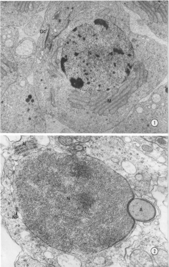
During the early spermatid phase, the nucleus resembles that of somatic cells with
electron-opaque chromatin (Fig. 1). Subsequently, there was a gradual condensation of this
chromatin, with an increase of its electron density (Fig. 2). As differentiation follows, the
nucleus begins to acquire a triangular configuration and chromatin fibrillar with a distinct
aspect were observed. In the nuclear material two relatively dense regions could be
distinguished: one near to the nuclear envelope, showing homogeneously condensed
chromatin attached to it, and the other in the nucleus central region, showing the chromatin
with a fibrillar aspect (Figs 3, 4). Structural changes of nuclear development have been
described for cricket (Cruz-Landim and Ferreira, 1976; Kierszenbaum and Tres, 1978), fruit-fly
(Tokuyasu, 1974; Quagio-Grassiotto and Dolder, 1988), and scorpion (Riess et al., 1977;
Werner and Bawa, 1988).
Our observations have demonstrated the peculiar organization of the nuclear material
during spermatid differentiation of Diabrotica speciosa. The fibrillar chromatin is organized in
the lamellae throughout the nucleus, from the periphery towards the center (Fig. 5). This
arrangement disappears in the mature sperm cell (Fig. 6), suggesting that chromatin
organization patterns may be characteristic of the spermatid differentiation stage. A
paracrystalline arrangement of nuclear material observed in spermatozoa of Coelomera lanio
(Fig. 7) has not been reported elsewhere. Previous studies carried out on cricket spermatids
have shown a sticklike chromatin arrangement (Cruz-Landim and Ferreira, 1976) while the
nucleus of scorpion spermatozoa present a lamellar arrangement of the chromatin (Riess et
al., 1977; Werner and Bawa, 1988), indicating the existence of a variety of chromatin
arrangements.
Electron-dense reaction product revealed by the ethanolic-phosphotungstic acid
technique (E-PTA) was observed in the nucleus of the spermatids, indicating the presence of
basic protein. The reaction occurs only in regions where chromatin condensation appears
incomplete (Figs 8, 10). This result is similar to those described in nucleus spermatids of the
cricket (Kierszenbaum and Tres, 1978) and fruit-fly (Quagio-Grassiotto and Dolder, 1988).
Sections stained with uranyl and lead staining showed an increase of chromatin contrast
during the process of condensation (Figs 9, 11). E-PTA reaction in the nuclear material reduced
with the chromatin condensation increase (Fig. 12). This may reflect the substitution of
histones for specific basic proteins in the chromatin, which permits a higher degree of
plates, localized in the implantation region of the flagellum, were also positive for basic protein
reaction.
During the beetles’ nuclear transformations, structures such as microtubules and
cytoplasmic membranes have been found surrounding the nucleus (Figs 3-7). This has been
similarly reported for several other insects (Kessel, 1966; Schrankel and Schwalm, 1974;
Tokuyasu, 1974). The presence of microtubules and cytoplasmic membranes in spermatids
with this precise orientation with respect to the nucleus, suggest that they may be involved
with nuclear elongation, since these structures disappear after this event is completed. The
different chromatin arrangement patterns may reflect specific intranuclear mechanisms. This
peculiar pattern of condensation observed in sperm chromatin may also offer sufficient
protection to the genome during its transportation to the oocyte.
Acknowledgements To Dr E. W. Kitajima of the Electron Microscope Laboratory of the
University of Brasilia for the use of the equipment and reagents. This work has been supported
References
Baccetti, B. 1972. Insect sperm cells. Adv. Insect Physiol., 9, 315-397. Baccetti, B. and Daccordi, M. 1988. Sperm structure and phylogeny of the
Chrysomelidae. In Biology of Chrysomelidae (eds. P. Jolivet, E. Petitpierre and T. H.
Hsiao), pp. 357-378. Academic Publishers, Kluwer.
Bloom, F. E. and Aghajanian, G. K. 1968. Fine structural and cytochemical analysis of
the staining of synaptic junctions with phosphotungstic acid. J. Ultrastruct. Res., 22,
361-371.
Burrini, A. G., Magnano, L., Magnano, A. R., Scala, C. and Baccetti, B. 1988.
Spermatozoa and phylogeny of Curculionoidea (Coleoptera). Int. J. Insect Morphol.
Embryol., 17, 1-50.
Cruz-Landim, C. and Ferreira, A. 1976. Aspectos ultraestruturais da diferenciação
nuclear durante a espermatogênese de Miogrillus sp (Orthoptera). Rev. Bras. Biol.,
36, 561-576.
Fawcett, D. W., Anderson, W. A. and Phillips, D. M. 1971. Morphogenetic factors
influencing the shape of the sperm head. Devel. Biol., 26, 220-251.
Kessel, R. G. 1966. The association between microtubules and nuclei during
spermiogenesis in the dragonfly. J. Ultrastruct. Res., 16, 293-304.
Kierszenbaum, A. L. and Tres, L. L. 1978. The packaging unit: a basic structural feature for the condensation of late cricket spermatid nuclei. J. Cell Sci., 33, 265-283.
Loir, M. and Courtens, J. L. 1979. Nuclear reorganization in ram spermatids. J.
Ultrastruct. Res.. 67, 309-324.
Loir, M. and Lanneau, M. 1978. Transformation of ram spermatid chromatin. Exp. Cell
Res., 115, 231-243.
Loir, M. and Lanneau, M. 1984. Structural function of the basic nuclear proteins in ram spermatids. J. Ultrastruct. Res., 86, 262-276.
McMaster-Kaye, R. and Kaye, J. S. 1976. Basic protein changes during the final stages of sperm maturation in the
house cricket. Exp. Cell Res., 97, 378-386.
Mello, M. L. S. 1987. Nuclear cytochemistry and polarization microscopy of the
spermatozoa of Triatoma infestans Klug. Z. Mikrosk. Anat. Forsch., 101, 245-250.
Phillips, D. M. 1970. Insect sperm: their structure and morphogenesis. J. Cell Biol., 44,
243-277. Quagio-Grassiotto, I. and Dolder, H. 1988. The basic nucleoprotein E-PTA
reaction during spermiogenesis of Ceratitis capitata (Diptera, Tephritidae). Cytobios,
53, 153-158.
Riess, R. W., Barker, K. R. and Biesele, J. J. 1978. Nuclear and chromosomal changes
during sperm formation in the scorpion, Centruroides vittatus (say). Caryologia, 31,
147-160.
Schrankel, K. R. and Schwalm, F. E. 1974. Structures associated with the nucleus during
chromatin condensation in Coelopa frigida (Diptera) spermiogenesis. Cell Tiss. Res..
153, 45-53.
Tokuyasu, K. T. 1974. Dynamics of spermiogenesis in Drosophila melanogaster IV.
Nuclear transformation. J. Ultrastruct. Res., 48, 284-303.
Werner. G. and Bawa, S. R. 1988. Spermatogenesis in the pseudoscorpion Diplotemnus


