This Accepted Author Manuscript is copyrighted and published by Elsevier. It is posted here by agreement between Elsevier and University of Brasilia. Changes resulting from the publishing process - such as editing, corrections, structural formatting, and other quality control
mechanisms - may not be reflected in this version of the text. The definitive version of the text was subsequently published in [Tissue and Cell, Volume 33, Issue 3, June 2001, Pages 286–293, doi:10.1054/tice.2001.0174].You may download, copy and otherwise use the AAM for non-commercial purposes provided that your license is limited by the following restrictions: (1) You may use this AAM for non-commercial purposes only under the terms of the CC-BY-NC-ND license.
(2) The integrity of the work and identification of the author, copyright owner, and publisher must be preserved in any copy.
(3) You must attribute this AAM in the following format: [agreed attribution language, including link to CC BY-NC-ND license + Digital Object Identifier link to the published journal article on Elsevier’s ScienceDirect® platform].
________________________________________________________________________
Este Manuscrito do Autor Aceito para Publicação (AAM) é protegido por direitos autorais e publicado pela Elsevier. Ele esta disponível neste Repositório, por acordo entre a Elsevier e a Universidade de Brasília. As alterações decorrentes do processo de publicação - como a edição, correção, formatação estrutural, e outros mecanismos de controle de qualidade - não estão refletidas nesta versão do texto. A versão definitiva do texto foi posteriormente publicado em [Tissue and Cell, Volume 33, Número 3, Junho de 2001, Páginas 286–293,
doi:10.1054/tice.2001.0174]. Você pode baixar, copiar e utilizar de outra forma o AAM para fins não comerciais , desde que sua licença seja limitada pelas seguintes restrições:
(1) Você pode usar este AAM para fins não comerciais apenas sob os termos da licença CC- BY- NC-ND.
(2) A integridade do trabalho e identificação do autor, detentor dos direitos autorais e editor deve ser preservado em qualquer cópia.
Nuclear changes and acrosome formation during spermiogenesis in
Euchistus heros (Hemiptera: Pentatomidae)
A.P. Fernandes G. Curia F.G.R. Francça S.N. Báo
Abstract
Ultrastructural and cytochemical studies were carried out on nuclear changes and acrosome formation during the spermiogenesis of the phytophagous bug Euchistus heros. The development of the nucleus involves changes in the shape and in degree of chromatin condensation: initially it is dispersed and with a low-electron density, then assumes a fibrillar arrangement and finally compacts in an electron-dense material. The acrosome is formed by the Golgi complex and presents unusual morphological features during its development. The reaction product of acid phosphatase, glucose-6-phosphatase and thiamine pyrophosphatase activities were detected during various stagesof acrosome development. In contrast, residues of α-N-acetylgalactosamine and basic proteinswere only reported in the intermediate and late stages of the differentiation process, respectively.
Keywords: bugs, insect, lectins, phosphatases, phytophagous, spermatozoon
Introduction The spermatozoon is a highly specialized cell which has many unusual
features. The main compartments of a typical insect spermatozoon consist of the head
(nucleus and acrosome) and the tail (axoneme and mitochondrial derivatives) (for reviews see
Phillips, 1970; Baccetti, 1972). Sperm nucleus development is characterized by the transition
from a spherical into a highly asymmetric configuration and by the chromatin conversion from
a dispersed to a very condensed state (Tokuyasu, 1974). The process of sperm chromatin
condensation occurs in a specific fashion which can be characteristic of both the
differentiation stage and species (Werner & Bawa, 1988).
The acrosome is essential for the recognition and penetration of the sperm within the
egg, leading to fertilization. This organelle is formed by the Golgi complex (Phillips, 1970;
Baccetti, 1972). The acrosome development begins with a spherical body, the proacrosomal
granule. This structure results from the fusion of vesicles produced by the Golgi complex, and
is gradually modified until it reaches its final shape. The size, shape and internal structure of
the mature acrosome are variable for the different animal species (Anderson & Personne,
1975).
The morphogenetic changes that occur during spermiogenesis involve the
participation of several enzymes (including phosphatases) and carbohydrate-rich molecules
(Yanagimachi, 1994). In recent published accounts on spermiogenesis, the detection of several
cytochemical and biochemical studies (Perotti & Riva, 1988; Báo et al., 1989; Báo & de Souza,
1992, 1994; Craveiro & Báo, 1995; Furtado & Báo, 1996; Cattaneo et al., 1997; Báo, 1997;
Fernandes & Báo, 1999; Pasini et al. 1999).
A cytochemical approach is useful to determine the functional role of the different
sperm elements in the movement and in the fertilization process, and particularly to detect
the role of enzymes and carbohydrate-rich molecules during the differentiation process of
these cells. We use ultrastructural and cytochemical techniques to analyze the morphological
changes of the nucleus and the acrosomal complex formation during the Euchistus heros
spermiogenesis. This insect is polyphagous, feeding on soybean, legumes, and on some species
of Solanaceae, Brassicaceae and Compositae (Panizzi, 1997), and is therefore considered a pest
of economically important crops throughout the world.
Materials and methods
The insects studied were adult male of the phytophagous bug E. heros(Hemiptera,
Pentatomidae), obtained from a laboratory colony reared at the National Center of Genetic
Resource (CENARGEN), Brasília, Brazil.
Transmission electron microscopy
Part of the material was fixed for 4 h at 48C in 2.5% glutaraldehyde, 4%
paraformaldehyde, 5 mM CaCl2 and 3% sucrose, in 0.1 M sodium cacodylate buffer, pH 7.3.
After fixation, the specimens were rinsed in the same buffer, and postfixed in 1% osmium
tetroxide, 0.8% potassium ferricyanide, and 5 mM CaCl2 in cacodylate buffer. The material was
dehydrated in a graded series of acetone (30±100%) and embedded in Spurr. Ultrathin
sections were stained with uranyl acetate and lead citrate. For the alcoholic phosphotungstic
acid method (E-PTA), the procedure used was that reported by Bloom and Aghajanian (1968).
Specimens were block stained with a solution of 3% PTA in absolute ethanol for 16 h at 48C.
The material was then embedded and sectioned as above described; their sections were
Enzyme cytochemistry
The testes were dissected and briefly fixed for 15 min at 48C in 1% glutaraldehyde
buffered with 0.1 M sodium cacodylate pH 7.2. After fixation, the specimens were washed
with buffer and incubated for 1 h at 378C in the following media:
1. Acid phosphatase activity: 0.1 M Tris-maleate buffer, pH 5.0, 7 mM cytidine-5-monophosphate, 2 mM cerium chloride and 5% sucrose (Pino et al., 1981);
2. Glucose-6-phosphatase activity: 5 mM glucose- 6-phosphate, 5 mM manganese chloride, 4 mM ceriumchloride,5%sucrose and0.1 MTris-maleate buffer, pH 6.5 (Robinson & Karnovsky, 1983).
3. Thiamine pyrophosphatase activity: 2.2 mM thiamine pyrophosphate, 5 mM manganese chloride, 4 mM cerium chloride, 5% sucrose and 0.1 M Trismaleate buffer, pH 7.2 (Angermuller & Fahimi, 1984). The controlsfor acid phosphatase, glucose- 6-phosphatase and thiamine pyro6-phosphatase activities were incubated in the same medium from which the speci®c substrates were omitted.
After incubation of the testes in one of the media as described above, the specimens were
washed with sodium cacodylate buffer and fixed again for 3 h at 48C in a solution containing
4% paraformaldehyde, 2% glutaraldehyde in 0.1 M sodium cacodylate buffer, pH 7.2. Then, the
specimens were washed in clean buffer, and postfixed in a solution containing 1% osmium
tetroxide, 0.8% potassium ferricyanide and 5 mM calcium chloride in 0.1 M sodium cacodylate
buffer. Subsequently, they were dehydrated in acetone and embedded in Spurr. Thin sections
were stained with uranyl acetate and lead citrate.
Carbohydrate detection
For lectin labeling, testes were fixed for 3 h at 48C in a solution of 4%
paraformaldehyde, 0.5% glutaraldehyde, 0.2% picric acid, 3.5% sucrose and 5 mM CaCl2 in 0.1
M sodium cacodylate buffer, pH 7.2. After several rinses in the same buffer, free aldehyde
groups were quenched with 50 mM ammonium chloride in 0.1 M sodium cacodylate buffer for
1 h, followed by block-staining in 2% uranyl acetate in 15% acetone for 2 h at 48C (Berryman &
Rodewald, 1990). Specimens were dehydrated in 30±90% acetone. Embedding was performed
in LRGold resin. Ultrathin sections were collected on nickel grids, pre-incubated in phosphate
buffered saline (PBS) containing 1.5% bovine serum albumin (PBS-BSA) and 0.01% Tween 20,
and subsequently incubated for 1 h at room temperature in the presence of Helix pomatia
agglutinin (HPA) gold-labeled in PBS-BSA pH 8.0 at a dilution of 1:10. After incubations the
and lead citrate. Controls consisted in the addition of 200±300 mM of the corresponding
monosaccharide to the incubation medium. The lectin used was obtained from Sigma Chemical
Company. The glycoprotein was labelled with colloidal gold particles (8±10 nm), according to
Roth (1983).
All observations were performed in a Jeol 100C transmission electron microscope.
Results The spermatids of Euchistus heros undergo specific morphofunctional modifications
during spermiogenesis. The acrosome and flagellum formation occurssimultaneously with the
nuclear transformations involving the shape and the degree of chromatin condensation.
Results The spermatids of Euchistus heros undergo specific morphofunctional
modifications during spermiogenesis. The acrosome and flagellum formation
occurssimultaneously with the nuclear transformations involving the shape and the degree of
chromatin condensation.
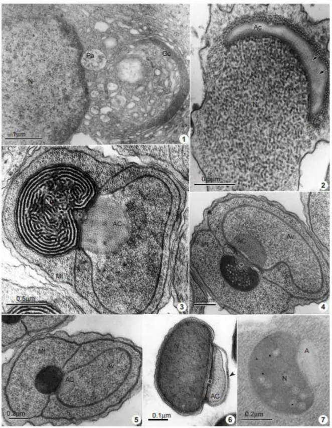
During the early spermatid stage, the nucleus resemblesthat ofsomatic cells and
presents electron dense areas of chromatin near the nuclear envelope (Fig. 1). Subsequently,
there is a gradual condensation of the nuclear chromatin with an increase in its electron
density, showing a granular aspect (Fig. 2). In the next stage, the nuclear chromatin shows a
fibrillar arrangement (Fig. 3), and then continues with its condensation process, that occurs
from the margin to the central portion of the nucleus. The chromatin may have a
paracrystalline aspect (Fig. 4) before becoming completely compact, homogeneous and
electron dense (Figs 5 & 6).
During the differentiation process, numerous microtubules can be observed
surrounding the nucleus (Figs 2±5). At the end of the spermatid elongation, the cytoplasmic
microtubules are eliminated (Fig. 6). Reaction products of the enzymatic activity as well as
basic proteins have not observed during the nuclear differentiation process. Nevertheless, few
a-N-acetylgalactosamine residues, showed by the gold-labeled HPA, appear in the nucleus
during late differentiation (Fig. 7).
The acrosome formation actively involves the Golgi complex. During the first stages of
spermiogenesis, numerous vesicles of the Golgi complex are observed (Fig. 1). These vesicles
join into a large proacrosomal vesicle (Fig. 1) which adheres to the nuclear envelope. At this
stage, the acid phosphatase activity is located at the level of the cisternae of the Golgi
complex, mainly on the cis and trans Golgi network; a diffuse weak reaction is also visible in
the proacrosomal granule (Fig. 8). The reaction product of glucose-6-phosphatase activity is
also observed in the Golgi complex, but only in the trans Golgi and trans Golgi network (Fig. 9).
Simultaneously with the chromatin condensation, and the nuclear and cellular
the anterior end of the nucleus. In early spermatids, the proacrosomal vesicle presents an
electron lucent cap appearance, surrounded by microtubules, and its content appears with a
tubular arrangement (Fig. 2). Posteriorly, it becomes a large three-layered structure, consisting
of: an electron dense inner cone which adheres to the nucleus; the acrosomal content, that
shows a tubular organization; and an outermost extra acrosomal vesicle, with a granular
aspect (Figs 3±5). The morphology of the acrosomal complex is maintained until the end of
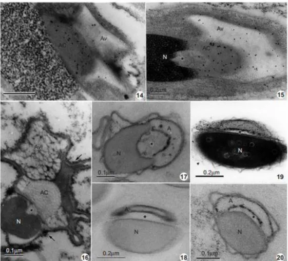
chromatin condensation. During this intermediate stage of development, acid phosphatase
activity is scattered in the acrosomal vesicle (Fig. 10), in the plasma membrane and in the
remnants of cytoplasm (Fig. 11). At the same stage, glucose-6-phosphatase activity is detected
scattered in the acrosomal vesicle (Fig. 12) and thiamine pyrophosphatase activity is present
on the membrane surrounding the acrosomal vesicle (Fig. 13). In sections from LRGold
embedded material, the presence of a-N-acetyl-galactosamine shown by the gold-labeled HPA
is initially evident in the acrosomal content, then it decreases progressively (Figs 14 & 15) and
disappears at the end of spermiogenesis (Fig. 7). The posterior domain of the acrosomal vesicle
regresses and the acrosomal complex appears more compact, assuming its ®nal shape. At a
later stage of spermiogenesis, acid phosphatase activity is detectable on the plasma
membrane in the acrosomal vesicle (Figs 16 & 19); a glucose- 6-phosphatase activitythat
appearsonlyonthe acrosomal membrane (Fig. 20). At this stage, a positive reaction of the
acrosomal membrane to the ethanolic phosphotungstic acid treatment is observed (Fig. 18).
Mature spermatozoa show a compact nucleus and two acrosomal regions: the inner one,
which appears electron lucent and without sugar residues or phosphatase activities, and the
outer one, whose membranes are positive to the glucose-6-phosphatase activity (Fig. 17).
The controls for enzymatic activities and detection of carbohydrate residues are
negative (not shown), demonstrating the specificity of the reactions.
Discussion
During spermiogenesis, the spermatids undergo specific morphofunctional
modifications which involve nuclear elongation, chromatin condensation, acrosomal formation
and flagellar development with axoneme and mitochondrial derivatives formation. Several
enzymes and glycoproteins may be involved in this remodeling, as well as in the chemical
changes that occur during this process.
Sperm nucleus development of E. heros is characterized by a transition from a
Heteropterans (Trandaburu, 1973; Itaya et al., 1980; Dolder, 1995; Fernandes & Báo, 1998).
The organization of the nuclear material during spermatid differentiation resembles that of
Acrosternum aseadum and Nezara viridula, previously reported by Fernandes and Báo (1998).
Although the E-PTA method has shown positive results in spermatids of beetles (Báo & Hamú,
1993) and fruit-fiies (Quagio-Grassiotto & Dolder, 1988). The presence of basic proteins has
not been reported during nucleus development of E. heros. Thus, other molecules other those
basic proteins participate in the chromatin condensation process in E. heros. Residues of
a-N-acetyl-galactosamine have been detected using gold-labeled HPA lectin on the late spermatid
nucleus of E. heros. Biochemical and cytochemical studies have demonstrated the presence of
sugar residues in intracellular compartments, mainly in the nucleus, associated with the dense
chromatin (Vannier-Santos et al., 1991; Báo & de Souza, 1992; Craveiro & Báo, 1995, Báo,
1997). Despite the fact that the role of nuclear glycoproteins is unclear, they seem to modulate
the physicochemical environment of the nucleoplasm and/
orparticipatedirectlyinlocalizedmolecularinteractionsat speci®c sites of the genome (Kan &
Figs 8 & 9 The reaction products of acid phosphatase and glucose-6-phosphatase activities, respectively, are associated with are Golgi, trans Golgi and trans Golgi network (arrowheads). Light reaction product of acid phosphatase activity (arrows) was observed on the proacrosomal granule (Pa). Nucleus (N); Golgi complex (Gc). X 32 000 and X 16 000, respectively. Figs 10 & 11 Localization of acid phosphatase. Note that the electron dense reaction product (arrows) in the intermediate stage of differentiation is present mainly on the acrosomal vesicle (Av), and scattered on the cytoplasm and plasma membrane. Acrosomal content (AC); inner cone (asterisks); nucleus (N). X 67 600; X 69 300, respectively.
Figs 14 & 15 Spermatids in intermediate stage of development showing an intense labelling by Helix pomatia agglutinin (HPA), mainly on the acrosomal content (AC) and on the acrosomal vesicle (Av). Inner cone (asterisk); nucleus (N). X 39 000 and X 52 000, respectively. Fig. 16 Localization of the acid phosphatase activity in the final stage of spermatid differentiation. The reaction product can be observed on the plasma membrane (arrows) and on the acrosomal vesicle (Av). Acrosomal content (AC); inner cone (asterisk); nucleus (N). X 112 000. Fig. 17 Localization of glucose-6-phosphatase activity. Note the presence of the reaction product on the acrosomal membrane (A). Inner cone (asterisk); nucleus (N). X 112 000. Figs 18±20 Detection of basic proteins by E-PTA, acid phosphatase and glucose-6-phosphatase activity, respectively, in the late spermatids. Note the presence of basic proteins and the enzymes on the acrosomal membrane (A). Nucleus (N); inner cone (asterisk). X 85 000; X 85 800 and X 145 200, respectively.
During the formation of the proacrosomal vesicles, the acid phosphatase and
glucose-6-phosphatase activities have been detected cytochemically in the Golgi complex, resembling
those in A. aseadum and N. viridula spermatids (Fernandes & Báo, 1999). On the contrary, in E.
heros, the acid phosphatase activity appears scattered in the proacrosomal vesicle. Since the
trans Golgi network is the site where proteins finally exit from the Golgi to reach their final
cellular sites (Griffiths & Simons, 1986; Grab et al., 1997), such as plasma membranes,
secretion granules and lysosomes, these enzymatic activities have been associated with the
Golgi complex. Indeed, these outcomes indicate an important role of these enzymes during
Despite the fact that the thiamine pyrophosphatase has been generally considered a
cytochemical marker for the trans side of the Golgi complex in many cell types (Cheetham et
al., 1971; Angermuller & Fahimi, 1984; Roth et al., 1985), this is true for E. heros, A. aseadum
and N. viridula spermatids (Fernandes & Báo, 1999), while it was not possible to detect this
enzyme in the Golgi complex.
The acrosome is a large secretory vesicle and carries a variety of hydrolytic enzymes,
stored in the form of proenzymes, as well as several proteases and glycosidases which are
essential for successful fertilization (for reviews see Yanagimachi, 1994). During the
intermediate stages of development, a significant concentration of phosphatases in the large
acrosomal vesicle of E. heros spermatids has been observed. The presence of thiamine
pyrophosphatase activity on the acrosomal vesicle membrane differs from the results obtained
from A. aseadum and N. viridula, where this enzyme was not detected in the acrosomal
components during initial and intermediate stages of differentiation (Fernandes & Báo, 1999).
These enzymes, at least in E. heros, seem to be involved in the remodelling and the enzymatic
condensation of the acrosome. This indicates that the acrosome has different responses to the
same enzyme during the differentiation process.
During early spermiogenesis, an intense presence of a-N-acetyl-galactosamine residues
on the acrosomal complex has been observed. Later on, these residues were not detected,
indicating that this carbohydrate is involved in the acrosome maturation but is not essential in
the final stages of spermiogenesis. Recent studies using gold-labeled lectins have shown that
glycoproteins of mosquito, blood-sucking bug, fruit-fly and beetle acrosomes contain different
specific sugars (Báo & de Souza, 1992; Perotti & Riva, 1988; Perotti & Pasini, 1995; Craveiro &
Báo, 1995) and that sugar residues are not uniformly distributed within the acrosome.
Furthermore, this distribution may be variable during the process of development. The
glycoconjugates are essential to provide specificity for the recognition and fusion of gametes
(Yanagimachi, 1994). Some previous cytochemical and biochemical investigations on the
spermatozoa of Drosophila suggest that the sperm plasma membrane can be characterized by
a-Man/ a-Glc residues concentrated in the acrosomal region (Perotti & Riva, 1988; Perotti &
Pasini, 1995; Cattaneo et al., 1997; Pasini et al., 1999). As these sugar residues could not be
found in the spermiogenetic process of E. heros, this indicates that other sugar residues may
be involved in acrosomal maturation and in the oocyte-recognition process.
In the last stages of differentiation, we observed both enzymatic and basic proteins on
the acrosomal membrane, but not in the acrosomal content. The internal tubular arrangement
of the acrosomal content of E. heros was observed both in the firststages of development and
water-strider Gerris (Tandler & Moriber, 1966; Werner & Werner, 1993), and the milkweed bug
Oncopeltus (Barker & Riess, 1966), A. aseadum and N. viridula (Fernandes & Báo, 1999). Such
paracrystalline material, however, is not present in the mature acrosome of Leptocoris (Itaya
et al., 1980) and Notonecta (Werner et al., 1988).
The elucidation of the enzymatic activities and the localization of carbohydrate
residues contributes to the enlightening of some particular aspects of spermiogenesis in E.
heros. Our results show that different species use different carbohydrate residues and
enzymes to control their own development, indicating that the presence and functional role of
carbohydrates and enzymes during the spermiogenetic process seems to be species-specific.
ACKNOWLEDGEMENTS We thank Dr M. Borges and Mr Hélio Moreira dos Santos for supplying
the insects. We thank Mrs Ruscaia Dias Teixeira for linguistic revision. This work was supported
by Coordenação o de Aperfeiçoamento de Pessoal de Nível Superior (CAPES) and Conselho
Nacional de Desenvolvimento Científico e Tecnológico (CNPq).
REFERENCES
Anderson, W.A. and Personne, P. 1975. The form and function of spermatozoa: a comparative view. In: Afzelius, B.A. (ed) The Functional Anatomy of the Spermatozoon, Pergamon Press, Oxford, pp 3-14.
Angermuller, S.L. and Fahimi, D.H. 1984. A new cerium-based method for cytochemical localization of thiaminepyrophosphatase in the Golgi complex of rat hepatocytes. Histochemistry, 80, 107-111.
Baccetti, B. 1972. Insect sperm cell. Adv. Insect Physiol., 9, 315-397.
Báo, S.N. 1997. Cytochemical localization of carbohydrate in the spermatid of Rhodnius prolixus (Hemiptera, reduviidae). Acta Microscopica, 6(1), 14-20.
Báo, S.N. and de Souza, W. 1992. Lectin binding sites on head structures of the spermatid and spermatozoon of the mosquito Culex quinquefasciatus (Diptera, Culicidae). Histochemistry, 98: 365-371.
Báo, S.N. and de Souza, W. 1994. Structural specialization in the flagellum of the spermatozoon of the bloodsucking bug (Rhodnius prolixux; Hemiptera, Reduviidae). Tissue & Cell, 26, 299-308.
Báo, S.N. and HamuÂ, C. 1993. Nuclear changes during spermiogenesis in two chrysomelid beetles. Tissue & Cell, 25(3), 439-445.
Báo, S.N., Quaggio-Grassiotto, I. and Dolder, H. 1989. Acrosome formation in Ceratitis capitata (Diptera, Tephritidae). Cytobios., 58, 93-100.
Berryman, M.A. and Rodewald, R.D. 1990. An enhanced method for post-embedding
immunocytochemical staining which preserves cell membranes. J. Histochem. Cytochem., 38, 159-170.
Bloom, F.E. and Aghajanian, G.K. 1968. Fine structural and cytochemical analysis of the staining of synaptic junctions with phosphotungstic acid. J. Ultrastruct. Res., 22, 361-371.
Cattaneo, F., Pasini, M.E. and Perotti, M.-E. 1997. Glycosidases are present on the surface of Drosophila melanogaster spermatozoa. Mol. Reprod. Devel., 48, 276-281.
Cheetham, R.D., MorreÂ, J.D., Pannek, C. and Friend, D.S. 1971. Isolation of a Golgi apparatus rich fraction from rat liver. IV. Thiamine pyrophosphatase. J. Cell Biol., 49, 899-905.
Craveiro, D. and Báo, S.N. 1995. Localization of carbohydrates in spermatids of three chrysomelidae beetles (Coleoptera, Chrysomelidae). Bio. Cell, 19(3), 195-202.
Dolder, H. 1995. Nuclear alterations during spermiogenesis of Triatoma infestans (Hemiptera, Reduviidae). In: Jamieson, B.G.M., Ausio, J. and Justine J.-L. (eds) Advances in Spermatozoal Phylogeny and Taxonomy. MeÂmoires du MuseÂum National D'Histore Naturelle, 166, 285-289.
Fernandes, A.P. and Báo, S.N. 1998. Spermiogenesis in phytophagous bug (Hemiptera, Pentatomidae): an ultrastructural study. J. Submicrosc. Cytol. Pathol., 30(4), 485-493.
Fernandes, A.P. and Báo, S.N. 1999. Ultrastructural localization of enzymatic activity during spermiogenesis in two phytophagous bugs (Hemiptera: Pentatomidae). Tissue & Cell, 31(3), 349-356.
Furtado, D.A. and Báo, S.N. 1996. Ultrastructural localization of phosphatases in sperm cells of the beetle Diabrotica speciosa (Coleoptera, Chrysomelidae). Cytobios., 88, 83-93.
Grab, D.J., Webster, P., Verjee, Y. and Lonsdale-Eccles, J. 1997.Golgi-associated
phosphohydrolases in Trypanosoma brucei brucei. Mol. Biochem. Parasitol., 86, 127-132.
Griffiths, G. and Simons, K. 1986. The trans Golgi network: sorting at exit site of the Golgi complex. Science, 234, 438-443. Itaya, P.W., Thompson, S.A. and Heidger, P.M. 1980. Fine structure of late stages of spermiogenesis in Leptocoris trivittata Say (Hemiptera: Corizidae). Inter. J. Insect Morphol. Embryol., 9, 135-145.
Kan, F.W.K. and Pinto da Silva, P. 1986. Preferential association of glycoproteins to the euchromatin regions of cross-fractured nuclei is revealed by fracture-label. J. Cell Biol, 102: 576-586.
Panizzi, A.R. 1997. Wild hosts pentatomids: ecological significance and role in their pest status on crops. Ann. Rev. Entomol., 42: 99-122.
Perotti, M.E. and Pasini, M.E. 1995. Glycoconjugates of the surface of spermatozoa of Drosophila melanogaster a qualitative and quantitative study. J. Exp. Zool., 271, 311-318.
Perotti, M.E. and Riva, A. 1988. Concanavalin A binding sites on the surface of Drosophila melanogaster sperm: a fluorescence and ultrastructural study. J. Ultrastruct. Res., 100, 173-182.
Phillips, D.M. 1970. Insect sperm: their structure and morphogenesis. J. Cell Biol., 44, 243-277.
Pino, R.M., Pino, L.C. and Bankston, P.W. 1981. The relationships between the Golgi apparatus, GERL, and lysosomes of fetal rat live Kupffer cells examined by ultrastructural phosphatase cytochemistry. J. Histochem. Cytochem., 29, 1061-1070.
Quaggio-Grassiotto, I. and Dolder, H. 1988. The basic nucleoprotein E-PTA reaction during spermiogenesis of Ceratitis capitata (Diptera, Tephritidae). Cytobios., 53, 153-158.
Robinson, J.M. and Karnovsky, M.J. 1983. Ultrastructural localization of several phosphatases with cerium. J. Histochem. Cytochem. 31, 1197-1208.
Roth, J. 1983. Application of lectin-gold complexes for electron microscopic localization of glycoconjugates on this sections. J. Histochem. Cytochem., 31, 987-999.
Roth, J., Taatjes, D.J., Lucocq, J.M., Weinstaein, J. and Paulson, J.C. 1985. Demonstration of an extensive trans-tubular network continuous with the Golgi apparatus stack that may function in glycosylation. Cell, 43, 287-295.
Tandler, B. and Moriber, L.G. 1966. Microtubular structures associated with the acrosome during spermiogenesis in the water-strider, Gerris remigis (Say). J. Ultrastruct. Res., 14, 381-404.
Trandaburu, V. 1973. Electron microscope observations on the spermiogenesis in Eurydema ventralis Kol. (Heteroptera-Pentatomidae). Anat. Anz. Bd., 133, 111-117.
Vannier-Santos, M.A., Saraiva, E.M.B. and de Souza, W. 1991. Nuclear and cytoplasmic lectin binding sites in promastigotes of Leishmania. J. Histochem. Cytochem., 39, 793-800.
Werner, G. and Bawa, S.R. 1988. Spermatogenesis in the pseudoscorpion Diplotemnus sp with reference to nuclear changes. J. Ultrastruct. Mol. Struct. Res., 98, 119-136.
Werner, G., Afzelius, B.A. and Mosler, B. 1988. Acrosome formation during spermiogenesis in Notonecta glauca L. (Heteroptera). J. Submicrosc. Cytol. Pathol., 20(1), 123-135.
Werner, G. and Werner, K. 1993. Changes in the nucleus, endoplasmic reticulum, Golgi apparatus, and acrosome during spermiogenesis in the water strider, Gerris najas Deg. (Heteroptera: Gerridae). Int. J. Insect Morphol. Embryol., 22(5), 521-534.