R E S E A R C H
Open Access
Feeding habits of mosquitoes (Diptera: Culicidae)
in an area of sylvatic transmission of yellow fever
in the state of São Paulo, Brazil
Luis Filipe Mucci
1*, Rubens Pinto Cardoso Júnior
2, Marcia Bicudo de Paula
3, Sirle Abdo Salloum Scandar
2,
Márcio Lunardeli Pacchioni
4, Aristides Fernandes
3and Cleide Aschenbrenner Consales
5Abstract
Background:The reintroduction of sylvatic yellow fever in the state of São Paulo after about six decades was confirmed in the Northwestern region in 2000, where in 2008 there also occurred an important epizootic. The purpose of this study was to investigate the feeding habits of culicids potentially involved in the sylvatic transmission of the virus in this region.
Methods:Specimens were collected in 24 forested localities at ground level with hand nets and mouth aspirators. Collections were made quarterly between October 2006 and July 2008 during daylight hours. Blood-meal identification was carried out in mosquitoes of the tribes Aedini, Mansoniini and Sabethini. The biotin/avidin sandwich ELISA was employed to determine six source types: bird, bovine, equine, rat, human and monkey.
Results:A total of 24,879 females of the three tribes were obtained, 245 (0.98%) of which were engorged. The presence of three different blood sources per engorged female was the predominant situation, and included 35.10% of the total of samples processed. Samples with two or four different sources were represented by 25.31% and 25.71%, of the specimens, respectively, while just 9.39% had only one type and 1.22%, five different sources. Aedes scapularis,Ae. serratus(Group),Psorophora albigenuandPs. feroxwere the most abundant species and accounted for about 95% of the engorged specimens. Of the principal vector species,Haemagogus janthinomys/
capricorniiwas found with bird, bovine and primate blood. These sources were predominant and alternated top
ranking as the most frequent source according to the mosquito species and collection site. In general, primate blood was the most prevalent source.
Conclusions:The human population of the region visits this ecotone frequently, which indicates the need for the periodical assessment of vaccination coverage against yellow fever. The frequency of non-human primate blood source in mosquito species that show minor vector importance in yellow fever virus transmission deserves attention. The eclectic feeding habits and some aspects of the interactions between potential vectors and reservoirs of yellow fever may be associated with the habitat fragmentation characteristic of the region. We recommend that further studies on the capacity and vector competence be performed on secondary vectors in extra-Amazonian region.
Keywords:Feeding habit, Sylvatic yellow fever, Neotropical mosquitoes, Non-human primate, Forest fragmentation
* Correspondence:lfmucci@gmail.com
1Laboratory of Biology and Ecology of Mosquitoes, Superintendence for
Endemic Disease Control, State Health Secretariat, Taubaté, São Paulo State, Brazil
Full list of author information is available at the end of the article
Background
The last 20 years have been characterized by the expan-sion of sylvatic transmisexpan-sion of yellow fever beyond the Amazonian biome in Brazil [1]. This situation represents a serious challenge for public health services, especially in relation to the improvement of surveillance systems, the defining of strategies for the immunization of the susceptible population and the adoption of preventive measures to avoid urban transmission [2-6].
Within this scenario, the hypotheses relating to the introduction and circulation of the virus have been called into question in view of the observation of the distinct as-pects of the localities affected in terms of vector fauna, isolation of viruses and the epidemiological or epizootic characteristics of the outbreaks. Generally speaking, non-human primates have always been considered the main reservoirs of the virus in the natural environment [1,7]. However, the participation of other mammals in the main-tenance of the virus and the influence of the double vector-reservoir role of the mosquitoes in the disease cycle are discussed [1,8,9]. Furthermore, it was recently sug-gested a potential mediation of human migrations in virus dispersion based on genetic divergence in Brazilian strains [10].
As to the vector importance of the Culicidae, Haema-gogus janthinomys and Hg. leucocelaenus are actually considered the main vectors of the yellow-fever virus in the country [1]. Other species such asHg. capricornii,Hg. albomaculatus, Hg. spegazzinii, Hg. tropicalis, Sabethes chloropterus, Sa. glaucodaemon, Sa. soperi, Sa. cyaneus, Sa. quasicyaneus, Ae. serratus, Aedes fulvus, Ae. scapu-laris, Psorophora albipesandPs. feroxhave generally been considered locally important or auxiliary vectors, and for some of these species, viral isolation was recorded only once [11-14].
Studying the Culicidae feeding habits has been proven fundamental in the elucidation of the yellow-fever epi-demiological chain [15-17]. The role of some of these vec-tors as a link in the interchange of viruses between humans and other primates has long since been demonstrated, but it calls for a re-investigation in order to update our know-ledge of regional“eco-epidemiologies”and expand research to cover other mosquito species and alternative feeding sources [18]. It is important to bear in mind that the dis-persal and availability of vertebrate hosts, including humans and domestic animals, have been undergoing significant changes by virtue of the increase in agricultural and cattle-raising activities and the degradation of forest environ-ments, which may be bringing adaptive pressure to bear on the subpopulations of these mosquitoes, raising the possi-bility of alterations related not only to the characteristics of their niches but also to their vector capacity [19].
In the present study we have sought to investigate the feeding habits of mosquitoes of the Aedini, Mansoniini
and Sabethini tribes collected in forest environments ad-jacent to the main rivers of the northwestern region of São Paulo. The state where the reintroduction of yellow fever was confirmed in 2000 after an absence of about six decades, and the location in 2008 of one of the great epizootic outbreaks recently registered in Brazil [5,20].
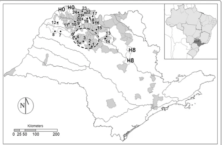
Coincidently, these epizootics were simultaneous with the period of the field collection, when there was in the region a series of communications on the deaths of monkeys (n = 242) with clinical and epidemiological confirmation (n = 65) and laboratory confirmation (n = 4) for yellow fever [21], as well as the occurrence of two hu-man cases in the municipalities of Luiz Antônio and São Carlos, toward the southeast (Figure 1).
Methods
A total of 24 collection sites were selected in the north-western region of the state of São Paulo, in 22 munici-palities spread over the river basins of the Tietê, São José dos Dourados and Turvo (Figure 1). The area covers the territory of 167 municipalities with a total population of about 3.3 million inhabitants, according to the IBGE cen-sus of 2010. The entire study area is included in the“area with vaccine recommendation for yellow fever” deter-mined by the Epidemiological Surveillance Center/Disease Control Coordination of the São Paulo State Health Secre-tariat. The estimated vaccine coverage for the period of 2003 to 2013 exhibits a predominance of subareas with rates between 60% and 90%, followed by those between 20% and 60%, and a few areas with rates over 90% [22].
The collections were undertaken within forests of sec-ondary succession characteristics, represented by frag-ments of gallery forest, forested savannah (Cerradão), semi-deciduous seasonal forests and phytophysiognomy of contact between these types [23]. Variations were ob-served in the herbaceous and bush strata, in the canopy height (between 8 and 25 meters), and in the size of the for-est fragments (from 6.50 to 2806.44 hectares) (Additional file 1). With regard to climate, except for the sites in the municipality of Riolândia which presented a humid sub-tropical climate (Cwa), the other sites were situated in the tropical savanna climate (Aw) in accordance with Köppen’s classification, both types having rainy sum-mers and dry winters [23].
to apprehend the specimens by approaching them with a hand-net catcher and mouth aspirator (Castro’s capturers). Individual protection equipment was used (a legionnaire’s cap, gloves and jacket) to avoid contact between the col-lectors and the mosquitoes, to ensure that the human blood found in the engorged females was not that of the collectors themselves. The specimens obtained were sacrificed in chloroform vapor and conditioned in ento-mological boxes each hour, when not engorged. The engorged females were placed individually in 1.5 mL cryotubes, preserved in the field in thermal boxes with ice and later conserved in a freezer at −20°C until they
could be identified and processed.
Identification was undertaken individually to the taxo-nomic species level, following Consoli and Lourenço-de-Oliveira [24], Forattini [25] and Lane [26] whereas the name abbreviations used were those of Reinert [27]. A cold table was used for the identification of the frozen ma-terial; and the engorged females belonging to the Aedini, Mansoniini and Sabethini tribes were selected for investi-gation of the blood source. In this study we consider that
Aedes serratusGroup includes females ofAe. serratusand
Ae. serratus/aenigmaticus.
The technique employed to determine the presence of ingested blood in the abdominal contents was the biotin/avidin sandwich enzyme-linked immunosorbent assay (ELISA) in accordance with the procedures used by Marassá et al. [17]. The six feeding sources tested were: bird, bovine, equine, human, monkey and rat. For counting the number of different blood sources, we consid-ered five major groups by Family level, grouping humans and monkeys, to enable comparison with results of other studies that do not distinguish these two types.
human (exclusively). Samples non-reactive to human IgG1 were considered to contain only monkey blood by the exclusion criterion, which indicates non-human pri-mates (NHP).
Results
A total of 25,395 mosquitoes of the Aedini, Mansoniini and Sabethini tribes were collected, 24,879 (97.97%) of which were female and 516 (2.03%) male, belonging to eight genera and 42 species, representing 45 taxa, including specimens identified to the subgenus level (Additional file 2). The females whose abdomens were distended with fresh blood, eggs, or mixed content numbered 358 (1.44%), 245 (0.98%) of which, with fresh blood (engorged), were selected and processed. Engorged females (samples) were obtained at 20 of the 24 sites investigated.
Of the principal vector species, only Hg. janthinomys/ capricorniiwas found engorged, despite having been less abundant than Hg. leucocelaenus (n = 147 × n = 346 fe-males). Of the 845 females of the genusSabethes, belong-ing to the followbelong-ing species: Sa. glaucodaemon(n = 690),
Sa. albiprivus(n = 100), Sa. gymnothorax(n = 27),Sa. tri-dentatus (n = 15), Sa. chloropterus (n = 9), Sa. undosus/ fabricii(n = 3) andSabethesspp.(n = 1) (Additional file 2), only two females of Sa. glaucodaemon presented a dis-tended abdomen, though none was engorged.
Aedes scapularis, Ae. serratus(Group),Ps. albigenuand
Ps. ferox were the most abundant species and accounted for about 95% of the engorged specimens. The rate of engorged females out of all females collected varied ac-cording to the collection sites for each species. For Ae. scapularisthe rate varied from 0.20 to 11.54%, forAe. ser-ratus(Group) from 0.49 to 5.36% (site 3 with 100% of the two specimens captured), for Ps. albigenu from 0.25 to 14.29%, forPs. feroxfrom 0.62 to 6.67% (Table 1) and for less abundant species from 0.14 to 2.27%, except for Ae. fulvus, which presented only one female collected and engorged (Additional file 3).
Of the 245 females engorged, eight did not react to any type of antibody used (3.27%). For the others, from one to five different blood sources were identified in each speci-men, with predominance of three (n = 86, 35.10%) and lesser frequencies of five sources (n = 3, 1.22%). Samples with two (n = 62) or four (n = 63) different sources repre-sented 25.31 and 25.71% of the total, respectively, and with only one blood source (n = 23), 9.39%. Among the most abundant species, the percentage for one source was of 11.92% forAe. scapularis, 7.37% forPs. albigenu, 5.00% for Ae. serratus (Group) and 11.11% for Ps. ferox. Only primates, NHP and bovine were identified in isolation. In the remaining samples 18 combinations of different types of blood sources were observed (Table 2).
Primate blood was in general predominant, being found in 94.29% of the samples (n = 231), with 64.50%
for primates (n = 158), 0.41% for humans exclusively (n = 1) and 29.39% for NHP exclusively (n = 72). Bird and bovine were practically equal, being present in 176 (71.84%) and 172 (70.20%) samples, respectively. Equine blood was observed in 77 specimens (31.43%) and rat blood, the least frequent source, in 16 females (6.53%) (Table 3).
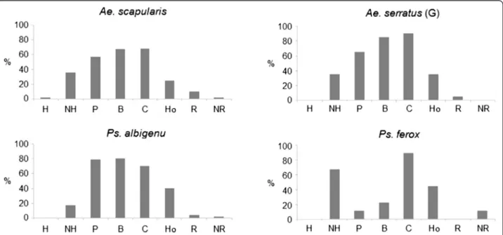
Among the most abundant species, Ps. albigenu pre-sented a larger number of multiple sources, proportion-ally, than did Ae. scapularis, the majority being of four different origins. The difference between primates and NHP was also greater in Ps. albigenu than inAe. scapu-laris, with a greater proportion of co-occurrence of hu-man and monkey for the first specie. Bird blood occurred with greater frequency in Ps. albigenu and Ae. serratus; bovine blood was proportionally most frequent inPs. ferox
and Ae. serratus. Psorophora ferox was also notable for presenting the greatest frequency of NHP blood and the lowest frequency of human blood among the more abundant species (Figure 2). The only specimen of Hg. janthinomys/capricorniipresented primate, bird and bo-vine blood, the most frequent combination among all the samples.
As to the sample distribution by hydrographic basin and collection site (Table 3), the greatest portion of them were obtained in the São José dos Dourados basin (n = 84) and at sites 19 to 24 of the Lower Turvo basin (n = 118). Sites with no samples occurred in the basins of the Tietê (4 and 5) and Middle and Upper Turvo (17 and 13, respectively).
In relation to the identification of sources, only rat blood was not found in samples from the Lower Turvo, which presented greater proportions of samples with equine (n = 51; 66.23%), bovine (n = 107; 62.21%), bird (n = 97; 52.11%) and human blood (n = 77; 48.43%); the frequency of source NHP was greater at these sites, though proportionally lower than the São José dos Dourados basin if the number of collection sites is taken into consideration (Table 3).
exclusively was registered at 12 of the 20 localities with samples (Table 3).
Discussion
The presence of engorged females is associated with microhabitats of resting places and digestion of blood, which do not necessarily coincide [28]. The feeding habit can be modified by geographical variations among dis-tinct subpopulations or by behavioral changes in the same subpopulation within one single reproductive sea-son [29]. Some Culicidae species bite one or more hosts repeatedly during each gonotrophic cycle, while others are less active [25]. There are cases in which this may also be observed within the same species under the influ-ence of meteorological variations, by infection with some parasite or even by the relative size of the specimens
[30-32]. In addition, the influence of the chosen types of collection methods must always be appreciated in the dis-cussion of results [33,34].
In this present study, the only collection technique used was applied at ground level, in forest fragments, the majority of which presented smaller areas compared to studies on fragmented landscapes [23]. The canopy heights of about 70% of these fragments attained less than fifteen meters (Additional file 1), being considered relatively low as compared to the heights established for the installation of arboreal platforms for collections in Amazonia, where the tops of the trees commonly reach 30 to 40 meters [35]. Nevertheless, some studies conducted in forest environments of the Savannah and Atlantic Forest biomes have shown more flexible behavior on the part of these mosquito species in their selection of the Table 1 Number of collected and engorged females by collection site and municipality
Municipality Collection site
Species
Ae. scapularis Ae. serratus(G) Ps. albigenu Ps. ferox Others
Col. Eng. %E/C Col. Eng. %E/C Col. Eng. %E/C Col. Eng. %E/C Col. Eng. %E/C
Novo Horizonte 1 1505 3 0.20 1 0 0.00 65 1 1.54 22 1 4.55 40 0 0.00
Sales 2 629 7 1.11 16 0 0.00 15 0 0.00 19 0 0.00 128 0 0.00
Ubarana 3 44 2 4.55 2 2 100 4 0 0.00 1 0 0.00 521 3 0.58
Zacarias 4 392 0 0.0 5 0 0.00 15 0 0.00 6 0 0.00 10 0 0.00
Barbosa 5 115 0 0.0 3 0 0.00 81 0 0.00 2 0 0.00 379 0 0.00
Buritama 6 226 3 1.33 3 0 0.00 12 0 0.00 5 0 0.00 5 0 0.00
Santo Antônio do Aracanguá
7 1125 10 0.89 0 0 0.00 1 0 0.00 1 0 0.00 11 0 0.00
Pereira Barreto 8 181 1 0.55 1 0 0.00 30 0 0.00 22 0 0.00 8 0 0.00
Sebastianópolis do Sul 9 828 19 2.29 59 0 0.00 705 24 3.40 58 0 0.00 12 0 0.00
Valentim Gentil 10 171 10 5.85 58 3 5.17 22 1 4.55 208 4 1.92 3 0 0.00
São João do Iracema 11 63 0 0.00 495 6 1.21 32 2 6.25 210 0 0.00 5 1 20.00
Marinópolis 12 674 8 1.19 1 0 0.00 14 2 14.29 5 0 0.00 527 4 0.76
Monte Azul Paulista 13 152 0 0.00 92 0 0.00 58 0 0.00 96 0 0.00 4 0 0.00
Paraíso 14 58 0 0.00 32 2 6.25 22 2 9.09 7 0 0.00 4 1 25.00
Guapiaçu 15 290 1 0.34 48 0 0.00 251 0 0.00 15 1 6.67 6 0 0.00
Nova Granada 16 50 0 0.00 128 0 0.00 235 1 0.43 41 0 0.00 115 2 1.74
Orindiuva 17 195 0 0.00 17 0 0.00 1494 0 0.00 9 0 0.00 540 0 0.00
Palestina 18 38 0 0.00 5 0 0.00 796 2 0.25 6 0 0.00 47 0 0.00
19 281 4 1.42 159 1 0.63 1338 18 1.35 141 0 0.00 142 0 0.00
Américo de Campos 20 412 3 0.73 144 1 0.69 244 6 2.46 118 1 0.85 15 1 6.67
Riolândia 21 52 6 11.54 538 3 0.56 596 19 3.19 279 0 0.00 70 0 0.00
22 73 2 2.74 163 4 2.45 292 12 4.11 101 1 0.99 22 0 0.00
Pontes Gestal 23 794 10 1.26 143 0 0.00 153 4 2.61 80 1 1.25 45 0 0.00
Cardoso 24 376 20 5.32 2 0 0.00 20 1 5.00 7 0 0.00 2 0 0.00
Total 8724 109 1.25 2085 22 1.06 6495 95 1.46 1459 9 0.62 2661 12 0.45
NHP: non-human primate; Col.: females collected, Eng.: females engorged and %E/C: percentage of females engorged out of all females collected.
Aedes serratus(G): Group includingAe. serratusandAe. serratus/aenigmaticus. Others corresponding to:Ae. fulvus,Ae. terrens,Coquillettidia venezuelensis,
heights of the tree strata at which they undertake their egg-laying and their feeding on hosts [16,36].
In view of this aspect, it was decided to apply the sam-pling effort to a horizontal rather than a vertical plane. According to the environmental characteristics of the study area, we considered that the collection technique whereby the collector moves around (allowing the gener-ation of a series of intrusion effects) results in the cover-age of a more varied gradient of microhabitats than the stationary technique on canopy platforms. Furthermore, distances of up to 1,100 meters, from the edge to the in-terior of the forests, were covered.
Despite this, no male specimens ofHg. janthinomysor
Hg. capricorniiwere obtained, which made it impossible
to determine which of the two species was more frequent in the area. In previous research undertaken in the same region, the collection of immature forms in the hollow spaces in trees in the municipality of Santa Albertina (SP, Brazil) permitted the identification of males ofHg. janthinomys, while Hg. capricornii was registered to-ward the southeast, in the municipality of Araraquara (SP, Brazil) [37,38].
It is worth observing that collection sites with the great-est relative abundances of Hg. janthinomys/capricornii
showed a proximity of about 25 kilometers (8 to 45 km) from the locations of the epizootic outbreaks ofAllouatta carayaconfirmed in 2008 [14,21]. At these sites, the forest fragments presented the greatest canopy heights (of about Table 2 Number of samples by combinations and numbers of types of blood source
Blood Source Species
Ae. scapularis Ae. serratus(G) Ps. albigenu Ps. ferox Others Total
n % n % n % n % n % n %
Primates 8 7.34 0 0.00 5 5.26 0 0.00 0 0.00 13 5.31
Primates + Bird 22 20.18 1 5.00 17 17.89 0 0.00 3 25.00 43 17.55
Primates + Bird + Cow 18 16.51 6 30.00 18 18.95 1 11.11 3 25.00 46 18.78
Primates + Bird + Horse 0 0.00 0 0.00 2 2.11 0 0.00 0 0.00 2 0.82
Primates + Cow + Horse 0 0.00 0 0.00 1 1.05 0 0.00 0 0.00 1 0.41
Primates + Bird + Cow + Horse 9 8.26 5 25.00 29 30.53 0 0.00 0 0.00 43 17.55
Primates + Bird + Cow + Rat 4 3.67 1 5.00 2 2.11 0 0.00 0 0.00 7 2.86
Primates + Cow + Horse + Rat 1 0.92 0 0.00 0 0.00 0 0.00 0 0.00 1 0.41
Primates + Bird + Cow + Horse + Rat 2 1.83 0 0.00 1 1.05 0 0.00 0 0.00 3 1.22
Human + Bird + Cow + Horse 1 0.92 0 0.00 0 0.00 0 0.00 0 0.00 1 0.41
NHP 2 1.83 1 5.00 2 2.11 0 0.00 1 8.33 6 2.45
NHP + Cow 7 6.42 2 10.00 5 5.26 2 22.22 1 8.33 17 6.94
NHP + Bird + Cow 12 11.01 2 10.00 4 4.21 0 0.00 0 0.00 18 7.35
NHP + Bird + Horse 1 0.92 0 0.00 0 0.00 0 0.00 0 0.00 1 0.41
NHP + Cow + Horse 12 11.01 0 0.00 2 2.11 3 33.33 0 0.00 17 6.94
NHP + Cow + Rat 0 0.00 0 0.00 1 1.05 0 0.00 0 0.00 1 0.41
NHP + Bird + Cow + Horse 1 0.92 2 10.00 3 3.16 1 11.11 1 8.33 8 3.27
NHP + Bird + Cow + Rat 4 3.67 0 0.00 0 0.00 0 0.00 0 0.00 4 1.63
Bird + Cow 0 0.00 0 0.00 1 1.05 0 0.00 0 0.00 1 0.41
Cow 3 2.75 0 0.00 0 0.00 1 11.11 0 0.00 4 1.63
Cow + Horse 1 0.92 0 0.00 0 0.00 0 0.00 0 0.00 1 0.41
1 source 13 11.93 1 5.00 7 7.37 1 11.11 1 8.33 23 9.39
2 sources 30 27.52 3 15.00 23 24.21 2 22.22 4 33.33 62 25.31
3 sources 43 39.45 8 40.00 28 29.47 4 44.44 3 25.00 86 35.10
4 sources 19 17.43 8 40.00 34 35.79 1 11.11 1 8.33 63 25.71
5 sources 2 1.83 0 0.00 1 1.05 0.00 0 0.00 3 1.22
Not reactive 2 1.83 0 0.00 2 2.11 1 11.11 8 66.67 8 3.27
Total 109 100 20 100 95 100 9 100 12 100 245 100
NHP: non-human primate.
Table 3 Number of samples by type of blood source, collection point and river basin Blood source River basin/Collection site
Tietê São José dos Dourados
1 2 3 4 5 6 7 8 Sub-T 9 10 11 12 Sub-T
Primate 4 7 7 0 0 1 9 0 28 33 2 6 5 46
Human 0 0 0 0 0 0 0 0 0 0 0 0 0 0
NHP 0 0 0 0 0 0 0 0 0 8 15 2 5 30
Bird 4 7 7 0 0 1 9 0 28 28 4 6 3 41
Cow 0 1 4 0 0 2 7 1 15 11 17 7 5 40
Horse 0 0 1 0 0 0 3 1 5 0 15 3 1 19
Rat 0 0 0 0 0 0 0 0 0 0 0 0 0 0
Not reactive 1 0 0 0 0 0 1 0 2 2 0 1 3 6
Number of Engorged♀ 5 7 7 0 0 3 10 1 33 43 18 9 14 84
Blood source River basin/Collection site Total
Turvo-Grande
13 14 15 16 17 18 19 20 21 22 23 24 Sub-T
Primate 0 3 1 2 0 2 18 9 22 13 11 3 84 158
Human 0 0 0 0 0 0 0 0 0 0 0 1 1 1
NHP 0 0 1 1 0 0 5 3 6 6 4 16 42 72
Bird 0 3 2 3 0 2 22 9 24 14 10 18 107 176
Cow 0 3 2 3 0 2 21 11 24 18 13 20 117 172
Horse 0 0 1 1 0 0 9 5 24 6 6 1 53 77
Rat 0 0 0 0 0 0 6 1 1 3 3 2 16 16
Not reactive 0 0 0 0 0 0 0 0 0 0 0 0 0 8
Number of Engorged♀ 0 3 2 3 0 2 23 12 28 19 15 21 128 245
NHP: non-human primate;♀= females.
25 m) and the largest areas of all the collection sites. In that setting the only engorged female ofHg. janthinomys/ capricorniifound was one of five specimens caught at site 20 and presented bird, primate and bovine blood, showing eclecticism as well as affinity for human blood, a fact also observed in relation toHg. janthinomys, but notHg. capri-cornii[15,16]. The forest fragment at that same location, one of the smallest, having an area of 12.1 hectares, was where Ae. scapularis, Ae. serratus/aenigmaticus and Ps. albigenuwere found with NHP blood (Additional file 1).
A synthesis of these results suggests that even though
Hg. janthinomys/capricornii is well-adapted to better preserved wild areas, it frequents the degraded, fragmen-ted environment of the region, maintaining its popula-tion on the basis of an original re-colonizapopula-tion of better preserved forests. This, together with the functional con-nectivity of these landscapes, reproduces dispersion sys-tems already reported for other animal groups [39].
Furthermore, the simultaneous finding of a female of
Hg. janthinomys/capricornii engorged with human and monkey bloods and of three other mosquito species with monkey blood in one of the smallest forest fragments indicates the possibility of the transmission of sylvatic yellow-fever in highly modified environments, a behavior also presented by ticks and Brazilian spotted fever in the Atlantic Forest and also discussed in relation to the epi-zootic outbreaks that occurred in São Paulo state in 2008/2009 [20,40]. That is to say, reflecting a process opposite to that of a dilution effect, by which the prob-ability of less specific contacts between vectors and hosts is increased by the fragmentation of their habitat [41]. The high percentage of multiple blood sources found in engorged females of the present study corrobo-rates that model.
The presence of only one blood source type in merely 9.39% of the specimens is much lower in comparison with the results of other research projects on wild species. Alencar et al. [15] found 60.6% of the females of Hg. janthinomyswith only one blood source in 11 localities of forested environments in five Brazilian states. In four well-preserved forested localities in central and southeastern Brazil, Alencaret al.[16] found 68.3% of the females of
Hg. leucocelaenusand 66.7% of those of Hg. capricornii
in the same condition. Moreover, dos Santos Silvaet al.
[42]– in six localities situated in units of environmental conservation, distributed in the Atlantic Forest, Pantanal and Cerrado biomes –found 74.7% of the females of 15 different Culicidae species with only one blood source.
Day [43] defined three strategies in the feeding behavior of the culicids: directed search, co-habitation and intense eclecticism. DespiteAe. scapularisandPs. albigenu appear-ing to come under the third definition, it is supposed to not be an intrinsic characteristic of that species, because it has also been observed among those less frequent ones.
On analyzing patterns of vector/host co-occurrence for a series of localities, Chaves et al. [19] concluded that the contact between a particular mosquito species and a host of a particular bird or mammal species depended more on the presence/absence of that host than on any intrinsic choice on the part of the vector.
The predominance of bird blood was observed by dos Santos Silva et al.[42] in all the 15 species of Culicidae obtained, some of them, such asAe. fulvus,Ae. scapularis, Ae. serratus, Ps. albigenu,Ps. feroxandMa. titillanshaving been investigated in this present study. For Hg. janthi-nomys,in nine out of 11 localities distributed throughout Brazil, the predominance of bird blood has been regis-tered, with rodent and human blood being of secondary importance [15]. In the state of Rio de Janeiro,Hg. capri-corniiandHg. leucocelaenusalso present a preference for birds, though in Goiás, rodents, humans and marsupials represent important sources forHg. leucocelaenus[16].
In the Ribeira Valley, in the southern portion of the state of São Paulo, research in five localities that formed an en-vironmental gradient of agricultural and cattle-raising ac-tivity, including residual Atlantic Forest, showed that only inAe. serratuswas bird, bovine and human blood found in the same proportions, with some tendency toward an association with birds in more highly preserved environ-ments. ForAe. scapularis,bovine blood was predominant, followed by equine and human (primate) and forPs. albi-genuandPs. ferox, human (primate) blood [44,45].
In the light of these findings, it may be inferred that birds serve as constant and frequent hosts for many Cu-licidae species, whereas the use of other blood sources, especially when predominant, depends more on specific situations. The idea that the finding of a particular blood source in female mosquitoes is the result of a balance between adaptive feeding affinity, the opportunity of-fered by the host and furthermore, on local environmen-tal conditions, as has been observed in North American mosquitoes [33,34].
When the four most abundant species are considered, it may be noted that only in Ps. ferox was bird blood relatively rare, whilst bovine blood was predominant. For
As to the primates, the group of greatest interest for the understanding of the wild cycle of yellow fever virus, coincidentally fewer NHP blood samples were detected at the sites closest to the epizootic area (1 to 6 localities) [14,21]. Conversely, at location 10, which corresponded to the single unit of environmental conservation among all sites of collection, the proportion of monkey blood samples was the greatest. Furthermore, no monkey deaths were registered in the municipality (Figure 1), which may constitute evidence of a dilution effect [42].
The fact that the human blood was found in samples from all the collection sites, except one, demonstrates the close proximity of humans to the wild ecotone. In five of these localities the residential estates were adja-cent to the forest fragments while at seven sites the farmhouses were located less than 800 meters from the forested areas. Ten of the localities lay beside great res-ervoirs while the others were situated on the banks of large rivers (Additional file 1). All these areas present attractions commonly used by the local population for recreational, sporting and fishing activities or for extract-ing clay and sand. The latter situation illustrates the oc-currence of the autochthonous human case in São Carlos (SP, Brazil) in 2008 [21].
These aspects reinforce the need to maintain continu-ous assessments of vaccination coverage [22]. On the other hand, considering the most abundant species col-lected during this study, it is also recommended that fur-ther research should be undertaken to ascertain ofur-ther aspects of the vector capacity and competence of sec-ondary vectors of the yellow fever virus.
Conclusions
In this study we observed a considerable proportion of non-human primate blood sources in mosquito species that show minor importance in yellow-fever virus trans-mission, which suggests that more attention should be dedicated to these species, especially in further studies on their vector competence and capacity.
Our results have also confirmed the proximity to or presence of human beings in the forest fragments of the collection sites at which samples were obtained. This finding indicates that the human population of the region frequents the wild ecotone, which indicates the need for periodic assessments of vaccination coverage against yel-low fever in this region of São Paulo state.
Finally, the finding of such highly eclectic feeding habits of mosquitoes in an area of ancient fragmentation in the Mata Atlântica and Brazilian Savannah may serve as a warning about the establishment of new co-evolution pro-cesses involving the vectors, hosts and viruses involved in the transmission of yellow fever and other arboviruses, which may consequently challenge us to revise our risk perceptions of these diseases.
Additional files
Additional file 1:Basic information on collection sites.
Additional file 2:Number of collected females and males of Aedini, Mansoniini and Sabethini tribes in the northwestern São Paulo state, Brazil, between 2006 and 2008.
Additional file 3:Number of collected and engorged females of the less abundant species.
Competing interests
The authors declare that they have no competing interests.
Authors’contributions
CAC, LFM and RPC Jr. were the lead researchers of this study. RPC Jr., SASS, MLP and CAC designed the study. CAC performed experiments and their analysis. RPC Jr. and MLP coordinated the entomological collections. MBP and AF performed entomological identification and taxonomical discussion. LFM processed the data and results. CAC, LFM, MBP and RPC Jr. participated in the analysis of the results and article writing. All authors read and approved the final manuscript.
Acknowledgments
The authors sincerely thank professor Almério de Castro Gomes (in memoriam) for the contributions in design of the study, Dr. Ana Maria Marassá for support in the execution of ELISA experiments and the agents team of Superintendência de Controle de Endemias (São Paulo State Health Secretariat) for the support in field collections.
Author details
1Laboratory of Biology and Ecology of Mosquitoes, Superintendence for
Endemic Disease Control, State Health Secretariat, Taubaté, São Paulo State, Brazil.2Regional Service 08, Superintendence for Endemic Disease Control, State Health Secretariat, São José do Rio Preto, São Paulo State, Brazil. 3Department of Epidemiology, School of Public Health, University of São
Paulo (USP), São Paulo, São Paulo State, Brazil.4Regional Service 09, Superintendence for Endemic Disease Control, State Health Secretariat, Araçatuba, São Paulo State, Brazil.5Diagnostics Section, Pasteur Institute, State Health Secretariat, São Paulo, São Paulo State, Brazil.
Received: 12 September 2014 Accepted: 3 March 2015
References
1. Vasconcelos PFC. Yellow fever in Brazil: thoughts and hypotheses on the emergence in previously free areas. Rev Saude Publica. 2010;44(6):1144–9. 2. Fortaleza CMCB, Rocha R, Aragão VDN, Almeida RAMB. Syndromic
surveillance and the reemergence of yellow fever in São Paulo State, Brazil, 2009. J Venom Anim Toxins incl Trop Dis. 2009;15(2):186–9 [http://www. scielo.br/scielo.php?pid=S1678-91992009000200002&script=sci_arttext] 3. Almeida MAB, Cardoso JC, Santos E, Fonseca DF, Cruz LL, Faraco FJC, et al.
Surveillance for yellow fever virus in non-human primates in Southern Brazil, 2001–2011: A tool for prioritizing human populations for vaccination. PLoS Negl Trop Dis. 2014;8(3):e2741.
4. Romano APM, Costa ZGA, Ramos DG, Andrade MA, Jayme V d S, Almeida MAB, et al. Yellow fever outbreaks in unvaccinated populations, Brazil, 2008–2009. PLoS Negl Trop Dis. 2014;8(3):e2740.
5. Chiaravalloti Neto F, Dibo MR, Barbosa AAC, Battigaglia M.Aedes albopictus
(S) na região de São José do Rio Preto, SP: estudo da sua infestação em área já ocupada peloAedes aegyptie discussão de seu papel como possível vetor de dengue e febre amarela. Rev Soc Bras Med Trop. 2002;35(4):351–7. 6. Mascheretti M, Tengan CH, Sato HK, Suzuki A, de Souza RP, Maeda M, et al.
Febre amarela silvestre: reemergência de transmissão no Estado de São Paulo, Brasil, 2009. Rev Saude Publica. 2013;47(5):1–9.
7. Araújo FAA, Ramos DG, Santos AL, Passos PHO, Elkhoury ANSM, Costa ZGA, et al. Epizootias em primatas não humanos durante reemergência do vírus da febre amarela no Brasil, 2007 a 2009. Epidemiol Serv Saude.
2011;20(4):527–36.
9. Mondet B, Vasconcelos PFC, Travassos da Rosa APA, Travassos da Rosa ES, Rodrigues SG, Travassos da Rosa JFS, et al. Isolation of yellow fever virus from nulliparousHaemagogus(Haemagogus)janthinomysin eastern Amazonia. Vector Borne Zoonotic Dis. 2002;2(1):47–50.
10. Vasconcelos PFC, Bryant JE, Travassos da Rosa APA, Tesh RB, Rodrigues SG, Barrett ADT. Genetic divergence and dispersal of yellow fever virus, Brazil. Emerg Infect Dis. 2004;10(9):1578–84.
11. Vasconcelos PFC, Quaresma JAS, Azevedo RSS, Martins LC. Febre amarela. In: Leão RNQ, Bichara CNC, Fraiha Neto H, Vasconcelos PFC, editors. Medicina Tropical e infectologia na Amazônia. Belém: Samauma Editorial; 2013. p. 549–67.
12. Segura MNO, Castro FC. Atlas de culicídeos na Amazônia brasileira: características especificas de insetos hematófagos da família Culicidae. Belém: Instituto Evandro Chagas; 2007.
13. Cardoso J d C, Almeida MAB, Santos ES, Fonseca DF, Sallum MAM, Noll CA, et al. Yellow fever virus inHaemagogus leucocelaenusandAedes serratus
mosquitoes, southern Brazil, 2008. Emerg Infect Dis. 2010;16(12):1918–24. 14. Moreno ES, Rocco IM, Bergo ES, Brasil RA, Siciliano MM, Suzuki A, et al.
Reemergence of yellow fever: detection of transmission in the State of São Paulo, Brazil, 2008. Rev Soc Bras Med Trop. 2011;44(3):290–6.
15. Alencar J, Lorosa ES, Dégallier N, Serra-Freire NM, Pacheco JB, Guimarães AE. Feeding patterns ofHaemagogus janthinomys(Diptera: Culicidae) in different regions of Brazil. J Med Entomol. 2005;42(6):981–5.
16. Alencar J, Marcondes CB, Serra-Freire NM, Lorosa ES, Pacheco JB, Guimarães AE. Feeding patterns ofHaemagogus capricorniiandHaemagogus leucocelaenus(Diptera: Culicidae) in two brazilian states (Rio de Janeiro and Goiás). J Med Entomol. 2008;45(5):873–6.
17. Marassá AM, Paula MB, Gomes AC, Consales CA. Biotin-avidin sandwich ELISA with specific human isotypes IgG1 and IgG4Culicidaemosquito blood meal identification from an epizootic yellow fever area in Brazil. J Venom Anim Toxins incl Trop Dis. 2009;15(4):696–706. http://www.scielo.br/scielo. php?pid=S1678-91992009000400008&script=sci_arttext.
18. Soper FL, Penna H, Cardoso E, Serafim Jr J, Frobisher M, Pinheiro J. Yellow fever withoutAëdes aegypti: study of a rural epidemic in the Valle do Chanaan, Espirito Santo, Brazil, 1932. Am J Epidemiol. 1933;18(3):555–87. 19. Chaves LF, Harrington LC, Keogh CL, Nguyen AM, Kitron UD. Blood feeding
patterns of mosquitoes: random or structured? Front Zool. 2010;7:3. doi:10.1186/1742-9994-7-3.
20. Moreno ES, Spinola RMF, Tengan CH, Brasil RA, Siciliano MM, Coimbra TLM, et al. Yellow fever epizootics in non-human primates, São Paulo state, Brazil, 2008–2009. Rev Inst Med Trop Sao Paulo. 2013;55(1):45–50.
21. Centro de Vigilância Epidemiológica“Prof. Alexandre Vranjac”. Casos de febre amarela silvestre em residentes do Estado de São Paulo, 2007–2008. Bol Epidemiol Paul. 2008;5(55):1–8.
22. Ministério da Saúde, Secretaria de Vigilância em saúde. Febre amarela: Aspectos epidemiológicos da febre amarela silvestre e a vigilância intensificada durante o período de monitoramento, Brasil, 2012/2013. Bol Epidemiol. 2014;45(7):1–10.
23. Necchi Júnior O, Branco LHZ, Casatti L, Castilho Noll MSM, Feres RJF, Noll FB, et al. Características da região noroeste do Estado de São Paulo e dos fragmentos florestais remanescentes estudados. In: Necchi Junior O, editor. Fauna e flora de fragmentos florestais remanescentes da região noroeste do Estado de São Paulo. Ribeirão Preto: Holos Editora; 2012. p. 15–36. 24. Consoli RAGB, Oliveira RL. Principais mosquitos de importância sanitária no
Brasil. Rio de Janeiro: Editora FIOCRUZ; 1994. p. 228.
25. Forattini OP. Culicidologia médica: identificação, biologia, epidemiologia, vol. 2. São Paulo: EDUSP; 2002. p. 864.
26. Lane J. Neotropical culicidae, vol. 2. São Paulo: EDUSP; 1953. p. 1112. 27. Reinert JF. Revised list of abbreviations for genera and subgenera of
Culicidae(Diptera) and notes on generic and subgeneric changes. J Am Mosq Control Assoc. 2001;17(1):51–5.
28. Achee NL, Grieco JP, Rejmankova E, Andre RG, Vanzie E, Polanco J, et al. Biting patterns and seasonal densities ofAnophelesmosquitoes in Cayo District, Belize, Central America, with emphasis onAnopheles darlingi. J Vector Ecol. 2006;31(1):45–57.
29. Molaei G, Andreadis TG, Armstrong PM, Bueno Jr R, Dennett JA, Real SV, et al. Host feeding pattern ofCulex quinquefasciatus(Diptera: Culicidae) and its role in transmission of West Nile virus in Harris County. Texas Am J Trop Med Hyg. 2007;77(1):73–81.
30. Greenberg JA, Lujan DA, Di Menna MA, Wearing HJ, Hofkin BV. Identification of blood meal sources inAedes vexansandCulex quinquefasciatusin Bernalillo County, New Mexico. J Insect Sci. 2013;13:75.
31. Maciel-de-Freitas R, Sylvestre G, Gandini M, Koella JC. The influence of dengue virus serotype-2 infection onAedes aegypti(Diptera: Culicidae) motivation and avidity to blood feed. PLoS One. 2013;8(6):e65252. 32. Farjana T, Tuno N. Multiple blood feeding and host-seeking behavior in
Aedes aegyptiandAedes albopictus(Diptera Culicidae). J Med Entomol. 2013;50(4):838–46.
33. Thiemann T, Nelms B, Reisen WK. Bloodmeal host congregation and landscape structure impact the estimation of female mosquito (Diptera: Culicidae) abundance using dry ice-baited traps. J Med Entomol. 2011;48(3):513–7.
34. Friesen KM, Johnson GD. Evaluation of methods for collecting blood-engorged mosquitoes from habitats within a wildlife refuge. J Am Mosq Control Assoc. 2013;29(2):102–7.
35. Pinto CS, Confalonieri UEC, Mascarenhas BM. Ecology ofHaemagogus sp.
andSabethes sp. (Diptera: Culicidae) in relation to the microclimates of the Caxiuanã National Forest, Pará, Brazil. Mem Inst Oswaldo Cruz. 2009;104(4):592–8.
36. Alencar J, Morone F, de Mello CF, Dégallier N, Lúcio PS, Serra-Freire NM, et al. Flight height preference for oviposition of mosquito (Diptera: Culicidae) vectors of sylvatic yellow fever virus near the hydroelectric reservoir of Simplicío, Minas Gerais, Brazil. J Med Entomol. 2013;50(4):791–5. 37. Tubaki RM, Menezes RMT, Vesgueiro FT, Cardoso Jr RP. Observations on
Haemagogus janthinomysDyar (Diptera: Culicidae) and other mosquito populations within tree holes in a gallery forest in the northwestern region of São Paulo state, Brazil. Neotrop Entomol. 2010;39(4):664–70.
38. Forattini OP, Gomes AC. Biting activity ofAedes scapularis(Rondani) and
Haemagogusmosquitoes in Southern Brazil (Diptera: Culicidae). Rev Saude Publica. 1988;22(2):84–93.
39. Pardini R, de Souza SM, Braga Neto R, Metzger JP. The role of forest structure, fragment size and corridors in maintaining small mammal abundance and diversity in an Atlantic forest landscape. Biol Conserv. 2005;124(2):253–66.
40. Ogrzewalska M, Uezu A, Jenkins CN, Labruna MB. Effect of forest fragmentation on tick infestations of birds and tick infection rates by
Rickettsiain the Atlantic forest of Brazil. Ecohealth. 2011;8(3):320–31. 41. Schmidt KA, Ostfeld RS. Biodiversity and the dilution effect in disease
ecology. Ecology. 2001;82(3):609–19.
42. dos Santos Silva J, Alencar J, Costa JM, Seixas-Lorosa E, Guimarães AE. Feeding patterns of mosquitoes (Diptera: Culicidae) in six Brazilian environmental preservation areas. J Vector Ecol. 2012;37(2):342–50. 43. Day JF. Host-seeking strategies of mosquito disease vectors. J Am Mosq
Control Assoc. 2005;21(4 Suppl):17–22.
44. Forattini OP, Gomes AC, Natal D, Kakitani I, Marucci D. Preferências alimentares de mosquitos Culicidae no Vale do Ribeira, São Paulo, Brasil. Rev Saude Publica. 1987;21(3):171–87.
45. Forattini OP, Gomes AC, Natal D, Kakitani I, Marucci D. Preferências alimentares e domiciliação de mosquitos Culicidae no Vale do Ribeira, São Paulo, Brasil, com especial referência aAedes scapularise aCulex (Melanoconion). Rev Saude Publica. 1989;23(1):9–19.
46. Duringan G, Siqueira MF, Franco GADC. Threats of the Cerrado remnants of the state of São Paulo, Brazil. Sci Agric (Piracicaba, Braz). 2007;64(4):355–63. 47. Casseb AR, Cruz AV, Jesus IS, Chiang JO, Martins LC, Silva SP, et al.
Seroprevalence of flaviviruses antibodies in water buffaloes (Bubalus bubalis) in Brazilian Amazon. J Venom Anim Toxins incl Trop Dis. 2014;20:9. doi:10.1186/1678-9199-20-9.