ISSN 0103-8478
Silvia de Araújo FrançaI Glauber de Souza MachadoII Paulo Roberto AndreoliIII Julio César Borges SantosI Roberto Maurício Carvalho GuedesI*
In-feed macrolide Leucomycin (Leucomag 30% PR) for prevention of proliferative
enteropathy in experimentally infected pigs
Ração medicada com Leucomicina (Leucomag 30% PR) para prevenção da enteropatia proliferativa em leitões experimentalmente infectados
ABSTRACT
The aim of this study was to evaluate the efficacy of orally administered leucomycin at 90 and 180ppm for the prevention of porcine proliferative enteropathy (PPE) in experimentally infected pigs. A total of 90 commercial five-week-old pigs were randomly assigned to receive leucomycin in feeding at 90 (T2), 180ppm (T3), or untreated (T1). All animals in the treated groups received medicated feed for 14 days starting one day before inoculation. Each pig was inoculated intragastrically with approximately 4.5x10⁹
Lawsonia intracellularis in the form of porcine intestinal mucosal homogenate. Body weight, feed consumption and clinic signs were evaluated throughout the study. Necropsies and gross evaluation of intestines were performed in all animals on day 23 post-inoculation (pi) or at death, and ileum samples were collected for immunohistochemistry (IHC) for L. intracellularis. Clinical presentation of the disease was more evident in the non-medicated group (T1) than in the medicated ones (T2, T3) between days 16 and 21pi. Average daily gain, average daily feed consumption and feed conversion efficiency were better in groups treated with either dose of leucomycin. The total intestine lesion length per group (T1, T2 and T3) was 869, 473 and 331cm, respectively. The majority of the animals (84.4%) were positive for L. intracellularis antigen in ileum sections stained by IHC. Under the conditions of this study, leucomycin administered in feed at 90 and 180ppm for 14 days was effective in improving performance of pigs inoculated with intestinal homogenate containing L. intracellularis.
Key words: swine, proliferative enteropathy, Lawsonia intracellularis, control, leucomycin.
RESUMO
Este experimento foi realizado com o objetivo de testar a eficiência da leucomicina no controle da EPS. Para isso, utilizaram-se 90 leitões de cinco semanas de idade, provenientes de granja sem histórico clínico de EPS. Os animais foram divididos em três grupos (tratamentos) de 30, sendo T1 o grupo dos inoculados e não medicados, T2 e T3 os inoculados e medicados com 90ppm e 180ppm de leucomicina, respectivamente. No dia zero do experimento, os 90 suínos foram inoculados com aproximadamente 4,5x10⁹ L.
intracellularis. A medicação foi utilizada nas rações dos grupos T2 e T3 somente do dia anterior até 13 dias após a inoculação com L. intracellularis. Ganho de peso, consumo de ração e sinais clínicos foram avaliados durante todo o experimento. Todos os leitões foram eutanasiados 23 dias pós-inoculação (pi). Os sinais clínicos foram mais evidentes nos animais do grupo T1 que nos do T2 e do T3 entre os dias 16 e 21. Ganho de peso diário, consumo diário de ração e conversão alimentar foram melhores nos grupos medicados com leucomicina. A extensão total das lesões intestinais por grupo (T1, T2 e T3) foram de 869, 473 e 331cm, respectivamente. A maioria dos animais (84,4%) teve marcação positiva para L. intracellularis
à IHC nas seções de íleo. Leucomicina nas doses de 90 e 180ppm por 14 dias foi eficiente para a melhora do desempenho de leitões inoculados com homogeneizado intestinal contendo
L. intracellularis.
Palavras-chave: suínos, enteropatia proliferativa, L. intracellularis, controle, leucomicina.
IEscola de Veterinária, Universidade Federal de Minas Gerais (UFMG). Av. Antônio Carlos 6627, Pampulha, 31270-901, Belo Horizonte, MG, Brasil. E-mail: guedes@vet.ufmg.br. *Autor para correspondência.
INTRODUCTION
Proliferative enteropathy (PE) is an infectious and contagious enteric disease that affects swine (LAWSON & GEBHART, 2000) and other animal species (COOPER et al., 1997a, b; HERBST et al., 2003; TOMANOVÁ et al., 2003). The etiologic agent of PE in all affected animal species is the obligate intracellular bacterium Lawsonia intracellularis (McORIST et al.,
1993, 1995).
PE has been diagnosed in all countries with relevant swine production, causing economic losses in different phases of the production system (CHANG et al., 1997; McORIST et al., 1997b; CHIRIBOGA et al., 1999). Clinically, the chronic form of disease causes losses due to reduction of average daily gain (ADG) and increased diarrhea in growing-finishing animals while the acute form is associated with mortality in young adults (replacement gilts, boars, or market age). Subclinical disease is characterized by decrease in performance in infected animals but no evident diarrhea (LAWSON & GEBHART, 2000).
Porcine proliferative enteropathy (PPE) can be experimentally reproduced using pure culture of L.
intracelullaris or by porcine intestinal mucosal
homogenate derived from affected animals (GUEDES & GEBHART, 2003a). Clinical signs may occur within seven to ten days pi in both challenge models (LAWSON & GEBHART, 2000; GUEDES et al., 2002, GUEDES & GEBHART, 2003b). It is important to note that subclinical infected animals (no outward clinical signs or diarrhea) may have the performance compromised (PARADIS et al., 2005).
Control of PPE usually consists of antimicrobials applied via feed, water and/or injectable. Recently, a modified-live vaccine has also become available for the control of this disease (KROLL et al., 2004). Medication delivered in feed has been the most frequently used control method. Leucomycin (Leucomag 30% PR), former known as Kitasamycin, produced by Streptomyces kitasatoensis(VEZINA et
al., 1979), is a macrolide registered by CEVA Sante Animale (São Paulo, SP). Several other macrolides have been demonstrated to be effective against L.
intracellularis, including tylosin (McORIST et al.,
1997a; LEE et al., 2001), josamycin (KYRIAKIS et al., 2002) and aivlosin (POMMIER et al., 2008). Macrolides, including leucomycin, inhibits bacteria protein synthesis and, consequently, the multiplication of the microrganism (PLUMB, 2002). Anecdotal evidence suggests that leucomycin is useful in the field for the prevention and control of PPE. However, there are no peer-reviewed clinical trials reporting the efficacy of
this molecule for controlling PPE. The purpose of this trial was to evaluate the efficacy of two concentrations of leucomycin for controlling PE in pigs experimentally inoculated with L. intracellularis.
MATERIALS AND METHODS
Animals, housing and feeding
This study was approved and conducted according to the Good Clinical Practice guidelines of the ethics committee for animal experimentation (CETEA) of the Universidade Federal de Minas Gerais. A total of 94 commercial mixed gender crossbred 35 days old pigs weighing between 5.160 and 8.350kg were used in this study. These animals were purchased from a local commercial swine farm free of Mycoplasma hyopneumoniae, Actinobacillus
pleuropneumoniae, toxigenicPasteurella multocida,
Brachyspira pilosicoli, B. hyodysenteriae,
herpesvirus of Aujeszky’s Disease, and with no clinical history of Salmonellosis or PPE. Brazil is free of porcine reproductive and respiratory syndrome (PRRS) virus and transmissible gastroenteritis virus.
The study was conducted in 15 elevated nursery pens measuring 1.4x1.4 meters (animal density of 0.33m2 pig-1), perforated plastic grating floors, with
artificial heating system, one nipple water and two hole deposit feeder, located in an experimental barn at the Veterinary School, Universidade Federal de Minas Gerais.
Study design
Ninety four pigs were identified by ear tags, weighed and allocated in pens with feed and water ad
libitum, at arrival, which occurred two days before the
inoculation. All animals were divided in three groups (T1, T2 and T3), with 30 pigs per group, using a complete random experiment design, balancing groups per weight, as follows: light pigs, moderate light, medium, moderate heavy and heavy. Five pens per treatment were allocated using a random digit table (MOORE & McCABE, 1999). Four outlier pigs, three too light and one too heavy were pull out from the trial. The three treatments were: no feed medication (T1); 90 ppm of leucomycin (T2); and 180ppm of leucomycin (T3) feed inclusion. Medicated feeds were administered from the day before the challenge (day –1) to day 13pi. All animals in all treatments received non medicated feed from day 13pi until the end of the study (day 23).
a flexible feeding tube with porcine intestinal mucosal homogenate of affected animals containing approximately 4.5x10⁹ L. intracellularis organisms.
Individual clinical observations based on the aspect of the feces (0, normal; 1, pasty; 2, watery with no blood; and 3, watery, blood tinged and/or with necrotic casts) were performed three times per week (Monday, Wednesday and Friday) throughout the study. All animals were weighted on days 13 and 23 (end of the study) and feed consumption was recorded throughout the study.
The study was terminated on day 23, when all pigs were humanly euthanised (electrocution and exsanguination) and PPE characteristic intestinal lesion incidence, severity and length were evaluated. Gross PE intestinal lesions were scored as follows: 0, normal; 1, mild to moderate edema and hyperemia of the mesentery and intestinal wall, as well as corrugated intestinal mucosa; 2, severe mesenteric and intestinal wall edema and hyperemia, as well as necrosis of the mucosal surface with formation of pseudodiphtheric membranes (necrotic enteritis); and 3, moderate to severe edema and hyperemia of the mesentery and intestinal wall, thick and corrugated mucosa, and blood clots in the intestinal lumen. Samples of ileum, 2cm from the ileocecal valve, were fixed by immersion in 10% neutral buffered formalin and processed for histology and immunohistochemistry (IHC).
Inocula
The inoculum was prepared from porcine intestines affected by histologically confirmed PE, following a previously described model (GUEDES et al., 2002; WINKELMAN et al., 2002; GUEDES & GEBHART, 2003a). Briefly, the scraped mucosa from affected intestines of several pigs was blended with sucrose-potassium-glutamate solution (SPG), 1:1w/v. All pigs received 40ml of the intestinal homogenate. Inoculum samples were sent for bacteriologic and parasitologic testing for detection of any other enteropathogenic confounding organisms, such as
Salmonella sp., Enteropathogenic Escherichia coli,
Eimeria sp.
Quantification of the inoculum was accomplished using immunoperoxidase staining (DAKO, K675; Dako Corporation, Carpinteria, California, USA) and a monoclonal antibody specific
for L. intracellularis organisms (GUEDES &
GEBHART, 2003b). Serial 1:10 dilutions of the inoculum in sterile phosphate buffered saline (PBS) were prepared. A fifteen-well glass slide was coated with 10μL of each dilution and dried at 37oC for 30 minutes.
Then, the slide was fixed with cold acetone, stained by
immunoperoxidase, and the numbers of L.
intracellularis were counted using a light microscope.
Histology and immunohistochemistry
The formalin fixed ileum samples were processed routinely for histology, embedded in paraffin, and sectioned 5μm thick. Ileum sections were stained using the immunohistochemistry method of labeled Streptavidin (DAKO, K675; Dako Corporation, Carpinteria, California, USA) with rabbit polyclonal antibodies to L. intracellularis(GUEDES & GEBHART,
2003b). The concentration of positive antigen labeled in the ileum sections was graded from 0, no positive antigen labeled to 4, all of the mucosa has positive antigen labeled. Both gross and IHC findings were evaluated by one pathologist, blinded to the treatment groups.
Statistical analysis
ADG, average daily feed consumption (ADFC) and feed conversion rate (FCR) among groups (T1, T2 e T3) were analysed using One-Way ANOVA, in a completely random design, comparing means by LSD test (Least Statistical Difference), being the pig the experimental unit for ADG and pen for ADFC and FCR. Kruskal-Wallis One-Way ANOVA was used to analyze gross lesions findings, being the pig the experimental unit. Crude lesion length data was adjusted by multiplying the level of severity of the lesions (ranging from 0 to 4) in each intestine segment by the length of the lesion in that segment in order to considered the severity of the lesions (ranging from 0 to 4) in each intestine segment by the length of the lesion in that segment (GUEDES & GEBHART, 2003a).
RESULTS
All fecal samples collected prior inoculation were negative by PCR. Each pig received a total of 4.5
x 109L. intracellularisorganisms. No other pathogen
was found in the inoculum used to challenge all pigs on day 0.
Clinical findings
Diarrhea was mild and isolated in animals from both medicated group, in contrast to the non-medicated (T1) that had severe and frequent diarrhea mainly from days 16 to 20pi. Two pigs from non-medicated group had bloody diarrhea on day 16pi. Other two pigs, one from non-medicated group (T1), 18 days pi, and the other from the group medicated with 90ppm of leucomycin (T2), 22 days pi, died. Both animals were very pale and had dry blood tinged feces in the perineal area.
Specifically between days 16 and 20pi, some animals from the non-medicated group (T1) were slightly lethargic and thin. Evident uneven body weight was observed among pigs of the non-medicated group, mainly in the last week of the experiment (data not shown).
Performance
The ADG difference between medicated and non-medicated groups demonstrated the positive effect of leucomycin (Table 1). Animals in the medicated groups (T2 and T3) were 2kg heavier then non-medicated ones (T1) by the end of the study. There was no difference among groups regarding the daily feed consumption; however, there was a significant reduction of feed consumption in group T1 (non-medicated) compared to T2 (90ppm of leucomycin) between days 14 and 23 (Table 1). FCR of T2 (90ppm) was intermediate between non-medicated (T1) and T3 (180ppm) results and not different, while T3 had a better (P<0.05) FCR than T1 (non-medicated) (Table 1). Pathology
Both animals that died in the last week of the study, one from the non-medicated group (day 18pi) and one from T2 (day 22pi) had gross intestinal lesions, 280 and 160cm long, respectively, typical of the acute form of proliferative enteropathy. These lesions were
characterized by dilated, thickened, turgid and hyperemic jejunum and ileum, with corrugated serosal surface and evident mucosal folds associated with the presence of formed blood clots in the lumen content. At the end of the trial, day 23pi, only one pig, from non-medicated group (T1) had necrotic lesions in the intestine (score 3) and all the other affected animals had score 1 lesions (Table 2). More animals from the non-medicated group (T1) had gross and longer lesions characteristic of PPE compared to medicated groups (T2 and T3) (Table 2). However, there was no difference among groups considering adjusted lesion length (lesion length x lesion score) (data not shown).
The great majority of the inoculated pigs had positive antigen labelling for L. intracellularisin
the ileum sections stained by IHQ (Table 2). One ileum sample from T1 and another from T3 were lost during sample collection and processing, and were not analysed.
DISCUSSION
A well-established challenge model was used in order to test the positive effect of leucomycin in controlling PPE (WINKELMAN et al., 2002; GUEDES & GEBHART, 2003a). All pigs were inoculated with porcine intestinal mucosal homogenate containing high concentration of L. intracellularis on the same day
and with the same dose to develop similar infection kinetics in all animals. The observation of two animals with blood diarrhea and another two that died in the third week pi with the acute hemorrhagic form of the disease are not common findings in this type challenge model. However, it indicates the potency of the inoculum.
The increased incidence of diarrhea in the third week pi, mainly between days 16 and 20, has been reported in different studies using either the pure
Table 1 - Average weight, in kg, average daily gain (ADG), in kg per day, average daily feed consumption (ADFC), in grams per day, and feed conversion rate (FCR) of non-medicated (T1), medicated with 90ppm (T2) and with 180ppm (T3) of leucomycin groups, in different times during the study.
Groups Wt day -2 Wt day 13 ADG –2 to 13 Wt day 23 ADG –2 to 23
T1 6.278 ± 0.812a* 11.602 ± 1.390a 0.355 ± 0.064a 8.458 ± 3.021a 0.339 ± 0.119a T2 6.068 ± 0.667a 11.843 ± 1.596ab 0.385 ± 0.080ab 10.474 ± 2.14b 0.419 ± 0.086b T3 6.227 ± 0.686a 12.267 ± 1.924b 0.403 ± 0.095b 10.972 ± 2.625b 0.438 ± 0.105b ADFC from -2 to 13 ADFC from 14 to 23 ADFC from -2 to 23 FCR from -2 to 23
T1 649.0 ± 59.9a* 547.7 ± 99.8ª 600.8 ± 61.8a 1.838 ± 0.328a
T2 670.9 ± 53.5a 699.0 ± 69.6b 684.3 ± 42.5b 1.632 ± 0.109ab
T3 661.2 ± 45.8a 635.0 ± 98.7ab 662.3 ± 62.3ab 1.448 ± 0.269b
culture or intestine homogenate challenge models (GUEDES et al, 2002; GUEDES & GEBHART, 2003a). This finding can be explained because the third week pi is reported as the period of peak of infection in the intestine (MacINTYRE et al., 2003; GEBHART & GUEDES, 2004). The result of the disease was successful, validating the challenge model, based on clinical signs, gross and microscopic findings (Table 2) of the non-medicated animals (T1).
Performance results are the main disease parameter to evaluate the efficacy of an antimicrobial molecule resulted from the chronic and subclinic form of proliferative enteropathy (McORIST & GEBHART, 2006). In this study, already on day 13pi, animals medicated with 180ppm (T3) of leucomycin were heavier and had better ADG than the non-medicated group (T1) (P<0.05) (Table 1). Considering all the studied period, both medicated groups had heavier animals and better ADG compared to T1. Despite the fact that both medicated groups had improved ADFC results compared to non-medicated one, this variable was only statistically superior in the group that received 90ppm of leucomycin (T2), considering the period from day 14 to 23 or the total period of study. However, FCR of animals receiving 180ppm of leucomycin (T3) was better (P<0.05) than non-medicated animals (Table 1). Either leucomycin concentration included in the diet the day before challenge was effective in improving performance results.
The efficacy of leucomycin was expected due to prior empirical findings in commercial farms and to the properties of other molecules in the pharmacological group. Macrolides, including leucomycin, can be bacteriostatic or bactericide
depending on the dose. This group of antimicrobial molecules is liposoluble and has great cell membrane penetration capacity. As a result, it can transpass the eukariotic lipidic cell membrane, important characteristic for its efficacy L. intracellularis as an obligate
intracellular bacterium. The majority of the leucomycin molecules is absorbed by intestine and excreted in their active form in the bile. Part of it is reabsorbed in the intestine and eliminated in the feces, which are important characteristics for controlling enteropathogenic agents (McORIST et al., 1997b; PLUMB, 2002).
Adjusted gross lesion and IHC results demonstrate that the majority of the animals were infected. Even though, there was no difference among groups regarding these too variables, gross lesion length was much longer in T1 group then the medicated groups (Table 2), and the two higher levels of infection (score 3 and 4 by IHC) were more frequently observed in non-medicated animals (Table 2). It seems that both levels of medication slightly ameliorate the level of infection.
CONCLUSION
Under the conditions of this study, leucomycin administered in feed at 90 and 180ppm for 14 consecutive days, beginning just before challenge, improved ADG, ADFC and FCR in treated pigs compared to unmedicated challenged controls.
ACKNOWLEDGEMENTS
RMCG was supported by fellowships from Conselho Nacional de Desenvolvimento Tecnológico e Científico (CNPq) (Brasília, Brazil).
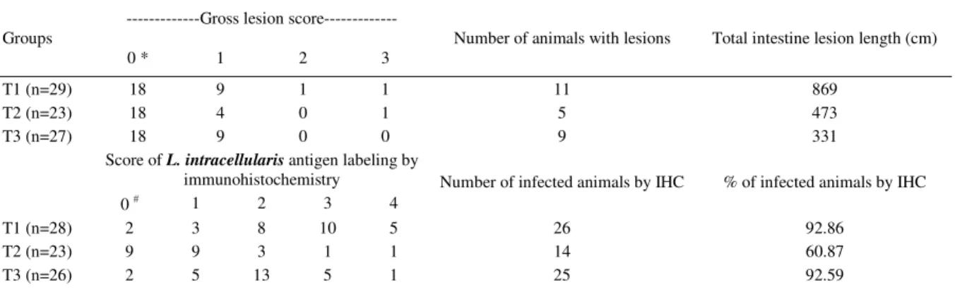
Table 2 - Length and severity of gross lesions characteristic of porcine proliferative enteropathy and intensity of L. intracellularis infection based on the immunohistochemistry (IHC) labeling in ileum fragments of pigs from groups unmedicated (T1), medicated with 90ppm (T2) and with 180ppm of leucomycin (T3) on day 23pi.
---Gross lesion score---Groups
0 * 1 2 3
Number of animals with lesions Total intestine lesion length (cm)
T1 (n=29) 18 9 1 1 11 869
T2 (n=23) 18 4 0 1 5 473
T3 (n=27) 18 9 0 0 9 331
Score of L. intracellularis antigen labeling by
immunohistochemistry
0 # 1 2 3 4
Number of infected animals by IHC % of infected animals by IHC
T1 (n=28) 2 3 8 10 5 26 92.86
T2 (n=23) 9 9 3 1 1 14 60.87
T3 (n=26) 2 5 13 5 1 25 92.59
REFERENCES
CHANG, W.L. et al. Prevalence of Lawsonia intracellularis in swine herds in Taiwan. Veterinary Record, v.141, p.103-104, 1997.
CHIRIBOGA, A.E.C.N. et al. Detection of Lawsonia intracellularis in faeces of swine from the main production regions in Brazil. Canadian Journal of Microbiology, v.45, p.230-234, 1999.
COOPER, D.M. et al. Comparison of the 16S ribosomal DNA sequence from the intracellular agent of proliferative enteritis in a hamster, deer, and ostrich with the sequence of a porcine isolate of Lawsonia intracellularis. International Journal of Systematic Bacteriology, v.47, p.635-639, 1997a. Available from: <http://ijs.sgmjournals.org/cgi/reprint/47/3/ 635>. Accessed on: Apr. 21, 2010.
COOPER, D.M., et al. Diagnosis of proliferative enteritis in frozen and formalin fixed, paraffin-embedded tissues from a hamster, horse , deer and ostrich using a Lawsonia intracellularis-specific multiplex PCR assay. Veterinary Microbiology, v.54, p.47-62, 1997b.
GEBHART, C.; GUEDES, R.M.C. Lawsonia intracellularis.
In: GYLES C.L. et al. (Eds.). Pathogenesis of bacterial infections in animals. 3.ed. Ames, Iowa: Blackwell, 2004. p.363-372.
GUEDES, R.M.C., GEBHART, C.J. Comparison of intestinal mucosal homogenate and pure culture of the homologous
Lawsonia intracellularis isolate in reproducing proliferative
enteropathy in swine. Veterinary Microbiology, v.93, p.159-166, 2003a.
GUEDES, R.M.C.; GEBHART, C.J. Preparation and characterization of polyclonal and monoclonal antibodies against Lawsonia intracellularis. Journal Veterinary Diagnotic Investigation, v.15, p.438-446, 2003b. Available from: <http://www.jvdi.org/cgi/reprint/15/5/438>. Accessed on: Apr. 21, 2010.
GUEDES, R.M.C. et al. Comparison of different methods for diagnosis of porcine proliferative enteropathy. Canadian Journal Veterinary Research, v.66, p.99-107, 2002. Available from: <http://www.ncbi.nlm.nih.gov/pmc/articles/ PMC226990/?tool=pubmed>. Accessed on: Apr. 21, 2010. HERBST, W. et al. Diagnosis of Lawsonia intracellularis using the polymerase chain reaction (PCR) in pigs with and without diarrhea and other animal species. Deutsch Tierarzti Wochenschr, v.110, p.361-364, 2003.
JONES, G.F. et al. Enhanced detection of intracellular organism of swine proliferative enteritis, Ileal symbiont intracellularis, in feces by polymerase chain reaction. Journal Clinical Microbiology v.31, p.2611-2615, 1993. Available from: < h t t p : / / j c m . a s m . o r g / c g i / r e p r i n t / 3 1 / 1 0 / 2611?view=long&pmid=8253956>. Accessed on: Apr. 26, 2010. KROLL, J.J. et al. Evaluation of protective immunity in pigs following oral administration of an avirulent live vaccine of
Lawsonia intracellularis. American Journal Veterinary Research, v.65, p.559-565, 2004.
KYRIAKIS, S.C. et al. The effect of josamycine on the control of ileitis in weaned piglets under field conditions. Journal Veterinary Pharmacology, v.25, p.279-284, 2002. LAWSON G.H.K.; GEBHART C.J. Proliferative enteropathy: review. Journal of Comparative Pathology, v.122, p.77-100, 2000. Available from: <http://www.sciencedirect.com/ science?_ob=MImg&_imagekey=B6WHW-45FC860-1R-1 & _ c d i = 6 8 6 science?_ob=MImg&_imagekey=B6WHW-45FC860-1R-1 & _ u s e r = 6 8 6 4 science?_ob=MImg&_imagekey=B6WHW-45FC860-1R-1 3 & _ p i i = S 0 0 2 1 9 9 7 5 9 9 9 0 3 4 7 X & _ o r i g = s e a r c h & _ c o v e r D a t e = 0 2 % 2 F 2 9 % 2 F 2 0 0 0 & _ s k = 9 9 8 7 7 9 9 9 7 & v i e w = c & w c h p = d G L b V z b -zSkzk&md5=7b692d6f717f01ae1f2d166826f720ec&ie=/ sdarticle.pdf>. Accessed on: Apr. 26, 2010. Doi:10.1053/ jcpa.1999.0347.
LEE, S.W. et al. Prevalence of porcine proliferative enteropathy and its control with tilosin in Korea. Journal Veterinary Science, v.2, p.209-212, 2001. Available from:
<http://pdf.medrang.co.kr/JVS/002/JVS002-03-08.pdf>. Accessed on: Apr. 26, 2010.
MacINTYRE, N. et al. Immunopathogenesis of experimentally induced proliferative enteropathy in pigs. Veterinary Pathology, v.40, p.421-432, 2003. Available from: <http:// vet.sagepub.com/content/40/4/421.full.pdf+html>. Accessed on: Apr. 26, 2010. doi: 10.1354/vp.40-4-421.
McORIST, S. et al. Reproduction of porcine proliferative enteropathy
with pure culture of Ileal symbiont intracellularis. Infection and Immunity, v.61, p. 286-4292, 1993. Available from: <http:// iai.asm.org/cgi/reprint/61/10/4286?view=long&pmid=8406817>. Accessed on: Apr. 26, 2010.
McORIST, S. et al. Characterization of Lawsonia intracellularis
gen. nov, sp nov, the obligately intracellular bacterium of porcine proliferative enteropathy. International Journal Systematic Bacteriology, v.45, p.520-525, 1995. Available from: <http:/ /ijs.sgmjournals.org/cgi/reprint/45/4/820>. Accessed on: Apr. 26, 2010.
McORIST, S. et al. Oral administration of tilosin phosphate for treatment and prevention of porcine proliferative enteropathy. American Journal Veterinary Research, v.58, p.136-139, 1997a.
McORIST, S. et al. Estimate of direct financial losses due to porcine proliferative enteropathy. Veterinary Record, v.140, p.579-581, 1997b.
McORIST, S.; GEBHART, C.J. Proliferative enteropathies. In: STRAW B.E. et al. (Eds.). Diseases of swine. 9.ed. Ames, Iowa: Blackwell, 2006. p.727-738.
PARADIS, M.A, et al. Subclinical ileitis produced by sequential dilutions of Lawsonia intracellularis in a mucosal homogenate challenge model. In: AMERICAN ASSOCIATON OF SWINE VETERINARIANS ANNUAL MEETING, 28., 2005, Toronto, Canada. Proceedings… Toronto: AASV, 2005. p.189-191. PLUMB, D.C. Veterinary drug handbook. 4.ed. Ames, Iowa: Iowa State, 2002. p. 993.
POMMIER, P. et al. Comparison of tylvalosin with tylosin for the control of subclinical iteitis in swine. Reviews Medical Veterinary, v.159, p.579-582, 2008.
TOMANOVÁ, K. et al. Lawsonia intracellularis in wild mammals in the Slovak Carpathians. Journal Wild Disease, v.39,
p.407-411, 2003. Available from: <http://www.jwildlifedis.org/cgi/ reprint/39/2/407>. Accessed on: Apr. 26, 2010.
WINKELMAN, N.L. et al. Lincomycin-medicated feed for the control of porcine proliferative enteropathy (ileitis) in swine. Journal Swine Health Production, v.10, p.107-111, 2002.
VEZINA, C. et al. Biosynthesis of kitasamycin (Leucomonycin) by Leucine analog-resistant mutants of Streptomyces kitasatoensis. Antimicrobial Agents and Chemotherapy,