IgE Sensitization Profiles Differ between
Adult Patients with Severe and Moderate
Atopic Dermatitis
Irene Mittermann1,2☯, Gustav Wikberg3☯, Catharina Johansson4, Christian Lupinek2,
Lena Lundeberg3, Reto Crameri5, Rudolf Valenta2, Annika Scheynius4*
1Christian Doppler Laboratory for the Development of Allergen Chips, Medical University of Vienna, Vienna, Austria,2Division of Immunopathology, Department of Pathophysiology and Allergy Research, Center for Pathophysiology, Infectiology and Immunology, Medical University of Vienna, Vienna, Austria,3Department of Medicine Solna, Karolinska Institutet, and Dermatology and Venereology Unit, Karolinska University Hospital, Stockholm, Sweden,4Department of Clinical Science and Education, Karolinska Institutet, and Sachs' Children and Youth Hospital, SE-118 83 Södersjukhuset, Stockholm, Sweden,5Swiss Institute of Allergy and Asthma Research (SIAF), University of Zurich, Davos, Switzerland
☯These authors contributed equally to this work. *[email protected]
Abstract
Background
Atopic dermatitis (AD) is a complex chronic inflammatory disease where allergens can act as specific triggering factors.
Aim
To characterize the specificities of IgE-reactivity in patients with AD to a broad panel of exogenous allergens including microbial and human antigens.
Methodology
Adult patients with AD were grouped according to the SCORAD index, into severe (n = 53) and moderate AD (n = 126). As controls 43 patients were included with seborrhoeic eczema and 97 individuals without history of allergy or skin diseases. Specific IgE reactivity was assessed in plasma using Phadiatop1, ImmunoCap
™, micro-arrayed allergens, dot-blotted recombinantMalassezia sympodialisallergens, and immune-blotted microbial and human proteins.
Results
IgE reactivity was detected in 92% of patients with severe and 83% of patients with moder-ate AD. Sensitization to cat allergens occurred most frequently, followed by sensitization to birch pollen, grass pollen, and to the skin commensal yeastM.sympodialis. Patients with severe AD showed a significantly higher frequency of IgE reactivity to allergens like cat (rFel d 1) and house dust mite (rDer p 4 and 10), toStaphylococcus aureus,M.sympodialis, a11111
OPEN ACCESS
Citation:Mittermann I, Wikberg G, Johansson C, Lupinek C, Lundeberg L, Crameri R, et al. (2016) IgE Sensitization Profiles Differ between Adult Patients with Severe and Moderate Atopic Dermatitis. PLoS ONE 11(5): e0156077. doi:10.1371/journal. pone.0156077
Editor:Michel Simon, CNRS-University of Toulouse, FRANCE
Received:February 8, 2016
Accepted:May 9, 2016
Published:May 26, 2016
Copyright:© 2016 Mittermann et al. This is an open access article distributed under the terms of the
Creative Commons Attribution License, which permits unrestricted use, distribution, and reproduction in any medium, provided the original author and source are credited.
Data Availability Statement:All relevant data are within the paper and its Supporting Information files.
and to human antigens. In contrast, there were no significant differences in the frequencies of IgE reactivity to the grass pollen allergens rPhl p 1, 2, 5b, and 6 between the two AD groups. Furthermore the IgE reactivity profile of patients with severe AD was more spread towards several different allergen molecules as compared to patients with moderate AD.
Conclusion
We have revealed a hitherto unknown difference regarding the molecular sensitization pro-file in patients with severe and moderate AD. Molecular profiling towards allergen compo-nents may provide a basis for future investigations aiming to explore the environmental, genetic and epigenetic factors which could be responsible for the different appearance and severity of disease phenotypes in AD.
Introduction
Atopic dermatitis (AD) is a complex chronic inflammatory skin disease with 15–30% of chil-dren and 2–10% of adults being afflicted [1]. The pathogenesis of AD is considered to result from a combination of a defective skin barrier and inappropriate immune responses with con-tribution of both genetic and environmental factors including microbial agents [2–4]. Allergens are important in the pathogenesis of AD since they can act as specific trigger factors. For exam-ple, exacerbations of AD can be observed in patients during allergen exposure[5] or upon ingestion of allergen-containing food [6]. AD has according to the presence or to the absence of detectable allergen-specific IgE antibodies been classified into either an extrinsic or an intrinsic type [7]. Still, another subgroup of patients with AD has an autoimmune IgE-medi-ated reactivity against auto-antigens in addition to sensitization against exogenous allergens [8,9].
In addition microorganisms play a role in eliciting and maintaining eczema in patients with AD [10]. One such microorganism isMalassezia, a commensal yeast that dominates the human fungal skin flora[11] but is also associated with a number of skin disorders, such as seborrhoeic eczema (SE), dandruff and AD [12].Malassezia sympodialisis one of the most fre-quently isolated species from the skin of both AD patients and healthy individuals [13,14]. Allergen specific IgE- and T-cell reactivity and/or positive atopy patch test reaction toM. sym-podialiscan be detected in around 50% of adult patients with AD[12]. These reactions are rarely found in other allergic diseases or skin disorders indicating a specific link between AD andMalasseziain a subset of patients [15]. So far there are 10M.sympodialisallergens, desig-nated Mala s 1 and Mala s 5–13, cloned, sequenced and characterized [16–22]. Four of the allergens lack homology to known proteins, whereas others have cross-reactivity to human homologues such as manganese superoxide dismutase [19] and thioredoxin [21]. The unique-ness of these allergens makes them highly interesting as diagnostic tools and to understand sen-sitization and cross-reactivity to human homologous antigens in AD.
There are several techniques for determining IgE sensitizations such as allergen-specific IgE serology, skin prick test, and when necessary oral food challenges. By introducing new methodologies for phenotype characterization of patients with AD it may be possible to develop personalized strategies for prevention and management to break, terminate or reverse the natural course of this chronic disease. One way of achieving this goal could be using micro-arrayed allergen molecules which allow measuring IgE reactivities in only a few
research program MeDALL. Work at SIAF was supported by the Swiss National Science Foundation (grant 320030_149978/1). The funders had no rule in study design, data collection and analysis, decision to publish, or preparation of the manuscript.
Competing Interests:RV has received research grants from BIOMAY AG, Vienna, Austria, and Thermo Fisher, Uppsala, Sweden, and serves as a consultant for BIOMAY AG, Vienna, Austria, Thermo Fisher, Uppsala, Sweden, and Fresenius Medical Care, Bad Homburg, Germany. The rest of the authors declare that they have no relevant conflicts of interest. This does not alter the authors’adherence to PLOS ONE policies on sharing data and materials.
microliters of serum or plasma to large panels of purified allergen molecules to identify dis-ease-causing allergens [23,24].
In this study we sought to perform a meticulous analysis of the IgE-reactivity profiles in AD patients with different severity to obtain information if and how molecular IgE sensitization profiles towards a comprehensive set of exogenous, microbial and endogenous allergens may be linked with different clinical phenotypes of AD including co-morbidity with respiratory symptoms.
Materials and Methods
Study population
All subjects (n = 319) were recruited from the Stockholm area and examined by a dermatolo-gist at the Dermatology and Venereology Unit, Karolinska University Hospital Stockholm, Sweden, during September until May to avoid the summer season. Patients with a clinical diag-nosis of AD or SE and healthy controls were included in the study (Table 1). Inclusion criteria for AD patients were diagnosis according to the UK working party [25], moderate to severe eczema, and skin lesions not only restricted to the hands. The severity of the eczema was assessed using the objective SCORAD index [26] and present or patient reported head and neck distribution was recorded. Exclusion criteria for the patients were other skin diseases than AD or SE, immune deficiencies, autoimmune diseases and ongoing malignant diseases. The history of patients reported respiratory allergic symptoms was documented as co-morbidity. The healthy controls were subjects who did not have clinical symptoms or history of allergy or skin disease. The study was approved by the Regional Ethical Review Board in Stockholm and all participants gave their written informed consent.
Preparation of protein/allergen extracts
M.sympodialis(ATCC strain 42132) and human A431 epithelial cell extracts were prepared as previously described [8,27].Staphylococcus aureus(ATCC 25923) was grown overnight in tryptic soy broth at 37°C and harvested by centrifugation at 3220 x g. The cells were washed twice with phosphate-buffered saline (PBS) and inactivated by boiling for 10 min. The cell pel-let was re-suspended in PBS and homogenized using an Ultra Turrax (IKA Labortechnik, Stau-fen, Germany) and stored at -20°C until use.
Recombinant allergens
Recombinant Mala s 1, rMala s 5–13 (S1 Table) were expressed inEscherichia coliBL21 (DE3) as his-tagged proteins and purified by inclusion body preparation and Ni2+-chelate chromatog-raphy [16–21]. Purity of proteins was assessed by SDS-PAGE and Coomassie Blue staining [28]. Protein concentrations were determined by Micro BCA Protein Assay (Pierce, Rockford, IL).
IgE serology
Total IgE, specific IgE to any of 11 common aeroallergen sources (Phadiatop1) andM. sympo-dialis-specific IgE (m70,) were measured in plasma using ImmunoCAP™(Phadia AB, Uppsala, Sweden).
For non-denaturing RAST-based dot blot assays, 0.2μg purified recombinant allergens
per centimeter slot of the preparative gel) ofM.sympodialis, human A431 epithelial cell line or
S.aureusextracts were boiled for 5 min with sodium dodecyl sulphate (SDS) sample buffer containing 5% v/v beta-mercaptoethanol, subjected to 12.5% SDS-PAGE and blotted onto nitrocellulose membranes [30,31]. A protein molecular weight marker (PageRuler prestained Protein Ladder Plus, Fermentas, St Leon-Rot, Germany) was used as standard. To define a cut-off for a positive/negative dot blot and immunoblot result human serum albumin (HSA) and serum from non-allergic patients were used for control purposes. In every experiment three sera from AD patients containing specific IgE toS.aureus,M.sympodialisand the human extract were included as positive control.
IgE-reactivity to 120 micro-arrayed allergen components was analyzed with a customized allergen-chip (Phadia Multiplexing, Thermo Fisher Scientific, Vienna, Austria) as previously described [24].
Statistical analyses
Descriptive statistics and group comparisons were computed using IBM SPSS Statistics, version 22 (IBM Corp., Armonk, NY, USA). For comparison of the two groups of patients suffering from moderate or severe AD, respectively, with respect to frequencies of reactivities to bacterial, yeast and human antigens and to micro-arrayed allergen molecules, two-sided asymptotic p-val-ues were calculated by Chi-square test. P-valp-val-ues0.05 were considered statistically significant.
Results
Demographic and clinical characterization of AD patients and controls
The 179 AD patients (56% females) had a median age of 28 years (range 18–65 years)
(Table 1). According to SCORAD 53 patients (30%) suffered from severe AD and 156 patients (87%) had present (n = 143) or previous (n = 13) head and neck distribution of their eczema. Debut of the eczema was similar in both groups; in the majority of the AD patients the debut
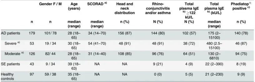
Table 1. Demographic and clinical characterization of AD patients and controls.
Gender F / M Age (years)
SCORADa) Head and neck distribution
Rhino-conjunctivitis and/or asthma
Total plasma IgE
b)122 kU/L
Total plasma IgE
b)(kU/L)
Phadiatop1
positivec)
n n median
(range)
median (range)
n (%) N (%) N (%) median
(range)
n (%)
AD patients 179 101/ 78 28 (18– 65)
34 (14–70) 156 (87) 144 (80) 102 (57) 175 (2– 15100)
140 (78)
Severea) 53 19 / 34 30 (18
– 65)
54 (41–70) 48 (91) 48 (91) 38 (72) 480 (2.5– 15100)
46 (87)
Moderatea) 126 82/ 44 28 (18
– 65)
31 (14–40) 108 (85) 96 (76) 64 (51) 130 (2–
6810)
94 (75)
SE patients 43 9 / 34 39 (18– 63)
NA NA 9 (21) 4 (9) 22 (2–390) 8 (19)
Healthy controls
97 59 / 38 35 (18– 65)
NA NA 0 (0) 5 (5) 21 (2–230) 9 (9)
a)Objective SCORAD [26], severe AD de
fined as SCORAD41
b)ImmunoCAP
™(Phadia AB), reference range 1.6–122 kU/L
c)Phadiatop1(Phadia AB), plasma IgE to any of 11 common aeroallergens, reference range0.35 kU/L
AD = atopic eczema, F = Female, M = Male, n = number of positive individuals, NA = not applicable, SE = seborrhoeic eczema
was during the first year (58% in the severe group and 57% in the moderate group) and 17% and 22%, respectively, after 5 years of age. The majority of AD patients (i.e., 144, 80%), had ever had rhino-conjunctivitis and/or asthma in addition to AD (i.e., 91% in the severe and 76% in the moderate AD group) whereas only 21% of the SE patients reported respiratory symp-toms (Table 1).
Total plasma IgE levels were elevated (>122 kU/L) in 57% of the patients, with higher
median IgE levels in the severe AD group (480 kU/L) than in the moderate AD group (130 kU/ L) (Table 1). Seventy-eight % of the AD patients had a positive Phadiatop1with a higher per-centage among the severe AD patients (87%) in comparison to the moderate AD patients (75%) (Table 1). Nine % of 43 individuals with SE had a total serum IgE level>122 kU/L and
19% had a positive Phadiatop1
(Table 1). Five % of the healthy controls (n = 97) had IgE levels higher than 122 kU/L and for 9% the Phadiatop1was positive (Table 1).
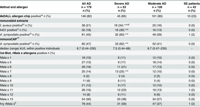
IgE reactivity to bacterial (
S
.
aureus)
, yeas
t (M
.
sympodialis
) and human
antigens was significantly more frequent in the severe AD group
IgE reactivity to nitrocellulose-blottedS.aureus,M.sympodialisand human antigens (epithelial cell-line A431) was analyzed using plasma samples from all 179 AD patients, 43 SE patients and 97 healthy controls. Twenty-one percent of the AD patients showed reactivity to antigens in theS.aureusextract (i.e., 34% in the severe AD group compared to 16% in the moderate AD group, p<0.01 (Table 2). Eighteen percent of the AD patients showed IgE reactivity to the
human cell extract. Again the percentage of reactivity was significantly higher (i.e., 30%) in severe than in moderate AD patients (i.e., 13%) (p<0.01) (Table 2).
Likewise, IgE reactivity against theM.sympodialisextract was detected significantly more often in severe (i.e., 60%) compared to moderate AD patients (i.e., 39%) with 45% of the 179 AD patients being positive on average (p<0.01) (Table 2). In addition, specific IgE toM.
sym-podialiswas assessed with the ImmunoCap™assay where 47% of the 179 AD patients, 62% with severe and 41% with moderate eczema (p<0.01) (Table 2andS1A–S1C Fig), whereas
none of the SE patients (Table 2) or healthy controls was positive in this assay (S2 Table). The median specific plasma IgE levels againstM.sympodialisof the severe AD patients was 7.5 kU/ L and of the moderate AD patients 6.7 kU/L (Table 2). The majority, 98% (n = 156) of AD patients with IgE reactivity toM.sympodialishad present or reported head and neck involve-ment (S2 Table). Notably, 8% in the group of 53 severe AD patients had a negative Phadiatop1 reaction, not elevated total serum IgE and no detectable IgE antibodies toM.sympodialis, m70 (S1B Fig), and among the 126 patients with moderate AD the corresponding figure was 19% (S1C Fig). There was a good agreement of results obtained with the immunoblot and Immuno-CAP™technology, only slightly higher positive results were observed with the CAP assay com-pared to the immunoblotting method (AD 47% comcom-pared to 45%, severe AD 62% versus 60%, moderate AD 41% versus 39%,Table 2).
No IgE-binding toS.aureusor human A431 extract was observed among the 43 SE patients, but one of them who was Phadiatop1positive, showed a weak IgE reactivity to one band in the
allergen extract-based ImmunoCAP™(47%) or Immunoblot assay (45%) (Table 2). IgE-reactiv-ity to rMala s 7 was significantly more frequently detected in severe compared to moderate AD patients (p<0.01,Table 2). rMala s 13 showed IgE reactivity in 38% of the severe and 27% of
the moderate AD patients, respectively, and was the most frequently detected Mala s allergen (Table 2). Among the 85M.sympodialisImmunoCAP™positive AD patients 52 of them (61%) reacted to rMala s 13 (S2 Table), therefore to be considered as a major allergen.
Only 1 patient with SE showed IgE reactivity to rMala s 11 (Table 2) and one healthy control to rMala s 13 (S2 Table).
A limited number of allergen sources contribute to a different extent to
sensitization in severe and moderate AD patients
Using the allergen chip [24] with 120 allergen components (S3 Table), 82% of the 179 AD patients showed IgE reactivity to at least one allergen, 85% of the severe and 80% of the moderate
Table 2. Frequencies of allergen-specific IgE reactivities in the AD and SE patients as tested by MeDALL allergen chip, Immunoblotting, Immuno-CAP™and RAST-based dot-blot assay.
All AD Severe AD Moderate AD SE patients
Method and allergen n = 179 n = 53 n = 126 n = 43
n (%) n (%) n (%) n (%)
MeDALL allergen chippositivea)n (%) 146 (82) 45 (85) 101 (80) 10 (23)
Immunoblot extracts
S.aureuspositiveb)n (%) 38 (21) 18 (34)**g) 20 (16) 0 (0)
A431 positivec)n (%) 32 (18) 16 (30)** 16 (13) 0 (0)
M.sympodialispositived)n (%) 81 (45) 32 (60)
** 49 (39) 1 (2)
ImmunoCAP™
M.sympodialispositivee)n (%) 85 (47) 33 (62)** 52 (41) 0 (0)
Median (range) kU/L within positive individuals 6.7 (0.44–230) 7.5 (0.44–68) 6.7 (0.47–230) -Dot-Blot, rMala s allergenspositive n (%)
rMala s 1 18 (10) 6 (11) 12 (10) 0 (0)
rMala s 5 27 (15) 9 (17) 18 (14) 0 (0)
rMala s 6 28 (16) 11 (21) 17 (13) 0 (0)
rMala s 7 25 (14) 13 (25)** 12 (10) 0 (0)
rMala s 8 4 (2) 2 (4) 2 (2) 0 (0)
rMala s 9 11 (6) 6 (11) 5 (4) 0 (0)
rMala s 10 21 (12) 9 (17) 12 (10) 0 (0)
rMala s 11 28 (16) 12 (23) 16 (13) 1 (2)
rMala s 12 14 (8) 6 (11) 8 (6) 0 (0)
rMala s 13 54 (30) 20 (38) 34 (27) 0 (0)
Any rMala sf) 78 (44) 31 (59) 47 (37) 1 (2)
a)MeDALL allergen chip, positive0.30 ISU b)Staphylococcus aureus
ATCC 25923 extract
c)A431 Human epithelial cell line extract d)Malassezia sympodialisATCC 42132 extract e)
ImmunoCAP™(Phadia AB),Malassezia sympodialisATCC 42132 extract (m70), positive0.35 kU/L
f)Individuals positive to one or more of the tested rMala s allergens g)Signi
ficant differences between severe and moderate AD patients are indicated**p<0.01 AD = atopic dermatitis, SE = seborrhoeic eczema
AD patients were positive, respectively, and 23% of the 43 patients with SE showed IgE-reactivity to micro-arrayed allergens (Table 2).
Fig 1Ashows a pie chart representation of the contribution of different allergen sources to IgE sensitization in severe and moderate AD patients. The chart was constructed based on the frequencies of IgE sensitizations towardsM.sympodialis,S.aureusand human cell extracts detected with immunoblotting (Table 2) and based on IgE-reactivity profiles towards 25 spe-cific allergen components determined with micro-arrayed allergen molecules (Table 3andS3 Table). As mentioned above the IgE sensitization toM.sympodialis,S.aureusand human anti-gens contributed significantly more to IgE sensitization in severe versus moderate AD (p<0.01,
Table 2). IgE sensitization to genuine cat allergens (rFel d 1 and/or rFel d 4) and house dust mite allergens (nDer p 1 and/or rDer p 2, rDer p 4, rDer p 5, rDer p 7, rDer p 10, rDer p 11, rDer p 14, rDer p 15, rDer p 18, rDer p 21, rDer p 23) also contributed more to IgE sensitiza-tion in severe versus moderate AD (Fig 1A). Birch pollen (rBet v 1) and mugwort (nArt v 1) contributed equally to IgE sensitization in severe and moderate AD but interestingly, the con-tribution of IgE sensitization to grass pollen (rPhl p 1 and/or rPhl p 2, rPhl p 5b, rPhl p 6) and dog allergens (rCan f 1 and/or rCan f 2, rCan f 4, rCan f 5, rCan f 6) was less in severe as com-pared to moderate AD (Fig 1A). IgE sensitization to other allergen sources was quite rare (S3 Table). Only 10% of all AD patients showed IgE reactivity to latex allergens (rHev b 6.01) and 2% to the mouldAlternaria alternata(rAlt a 1). IgE reactivities to recombinant food allergens, e.g., apple (rMal d 1, 42%), peach (rPru p 1, 18%), hazelnut (rCor a 1.0401, 34%) or soy (rGly m 4, 16%) were due to IgE cross-reactivity to the major birch pollen allergen Bet v 1 (52%) (S3 Table). Other plant-derived food allergens, e.g., hazelnut (rCor a 8), kiwi allergens (nAct d 1, nAct d 5) or wheat allergens (rTri a 19.0101) were recognized by only 4%, or less of the AD patients. IgE to animal-derived food allergens could be detected in only a small percentage of the AD patients, e.g., 7% of all AD patients showed IgE reactivity to egg allergens and cow`s milk allergens were recognized by 2% of the patients. The frequencies of IgE reactivity to each of the individual micro-arrayed allergens are reported inS3 Table.
In the group of 43 SE-patients, 14% showed IgE-reactivity to birch pollen (rBet v 1), 14% to grass pollen (rPhl p 1), 5% to house dust mite (rDer p 2) and 2% to dog allergens (rCan f 1) (S3 Table). Plasma samples from 12 Phadiatop1negative healthy controls were tested for control purposes and they were all negative (S3 Table).
Selective spreading in the molecular sensitization profiles of severe as
compared to moderate AD patients
The frequencies of IgE reactivity to the individual allergen molecules are shown inTable 3for the most frequently recognized allergen sources and for each of the tested components inS3 Table. For most of the allergens we observed that they were recognized more frequently by patients with severe as compared to patients with moderate AD (Table 3,S2A, S2B and S2D–
S2H Fig). For certain allergens (e.g. rFel d 1, rDer p 4, and rDer p 10) a significantly (p<0.05)
more frequent IgE recognition was found for the severe AD patients (Table 3). Interestingly, genuine grass pollen allergens (i.e., rPhl p 1, rPhl p 2, rPhl p 5b, and rPhl 6) did not follow this trend because they were recognized with a similar frequency in both patient groups (Table 3,
S2C Fig). Only the cross-reactive carbohydrate marker allergen from grass pollen (i.e., nPhl p 4) was more frequently recognized by severe AD patients (p<0.05,Table 3,S2C Fig). The same
pattern was observed when the patients were grouped into severe and moderate AD with patient reported history of respiratory allergic symptoms, with the exception that a significant difference (p<0.05) was reached for a higher IgE reactivity to rPhl p 2 in the moderate AD
Fig 1. Pie charts showing the contribution of (A), allergen sources and (B), individual allergen components to IgE sensitization.Each chart represents 100% of IgE reactivities detected in plasma from all AD patients of the respective group, severe AD (left chart) and moderate AD (right chart), in (A)to six allergen sources using the MeDALL allergen-chip (ISU0.3) and to extracts ofM.sympodialis,S.aureusand the human cell line A431 using immunoblotting, and in (B)to 25 allergen molecules using the MeDALL allergen-chip (ISU0.3). The sizes of the segments represent the proportion of the respective in (A), allergen source and in (B), allergen molecule among all recognized. Allergen sources/molecules start a 12 o’clock of the pie chart and continue clock-wise as listed with the color code.
Table 3. Frequencies and intensities of IgE reactivity to purified marker allergens in AD patients detected by allergen chip technology.
No. Allergen All AD Severe AD
Moderate AD
Severe AD Moderate AD Allergen family, Function, CCD
Severe AD Moderate AE
median (range)
median (range)
Rhinoconjuctivitis Rhinoconjuctivitis
n = 179 n = 53 n = 126 IgE (ISU)a) IgE (ISU) and/or asthma and/or asthma
n (%) n (%) n (%) n = 47 n (%) n = 96 n (%)
1 rFel d 1 93 (52) 36 (68)** 57 (45) 4.3 (0.31– 134.17)
4.14 (0.35– 127.92)
Uteroglobin 36 (77)** 51 (53)
2 rBet v 1 93 (52) 33 (62) 60 (48) 12.83 (0.33– 120.37)
8.96 (0.34– 102.94)
PR-10 32 (68) 51 (53)
3 rPhl p 1 79 (44) 24 (45) 55 (44) 2.11 (0.31– 48.74)
2.43 (0.31– 56.91)
β-Expansin 23 (49) 45 (47)
4 nPhl p 4 51 (29) 22 (42)* 29 (23) 1.53 (0.31– 16.6)
1.25 (0.39– 26.31)
Grass-group 4, CCD 21 (45)* 26 (27)
5 rPhl p 5b 47 (26) 12 (23) 35 (28) 4.83 (0.61– 28.58)
3.41 (0.32– 86.9)
Grass-group 5 11 (23) 32 (33)
6 rCan f 1 43 (24) 16 (30) 27 (21) 14.28 (0.44– 111.13)
6.91 (0.61– 121.54)
Lipocalin 16 (34) 25 (26)
7 rFel d 4 43 (24) 15 (28) 28 (22) 9.32 (0.45– 118.56)
2.9 (0.34– 55.56)
Lipocalin 15 (32) 27 (28)
8 rCan f 5 40 (22) 14 (26) 26 (21) 2.29 (0.54– 28.17)
3.62 (0.35– 27.48)
Arginine Esterase 14 (30) 23 (24)
9 rPhl p 6 35 (20) 12 (23) 23 (18) 0.89 (0.38– 14.56)
2.66 (0.36– 33.03)
Grass-group 5/6 11 (23) 21 (22)
10 nArt v 1 35 (20) 12 (23) 23 (18) 1.14 (0.34– 43.7)
1.39 (0.32– 38.99)
Defensin-like protein 12 (26) 21 (22)
11 rDer p 2 34 (19) 13 (25) 21 (17) 33.79 (0.57– 131.74)
5.18 (0.35– 103.86)
Group 2 mite-allergen
12 (26) 20 (21)
12 rPhl p 2 31 (17) 6 (11) 25 (20) 9.72 (0.74– 27.15)
5.59 (0.36– 33.3)
Grass-group 2/3 5 (11)* 24 (25)
13 rCan f 6 26 (15) 10 (19) 16 (13) 2.11 (0.4– 14.01)
1.72 (0.3– 7.25)
Lipocalin 10 (21) 15 (16)
14 nDer p 1 25 (14) 11 (21) 14 (11) 4.99 (0.62– 18.63)
0.88 (0.4– 31.58)
Group 1 mite-allergen
10 (21) 13 (14)
15 rCan f 2 25 (14) 9 (17) 16 (13) 1.03 (0.4– 49.04)
2.09 (0.34– 41)
Lipocalin 9 (19) 15 (16)
16 rDer p 4 18 (10) 10 (19)* 8 (6) 0.87 (0.32– 3.57)
0.71 (0.32– 4.87)
Group 4 mite-allergen
9 (19)* 7 (7)
17 rDer p 23
18 (10) 7 (13) 11 (9) 29.98 (0.7– 98.11)
14.16 (1.69– 43.88)
Chitin-binding domain
6 (13) 11 (12)
18 rCan f 4 14 (8) 6 (11) 8 (6) 8.65 (0.45– 64.14)
12.25 (0.39– 25.9)
Lipocalin 6 (13) 7 (7)
19 rDer p 10
12 (7) 8 (15)** 4 (3) 0.49 (0.3– 3.8)
2.57 (0.44– 3.47)
Tropomyosin 7 (15)* 3 (3)
20 rDer p 5 11 (6) 6 (11) 5 (4) 1.48 (0.44– 93.01)
2.47 (0.34– 87.36)
Group 5 mite-allergen
6 (13) 4 (4)
21 rDer p 7 9 (5) 4 (8) 5 (4) 1.82 (0.37– 3.71)
11.65 (0.75– 22.5)
Group 7 mite-allergen
4 (9) 4 (4)
22 rDer p 11
8 (4) 4 (8) 4 (3) 0.42 (0.3– 1.62)
0.86 (0.31– 1.09)
Group 11 mite-allergen
3 (6) 4 (4)
23 rDer p 15
5 (3) 3 (6) 2 (2) 0.33 (0.32– 16.44)
0.7 (0.68– 0.71)
Chitin-binding domain
3 (6) 2 (2)
24 rDer p 21
6 (3) 3 (6) 3 (2) 2.52 (0.86– 71.48)
47.28 (11.23– 80.02)
Group 21 mite-allergen
3 (6) 3 (3)
The levels of allergen-specific IgE to the individual allergens expressed in ISU varied between severe and moderate AD patients. For some allergens (e.g., rFel d 1) the IgE levels were comparable and for others severe AD patients showed higher allergen-specific IgE levels (e.g., rFel d 4, rDer p 2, rDer p 23) (Table 3).
An interesting finding was that the IgE reactivity profile of patients with severe AD was more spread towards several different allergen molecules as compared to patients with moder-ate AD (Fig 1B). The latter becomes visible when the contribution of the individual allergens to IgE sensitization is displayed in the form of a pie chart where it becomes obvious that the seg-ment of the 12 house dust mite allergens occupy a larger part, 26%, of the pie in severe AD patients compared to 17% in moderate AD patients (Fig 1B). In contrast the contribution of grass pollen allergens (i.e., rPhl p 1, rPhl p 2, rPhl p 5b and rPhl p 6) is lower in severe AD patients 19% compared to 29% in the moderate AD group (Fig 1B).
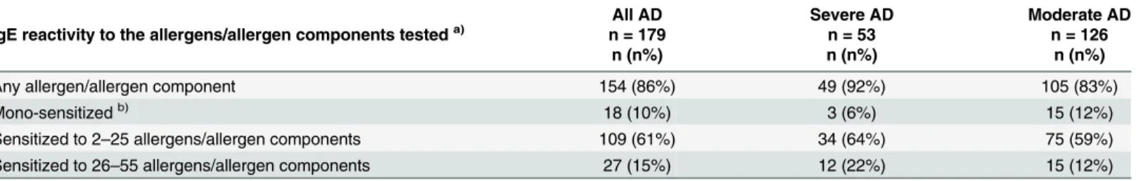
Summary of allergen-specific IgE reactivities in the AD patients
Altogether, IgE reactivity as tested to 120 allergen components (MeDALL allergen chip), and toM.sympodialis,S.aureusand human epithelial cell extracts (immunoblotting assays) was detected in 92% of patients with severe and 83% of patients with moderate AD (Table 4). Six %
Table 3. (Continued)
No. Allergen All AD Severe AD
Moderate AD
Severe AD Moderate AD Allergen family, Function, CCD
Severe AD Moderate AE
median (range)
median (range)
Rhinoconjuctivitis Rhinoconjuctivitis
n = 179 n = 53 n = 126 IgE (ISU)a) IgE (ISU) and/or asthma and/or asthma
n (%) n (%) n (%) n = 47 n (%) n = 96 n (%)
25 rDer p 18
6 (3) 2 (4) 4 (3) 3.76 (1.85– 5.67)
1.21 (0.32– 8.22)
Chitin-binding domain
2 (4) 4 (4)
26 rDer p 14
4 (2) 1 (2) 3 (2) 0.68 (0.68) 0.56 (0.39– 1.28)
Vitellogenin 1 (2) 3 (3)
36 (77)
a)MeDALL allergen chip, reference range0.3 ISU
Significant differences between severe and moderate AD patients are indicated*p<0.05,**p<0.01. Taken into account the number of tested variables (n = 26) the expected number of allergens to be significant at p level<0.05 by chance is 1.3.
AD = atopic dermatitis, CCD = cross-reactive carbohydrate determinates, n = natural, r = recombinant
doi:10.1371/journal.pone.0156077.t003
Table 4. Summary of allergen-specific IgE reactivities in the AD patients.
All AD Severe AD Moderate AD
IgE reactivity to the allergens/allergen components testeda) n = 179 n = 53 n = 126
n (n%) n (n%) n (n%)
Any allergen/allergen component 154 (86%) 49 (92%) 105 (83%)
Mono-sensitizedb) 18 (10%) 3 (6%) 15 (12%)
Sensitized to 2–25 allergens/allergen components 109 (61%) 34 (64%) 75 (59%)
Sensitized to 26–55 allergens/allergen components 27 (15%) 12 (22%) 15 (12%)
a)Tested for IgE reactivity to 120 allergen components (MeDALL allergen chip), and toM.sympodialis,S.aureusand human epithelial cell extracts
(immunoblotting).
b)Mono-sensitised severe AD patients:M.sympodialis(n = 3)
Mono-sensitised moderate AD patients: Grass pollen (n = 2), birch pollen (1), dog (1), cat (5), house dust mite (1),M.sympodialis(4), wasp venom (1).
(n = 3) of the severe and 12% (n = 15) of the moderate AD patients were mono-sensitized. The three patients in the severe group were all mono-sensitized toM.sympodialiswhereas the sen-sitization profile in the moderate group was spread over seven different allergen sources. The majority of the patients were sensitized to 2–25 allergens/allergen components (Table 4). The maximum number of allergens/allergen components detected in a patient in the severe group was 55 and in the moderate group 48.
Discussion
In this study we used a comprehensive panel of allergen molecules from exogenous allergen sources, microbes and autoantigens to characterize the molecular IgE reactivity profile of AD patients with defined clinical phenotypes. We found that patients with severe differed from patients with moderate AD regarding several characteristics. First, severe AD patients were characterized by the fact that they reacted to a larger panel of environmental allergens than patients with moderate AD. Interestingly, the spreading of the IgE recognition profiles in the severe AD patients included allergens from certain environmental allergen sources such as house dust mite, cat, and birch pollen allergens but not the major timothy grass pollen aller-gens (Phl p 1, Phl p 5, Phl p 2 and Phl p 6) (Fig 1). This finding would suggest that grass pollen allergens are less important as trigger factors for AD compared to birch pollen and indoor allergens in the studied population. The difference between birch pollen and grass pollen is unexpected because grass pollen contains several very potent allergens whereas birch contains only one major allergen (Bet v 1). Furthermore, the grass pollen season lasts longer than the birch pollen season and allergen loads are typically higher for grass pollen [32,33]. Since the route of exposure should be the same for both pollens it is quite conceivable that other factors play a role such as climatic effects where low humidity and cold tempera-tures negatively affect skin barrier functions and increase the risk of dermatitis [34]. We therefore hypothesize that an impaired skin barrier of patients after the usually long winter periods in Sweden before the birch pollen season may be responsible for the fact that birch pollen is associated more with severe AD whereas the skin barrier may regenerate towards the later grass pollen season.
Secondly, patients suffering from severe AD also differed from patients with moderate AD regarding their recognition of microbial“allergens”and autoantigens. IgE-reactivity to skin-associated microorganisms likeM.sympodialisorS.aureusas well as to human antigens was significantly higher in the severe AD group (Table 2). The percentage in all AD patients who showed IgE-reactivity to theM.sympodialisextract, as detectable with ImmunoCap™or immu-noblot analyses, was 47–45%, which is in agreement with previous studies as is the high fre-quency of head and neck involvement, particularly in the severe AD patients (S2 Table) [15,35–37].
quite possible that IgE recognition of human homologous allergens may have been triggered by sensitization to cross-reactive microbial allergens.
Different explanations for the broader and more spread allergen profile recognized by the severe AD patients may be considered. On the one hand it is possible that the severity of AD may depend on the number of different allergen molecules recognized and thus the sum of rec-ognized allergens may play a role as has been recently reported for respiratory allergy to house dust mites: Children with more severe respiratory manifestations (i.e., asthma plus rhinitis) exhibited a broader IgE-recognition towards individual house dust mite allergens than children suffering only from rhinitis [39]. This is similar to the findings made: We also found a broader recognition of house dust mite allergens in the severe AD group as compared to the moderate AD group (Table 3andFig 1). In this context, it has also been discovered that AD patients dif-fer regarding their molecular recognition profiles of house dust mite allergens compared to patients suffering only from house dust mite-associated respiratory allergy [40].
Alternatively, one may consider also other explanations. For example it may be considered that patients with more severe AD and broader IgE recognition profiles may be more“atopic” than the patients with moderate AD. It has been reported that patients with poly-sensitization to respiratory allergens differed from patients with oligo/monosensitization by their produc-tion of IL-4 and Th2 driving cytokines [41]. In this scenario which was recently suggested as a hypothesis [42], a more“atopic”genetic make-up would be responsible for poly-sensitization and for a more severe disease. This is in agreement with our study where besides having higher IgE levels, more broadly spread sensitization pattern and higher frequency of IgE-reactivity, 91% of the AD patients in the severe group reported co-morbidity with respiratory allergic symptoms compared with 76% in the moderate AD group (Table 1). Finally, one must con-sider epigenetic modifications as additional factors for the development of different disease phenotypes.
Another highly interesting subgroup of AD patients are those with a clinical picture of AD but who lack detectable IgE antibodies to known allergens and have normal total serum IgE levels. In the present study including all allergen components tested 13% of the AD patients were found in this category (8% of patients with severe and 16% of patients with moderate AD). At present, one may therefore define this group as“intrinsic”but it is possible that the corresponding allergens triggering the skin inflammation in this group are not yet defined. Alternatively, it is quite possible that these patients suffer from non-IgE-associated forms of AD [43].
In summary, our study revealed a hitherto unknown difference regarding the molecular sen-sitization profile in patients with severe and moderate AD. Molecular profiling towards aller-gen components may thus provide a basis for future investigations aiming to explore the environmental, genetic and epigenetic factors which could be responsible for the different appearance and severity of disease phenotypes in AD leading to a platform for future preven-tion and treatment strategies of different AD subgroups.
Supporting Information
S1 Fig. Concordance between total serum IgE, positive Phadiatop1and specific serum IgE toM.sympodialisextract using ImmunoCAP™.(A) 179 AD patients, (B) 53 severe AD patients, and (C) 126 moderate AD patients.
(TIF)
mould allergens in patients with severe (s), moderate (m) atopic dermatitis (AD) or sebor-rhoeic eczema (SE) measured by allergen chip technology. Displayed are percentages (y-axes) of sera containing IgE in the range of 0.3 to 0.99 ISU (white boxes), 1 to 15 ISU (grey) and>15
ISU (black) to the respective allergens (x-axes). (TIF)
S1 Table. RecombinantM.sympodialisallergens used in this study.
(PDF)
S2 Table.M.sympodialisspecific IgE reactivity in AD patients and controls.
(PDF)
S3 Table. Frequencies of IgE reactivity to purified allergens in AD patients and controls detected by allergen chip technology.
(PDF)
Acknowledgments
We thank MSc Anna Andersson, Karolinska Institutet, Stockholm, for technical assistance and Dr Nathalie Acevedo, Karolinska Institutet, for statistical analysis. We also thank Nurse Anna-Kerstin Andersson, Nurse Ingrid Eriksson, Dr Maria Tengvall-Linder, Dr Maria Karlsson, and Dr Tahereh Taklif, Karolinska University Hospital, Stockholm, for skillful patient handling.
Author Contributions
Conceived and designed the experiments: IM GW CJ RV AS. Performed the experiments: IM. Analyzed the data: IM GW CJ CL RC RV AS. Contributed reagents/materials/analysis tools: GW CJ LL RC RV AS. Wrote the paper: IM GW RV AS. Contributed to and approved the final version of the manuscript: IM GW CJ CL LL RC RV AS.
References
1. Bieber T. Atopic dermatitis. N Engl J Med 2008; 358: 1483–1494. doi:10.1056/NEJMra074081PMID: 18385500
2. Schneider L, Tilles S, Lio P, Boguniewicz M, Beck L, Lebovidge J, et al. Atopic dermatitis: A practice parameter update 2012. J Allergy Clin Immunol 2013; 131: 295–299 e227. doi:10.1016/j.jaci.2012.12. 672PMID:23374261
3. Caubet JC, Eigenmann PA. Allergic triggers in atopic dermatitis. Immunol Allergy Clin North Am 2010; 30: 289–307. doi:10.1016/j.iac.2010.06.002PMID:20670814
4. Leung DY, Guttman-Yassky E. Deciphering the complexities of atopic dermatitis: shifting paradigms in treatment approaches. J Allergy Clin Immunol 2014; 134: 769–779. doi:10.1016/j.jaci.2014.08.008 PMID:25282559
5. Werfel T, Heratizadeh A, Niebuhr M, Kapp A, Roesner LM, Karch A, et al. Exacerbation of atopic der-matitis on grass pollen exposure in an environmental challenge chamber. J Allergy Clin Immunol 2015; 136: 96–103 e109. doi:10.1016/j.jaci.2015.04.015PMID:26044854
6. Reekers R, Busche M, Wittmann M, Kapp A, Werfel T. Birch pollen-related foods trigger atopic dermati-tis in patients with specific cutaneous T-cell responses to birch pollen antigens. J Allergy Clin Immunol 1999; 104: 466–472. PMID:10452773
7. Tokura Y. Extrinsic and intrinsic types of atopic dermatitis. J Dermatol Sci 2010; 58: 1–7. doi:10.1016/ j.jdermsci.2010.02.008PMID:20207111
8. Natter S, Seiberler S, Hufnagl P, Binder BR, Hirschl AM, Ring J, et al. Isolation of cDNA clones coding for IgE autoantigens with serum IgE from atopic dermatitis patients. FASEB J 1998; 12: 1559–1569. PMID:9806765
10. Akdis CA, Akdis M, Bieber T, Bindslev-Jensen C, Boguniewicz M, Eigenmann P, et al. Diagnosis and treatment of atopic dermatitis in children and adults: European Academy of Allergology and Clinical Immunology/American Academy of Allergy, Asthma and Immunology/PRACTALL Consensus Report. Allergy 2006; 61: 969–987. PMID:16867052
11. Findley K, Oh J, Yang J, Conlan S, Deming C, Meyer JA, et al. Topographic diversity of fungal and bac-terial communities in human skin. Nature 2013; 498: 367–370. doi:10.1038/nature12171PMID: 23698366
12. Saunders CW, Scheynius A, Heitman J. Malassezia fungi are specialized to live on skin and associated with dandruff, eczema, and other skin diseases. PLoS Pathog 2012; 8: e1002701. doi:10.1371/ journal.ppat.1002701PMID:22737067
13. Jagielski T, Rup E, Ziolkowska A, Roeske K, Macura AB, Bielecki J. Distribution of Malassezia species on the skin of patients with atopic dermatitis, psoriasis, and healthy volunteers assessed by conven-tional and molecular identification methods. BMC Dermatol 2014; 14: 3. doi:10.1186/1471-5945-14-3 PMID:24602368
14. Sandstrom Falk MH, Tengvall Linder M, Johansson C, Bartosik J, Back O, Sarnhult T, et al. The preva-lence of Malassezia yeasts in patients with atopic dermatitis, seborrhoeic dermatitis and healthy con-trols. Acta Derm Venereol 2005; 85: 17–23. PMID:15848985
15. Casagrande BF, Fluckiger S, Linder MT, Johansson C, Scheynius A, Crameri R, et al. Sensitization to the yeast Malassezia sympodialis is specific for extrinsic and intrinsic atopic eczema. J Invest Dermatol 2006; 126: 2414–2421. PMID:16778796
16. Schmidt M, Zargari A, Holt P, Lindbom L, Hellman U, Whitley P, et al. The complete cDNA sequence and expression of the first major allergenic protein of Malassezia furfur, Mal f 1. Eur J Biochem 1997; 246: 181–185. PMID:9210481
17. Lindborg M, Magnusson CG, Zargari A, Schmidt M, Scheynius A, Crameri R, et al. Selective cloning of allergens from the skin colonizing yeast Malassezia furfur by phage surface display technology. J Invest Dermatol 1999; 113: 156–161. PMID:10469297
18. Rasool O, Zargari A, Almqvist J, Eshaghi H, Whitley P, Scheynius A. Cloning, characterization and expression of complete coding sequences of three IgE binding Malassezia furfur allergens, Mal f 7, Mal f 8 and Mal f 9. Eur J Biochem 2000; 267: 4355–4361. PMID:10880958
19. Andersson A, Rasool O, Schmidt M, Kodzius R, Fluckiger S, Zargari A, et al. Cloning, expression and characterization of two new IgE-binding proteins from the yeast Malassezia sympodialis with sequence similarities to heat shock proteins and manganese superoxide dismutase. Eur J Biochem 2004; 271: 1885–1894. PMID:15128298
20. Zargari A, Selander C, Rasool O, Ghanem M, Gadda G, Crameri R, et al. Mala s 12 is a major allergen in patients with atopic eczema and has sequence similarities to the GMC oxidoreductase family. Allergy 2007; 62: 695–703. PMID:17313403
21. Limacher A, Glaser AG, Meier C, Schmid-Grendelmeier P, Zeller S, Scapozza L, et al. Cross-reactivity and 1.4-A crystal structure of Malassezia sympodialis thioredoxin (Mala s 13), a member of a new pan-allergen family. J Immunol 2007; 178: 389–396. PMID:17182577
22. Gioti A, Nystedt B, Li W, Xu J, Andersson A, Averette AF, et al. Genomic insights into the atopic eczema-associated skin commensal yeast Malassezia sympodialis. MBio 2013; 4: e00572–00512. doi:10.1128/mBio.00572-12PMID:23341551
23. Harwanegg C, Laffer S, Hiller R, Mueller MW, Kraft D, Spitzauer S, et al. Microarrayed recombinant allergens for diagnosis of allergy. Clin Exp Allergy 2003; 33: 7–13. PMID:12534543
24. Lupinek C, Wollmann E, Baar A, Banerjee S, Breiteneder H, Broecker BM, et al. Advances in allergen-microarray technology for diagnosis and monitoring of allergy: the MeDALL allergen-chip. Methods 2014; 66: 106–119. doi:10.1016/j.ymeth.2013.10.008PMID:24161540
25. Williams HC, Burney PG, Hay RJ, Archer CB, Shipley MJ, Hunter JJ, et al. The U.K. Working Party's Diagnostic Criteria for Atopic Dermatitis. I. Derivation of a minimum set of discriminators for atopic der-matitis. Br J Dermatol 1994; 131: 383–396. PMID:7918015
26. Kunz B, Oranje AP, Labreze L, Stalder JF, Ring J, Taieb A. Clinical validation and guidelines for the SCORAD index: consensus report of the European Task Force on Atopic Dermatitis. Dermatology 1997; 195: 10–19.
27. Selander C, Engblom C, Nilsson G, Scheynius A, Andersson CL. TLR2/MyD88-dependent and -inde-pendent activation of mast cell IgE responses by the skin commensal yeast Malassezia sympodialis. J Immunol 2009; 182: 4208–4216. doi:10.4049/jimmunol.0800885PMID:19299719
29. Pahr S, Selb R, Weber M, Focke-Tejkl M, Hofer G, Dordic A, et al. Biochemical, biophysical and IgE-epitope characterization of the wheat food allergen, Tri a 37. PLoS One 2014; 9: e111483. doi:10. 1371/journal.pone.0111483PMID:25368998
30. Reginald K, Westritschnig K, Linhart B, Focke-Tejkl M, Jahn-Schmid B, Eckl-Dorna J, et al. Staphylo-coccus aureus fibronectin-binding protein specifically binds IgE from patients with atopic dermatitis and requires antigen presentation for cellular immune responses. J Allergy Clin Immunol 2011; 128: 82–91 e88. doi:10.1016/j.jaci.2011.02.034PMID:21513970
31. Towbin H, Staehelin T, Gordon J. Electrophoretic transfer of proteins from polyacrylamide gels to nitro-cellulose sheets: procedure and some applications. Proc Natl Acad Sci U S A 1979; 76: 4350–4354. PMID:388439
32. Schappi GF, Suphioglu C, Taylor PE, Knox RB. Concentrations of the major birch tree allergen Bet v 1 in pollen and respirable fine particles in the atmosphere. J Allergy Clin Immunol 1997; 100: 656–661. PMID:9389296
33. Schappi GF, Taylor PE, Pain MC, Cameron PA, Dent AW, Staff IA, et al. Concentrations of major grass group 5 allergens in pollen grains and atmospheric particles: implications for hay fever and allergic asthma sufferers sensitized to grass pollen allergens. Clin Exp Allergy 1999; 29: 633–641. PMID: 10231323
34. Engebretsen KA, Johansen JD, Kezic S, Linneberg A, Thyssen JP. The effect of environmental humid-ity and temperature on skin barrier function and dermatitis. J Eur Acad Dermatol Venereol 2016; 30: 223–249. doi:10.1111/jdv.13301PMID:26449379
35. Johansson C, Sandstrom MH, Bartosik J, Sarnhult T, Christiansen J, Zargari A, et al. Atopy patch test reactions to Malassezia allergens differentiate subgroups of atopic dermatitis patients. Br J Dermatol 2003; 148: 479–488. PMID:12653739
36. Zargari A, Eshaghi H, Back O, Johansson S, Scheynius A. Serum IgE reactivity to Malassezia furfur extract and recombinant M. furfur allergens in patients with atopic dermatitis. Acta Derm Venereol 2001; 81: 418–422. PMID:11859945
37. Sonesson A, Bartosik J, Christiansen J, Roscher I, Nilsson F, Schmidtchen A, et al. Sensitization to skin-associated microorganisms in adult patients with atopic dermatitis is of importance for disease severity. Acta Derm Venereol 2013; 93: 340–345. doi:10.2340/00015555-1465PMID:23073977 38. Balaji H, Heratizadeh A, Wichmann K, Niebuhr M, Crameri R, Scheynius A, et al. Malassezia
sympodia-lis thioredoxin-specific T cells are highly cross-reactive to human thioredoxin in atopic dermatitis. J Allergy Clin Immunol 2011; 128: 92–99 e94. doi:10.1016/j.jaci.2011.02.043PMID:21489611 39. Resch Y, Michel S, Kabesch M, Lupinek C, Valenta R, Vrtala S. Different IgE recognition of mite
aller-gen components in asthmatic and nonasthmatic children. J Allergy Clin Immunol 2015.
40. Banerjee S, Resch Y, Chen KW, Swoboda I, Focke-Tejkl M, Blatt K, et al. Der p 11 is a major allergen for house dust mite-allergic patients suffering from atopic dermatitis. J Invest Dermatol 2015; 135: 102–109. doi:10.1038/jid.2014.271PMID:24999597
41. Lagier B, Pons N, Rivier A, Chanal I, Chanez P, Bousquet J, et al. Seasonal variations of interleukin-4 and interferon-gamma release by peripheral blood mononuclear cells from atopic subjects stimulated by polyclonal activators. J Allergy Clin Immunol 1995; 96: 932–940. PMID:8543752
42. Bousquet J, Anto JM, Wickman M, Keil T, Valenta R, Haahtela T, et al. Are allergic multimorbidities and IgE polysensitization associated with the persistence or re-occurrence of foetal type 2 signalling? The MeDALL hypothesis. Allergy 2015; 70: 1062–1078. doi:10.1111/all.12637PMID:25913421 43. Campana R, Moritz K, Marth K, Neubauer A, Huber H, Henning R, et al. Frequent occurrence of T