UNIVERSIDADE DA BEIRA INTERIOR
Ciências da Saúde
Obtenção de vacina de DNA plasmídico HPV-16
E6/E7 e avaliação da sua imunogenicidade in vitro
e in vivo
Ana Margarida Cardoso Valério de Almeida
Dissertação para obtenção do Grau de Mestre em
Ciências Biomédicas
(2º ciclo de estudos)
Orientadora: Profª. Doutora Ângela Sousa Coorientadora: Profª. Doutora Fani Sousa
ii
iii
‘Imagination will often carry us to worlds that never were. But without it we go nowhere’ -Carl Sagan
v
To the most important person in my life, my mother. I love you.
vii
Acknowledgements
Firstly, I would like to express my profound gratitude to Professor Doctor Ângela Sousa and Professor Doctor Fani Sousa for all the support and guidance that both were able to provide me throughout this year. Without your expertise, knowledge, helpfulness and faith I would not be able to progress as far as I did.
To Professor Doctor João Queiroz, for being actively involved in this work, scientifically
supporting this project and contributing with useful ideas.
To University of Beira Interior, especially to the Health Sciences Research Centre for providing the adequate conditions for the development of this research project, and to Bia Separations company for kindly providing the monolithic support used in this project.
To Patrícia Pereira, whose precious patience and assistance allowed me to overcome the daily hurdles of research. I sincerely thank you for all the support and all the knowledge you shared with me, without you I would not been able to accomplish half of what I did this year.
To Joana Tomás, who patiently guided me through the transfection studies, helping whenever she could. Your availability and help was very important for the development of this work. To Susana Ferreira and Adriana Afonso, for helping me to better understand the experimental design tool and to interpret the experimental design data.
To all the people from Health Sciences Research Centre of the University of Beira Interior, especially to the Biotechnology and Biomolecular Sciences group, for all the help and advices provided.
To my lab colleagues, thank you for all the funny moments, the long talks and the knowledge we were able to share throughout this year.
To my mother, who was able to support me all my life, believing in me everytime. If it was not for you I would not be able to be here. I love you and I sincerely thank you for all you did for me, from the bottom of my heart.
To all of my friends, specially to Debbie, Maddie, Fani, Seni, Luís, Tixa, João Filipe, Daniela, Andreia, Duarte, Diogo, Henrique, Marília, Luís Daniel, André, Ana Catarina, Catarina Chendo, Catarina Nascimento, Mariana, for your endless friendship and for putting up with me throughout all these years. You are my chosen family, thank you.
viii
ix
Resumo Alargado
A constante evolução da ciência tem permitido uma melhor partilha de conhecimentos na área da tecnologia do DNA recombinante, fornecendo um melhor conhecimento da informação contida nos genes e o impacto que alterações nesses genes poderão ter no organismo. A descodificação do genoma humano aliada ao progresso obtido no desenvolvimento de variados vetores de transporte de informação genética permitiu a evolução de terapias baseadas na entrega de genes terapêuticos, como a terapia génica e as vacinas de DNA. O desenvolvimento destas terapias trouxe uma nova esperança para o tratamento de certas patologias que, até então, permaneciam como intratáveis. Vetores biológicos e não biológicos têm evoluído largamente nos últimos anos, no entanto, a toxicidade demonstrada pela maioria dos vetores biológicos tem levado a um aumento de utilização de vetores não biológicos.
O DNA plasmídico destaca-se entre os diversos vetores genéticos devido à simplicidade da sua produção, obtenção, baixo custo e ausência de toxicidade. As vantagens deste vetor têm levado a que a sua utilização como vacina de DNA tenha aumentado nos últimos anos, tornando-o o vetor de escolha na maioria dos estudos de investigação. As vacinas de DNA têm como modo de atuação a expressão de proteínas antigénicas com o objetivo de induzir uma resposta imunitária direcionada para essas mesmas proteínas, permitindo a prevenção e/ou tratamento de infeções virais e bacterianas. Torna-se imperativo o desenvolvimento de tecnologias que permitam a produção e purificação destes vetores, obtendo a maior percentagem de recuperação e pureza possíveis do plasmídeo na sua forma biologicamente ativa, a isoforma superenrolada (sc). A área da cromatografia tem progredido bastante no desenvolvimento de estratégias eficazes de purificação de plasmídeo, permitindo o aumento de produtividade e obtenção deste vetor e diminuindo eventuais custos associados à sua produção.
O Vírus do Papiloma Humano (HPV) é um vírus sexualmente transmissível que se encontra atualmente associado ao desenvolvimento de massas tumorais devido à produção de duas proteínas oncogénicas, oncoproteínas E6 e E7, capazes de alterar o ciclo de proliferação celular e de provocar o crescimento anormal de células do organismo infetado. A tecnologia de vacinas de DNA apresenta-se assim como uma terapia promissora para infeções provocadas pelo HPV, através da indução de uma resposta imunitária contra as proteínas referidas. Recentemente, o nosso grupo de investigação conseguiu desenvolver de forma eficaz a produção e purificação da vacina de DNA sc HPV-16 E6/E7 através da utilização de um monolito modificado com ligandos de arginina, tirando partido dos princípios básicos da cromatografia de afinidade. Contudo, a recuperação do plasmídeo não foi a esperada, tendo sido apenas recuperado 39% da molécula alvo.
x
O Desenho experimental é uma ferramenta estatística que, através da escolha correta dos fatores a serem avaliados, bem como os seus intervalos em estudo, permite a otimização de respostas de um sistema experimental. Deste modo, através do design experimental foi feita uma otimização ao sistema de purificação da vacina de DNA sc HPV-16 E6/E7 de modo a garantir um aumento de recuperação da molécula, mantendo o elevado nível de pureza. Com esse intuito, após uma avaliação inicial dos fatores e dos intervalos a serem usados, o design ‘Central Composite Face’ (CCF) foi utilizado para delinear um conjunto de experiências cromatográficas de modo a encontrar o ponto ótimo para a percentagem de recuperação do plasmídeo ser maximizada. A otimização foi bem-sucedida, permitindo a obtenção de uma percentagem de recuperação de cerca de 83%, mantendo-se a percentagem de 100% para a pureza.
Após a otimização da estratégia de purificação, estudos de transfeção in vitro foram realizados de modo a avaliar a capacidade de transfeção celular e consequente expressão da proteína codificada pelo gene-alvo contido na vacina de DNA. Células CHO-1, isoladas a partir de tecido ovárico de rato chinês, foram cultivadas e transfetadas com a isoforma sc purificada através da estratégia otimizada com o monolito de arginina, bem como com a isoforma circular aberta (oc) e DNA plasmídico obtido através de um kit comercial, de modo a avaliar qual a melhor estratégia para transfeção. Através das técnicas de western blot e imunocitoquímica foi possível verificar que a entrada do pDNA nas células eucarióticas ocorreu com sucesso (processo de transfeção), observando-se um aumento significativo de expressão génica das proteínas E6 e E7 em comparação ao grupo de controlo (células não transfetadas). A avaliação da expressão génica da proteína E6 dos diferentes tipos de plasmídeos utilizados permitiu verificar que o aumento de expressão desta proteína foi mais significativo com a amostra de plasmídeo sc purificado pelo monolito de arginina, concluindo-se que de facto a isoforma sc induz uma maior eficiência de transfeção.
Palavras-chave
Desenho experimental; Cromatografia de afinidade; transfeção; plasmídeo HPV16-E6/E7 superenrolado; vacinas de DNA.
xi
xiii
Abstract
The infection by Human Papilloma Virus (HPV) is associated with the development of different tumours, in particular the cervical cancer. Oncoproteins E6 and E7, produced by this virus, are responsible for the disturbance of the cell cycle, through interaction with several proto-oncogenes, leading to uncontrolled proliferation of the infected host cells. Therefore, the development of a suitable therapy against HPV infection with these oncoproteins is a promising strategy. DNA vaccines arise as a potential therapeutic solution in cancer treatment, being able to trigger a strong immune response against the target antigen, normally expressed by the infected cells. The purification of supercoiled (sc) plasmid HPV16 E6/E7 DNA vaccine with the arginine monolith was recently developed by our research group. In spite of achieving 100% purity, only 39% of the target molecule was recovered. Experimental design is a new tool able to project several experiments, by evaluating and combining different factors, with the intent of improving and optimizing a given experiment. Through the use of Composite Central Face design and the choice of three factors to be evaluated, such as binding step, washing step and pH, different experiments were performed in order to achieve the optimal range for the sc HPV16 E6/E7 purity and recovery. The aim was successfully achieved with 83% of recovery and 100% of purity. Thereafter, transfection studies were performed in order to evaluate the plasmid DNA (pDNA) vaccine efficiency. Several plasmid samples obtained from different purification methods were tested: plasmid purified by a commercial kit, open circular isoform (oc) and sc isoform purified by our optimized strategy with the arginine monolith. After 72 hours of transfection, the expression of E6 protein in CHO-1 cells was evaluated through immunocytochemistry. Through immunofluorescence comparison, higher E6 protein expression was detected by sc pDNA, showing a significant increase, when compared to control group. On the other hand, pDNA purified with the commercial kit and oc pDNA had no significant immunofluorescence different in comparison with control group. These data suggest that the sc pDNA obtained by our optimized purification strategy is able to efficiently transfect cells and express the target proteins, encouraging us to proceed to in vivo studies in order to evaluate the immunogenicity of this DNA vaccine.
Keywords
Affinity chromatography; Experimental design; supercoiled HPV16 E6/E7 plasmid; DNA vaccines; Transfection.
xv
Table of Contents
CHAPTER 1 - INTRODUCTION ... 1
1.1 DNA-BASED THERAPY ... 1
1.1.1 Gene therapy ... 1 1.1.2 DNA vaccines ... 21.1.3 Biological vs non-biological DNA therapy approaches ... 4
1.1.3.1 Biological approaches ... 4
1.1.3.2 Non-biological approaches ... 5
1.2 HUMAN PAPILLOMAVIRUS ... 9
1.2.1 Human papillomavirus molecular biology ... 10
1.2.2 Papillomavirus E6 oncoprotein ... 11
1.2.3 Papillomavirus E7 oncoprotein ... 12
1.2.4 Preventive and therapeutic vaccination ... 13
1.3 PLASMID DNA TECHNOLOGY ... 14
1.3.1 Construction, production and primary isolation of pDNA ... 14
1.3.2 Plasmid DNA purification ... 16
1.3.3.1 Monoliths: a new chromatographic support technology ... 19
1.3.4 Design of experiments ... 20
1.3.5 Plasmid application ... 21
xvi
CHAPTER 3 - MATERIALS AND METHODS ... 27
3.1 PRODUCTION ... 27
3.2 PURIFICATION ... 27
3.2.1 Alkaline lysis with NZYTech kit ... 27
3.2.2 Modified alkaline lysis ... 27
3.2.3 Affinity chromatography ... 28
3.2.3.1 Agarose gel electrophoresis ... 29
3.2.4 Design of experiments ... 29
3.2.4.1 Supercoiled pDNA quantification ... 29
3.3 CELL CULTURE ... 30
3.3.1 Transfection ... 30
3.3.2 Protein extraction ... 31
3.4 WESTERN BLOT ... 31
3.4.1 Protein quantification ... 31
3.4.2 Polyacrylamide gel electrophoresis ... 32
3.4.3 Electroblotting ... 32
3.5 IMMUNOCYTOCHEMISTRY ... 33
3.5.1 Immunofluorescence analysis ... 33
CHAPTER 4 – RESULTS AND DISCUSSION ... 35
4.1 EXPERIMENTAL DESIGN ... 35
4.1.1 Preliminary tests for threshold choice ... 35
xvii
4.1.2.1 Goodness of fit ... 46
4.1.2.2 Residual plots ... 47
4.1.2.3 Predicted versus Actual plots ... 48
4.1.2.4 Main effects ... 48
4.1.2.5 Surface and contour plots ... 50
4.1.2.6 Analysis of variance ... 53 4.1.3 Model validation ... 56
4.2 – TRANSFECTION EFFICIENCY ... 58
4.2.1 Western blot... 59 4.2.2 – Immunocytochemistry ... 60 4.2.2.1 E6 immunofluorescence quantification ... 62 4.2.2.2 E7 immunofluorescence quantification ... 64CHAPTER 5 – CONCLUSIONS AND FUTURE PERSPECTIVES ... 69
xviii
xix
List of Figures
CHAPTER 1- Introduction
Figure 1. Schematic representation of APC pathways ... 3
Figure 2. Vectors used in DNA-based therapy trials. ... 4
Figure 3. HPV genome structure. ... 10
Figure 4. Evolution of infection by HPV. ... 11
Figure 5. Degradation of p53 in the presence of HPV 16 E6. ... 12
Figure 6. Schematic representation of HPV-16 E6/E7 pDNA ... 15
Figure 7. Production, recovery and purification of pDNA in order to achieve high pharmaceutical grade for application ... 16
Figure 8. Schematic representation of arginine modified monolithic support ... 20
Figure 9. Schematic representation of CCF design ... 21
CHAPTER 3 – Materials and methods
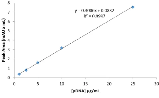
Figure 10. Calibration curve with plasmid DNA standards (1-25 μg/mL) ... 30Figure 11. Calibration curve with protein standards (0.2-10 μg/μL) ... 32
CHAPTER 4 – Results and discussion
Figure 12. Chromatographic profile of the E. coli clarified lysate sample injected in the arginine monolith and agarose electrophoresis of the different peaks obtained ... 36Figure 13. Evaluation of the flow rate effect in sc pDNA purification ... 37
Figure 14. pH influence on the sc pDNA purification with the arginine monolith ... 38
Figure 15. Example of the chromatographic profiles obtained in the analysis with the CIMacTM analytical column. ... 41
xx
Figure 17. Residual plot or recovery and purity ... 47
Figure 18. Predicted versus Actual plot for recovery and purity ... 48
Figure 19. Surface plot and contour plot for recovery response ... 51
Figure 20. Surface plot and contour plot for purity response ... 52
Figure 21. Chromatographic replicate runs for model validation of the optimal point ... 57
Figure 22. Western Blot for E6 and E7 proteins ... 59
Figure 23. CHO-1 cells immunocytochemistry images for E6 staining ... 61
Figure 24. CHO-1 cells immunocytochemistry images for E7 staining ... 62
Figure 25. E6 protein immunofluorescence comparison between the different groups (pvalue<0.001) ... 64
Figure 26. E7 protein Immunofluorescence comparison between the CT group and sc pDNA transfected group (pvalue<0.05) ... 66
xxi
xxiii
List of Tables
CHAPTER 1- Introduction
Table 1. DNA-based therapy clinical trials ... 2
Table 2. Classification of several HPVs. ... 9
Table 3. Specifications for pDNA to be considered as safe for administration. ... 17
CHAPTER 4 – Results and discussion
Table 4. Chromatographic conditions for pH range assessment ... 39
Table 5. Chosen factors versus levels of CCF design ... 42
Table 6. CCF design for three levels ... 43
Table 7. Designed experiments according to CCF design ... 43
Table 8. Responses of sc pDNA recovery and purity obtained for each run defined by CCF design ... 45
Table 9. Statistical coefficients of the model ... 46
Table 10. Summary of the main effects ... 49
Table 11. ANOVA table for recovery response ... 54
Table 12. ANOVA table for purity response... 55
Table 13. Optimal range for recovery and purity maximization ... 56
Table 14. Responses obtained from the optimization/validation of the model ... 58
Table 15. Confidence intervals for both responses ... 58
Table 16. Immunofluorescence for CT, kit, oc and sc pDNA transfected groups for E6
protein (n=3) ... 63
Table 17. Immunofluorescence for CT, kit, oc and sc pDNA transfected groups for E7
xxiv
xxv
List of Acronyms
ANOVA Analysis of variance
µg Microgram
µL Microliter
µm Micrometer
APCs Antigen-presenting cells
BCA Bicinchoninic acid assay
BSA Bovine Serum Albumin
CCF Central Composite Face
ºC Centigrade
CI Confidence intervals
CIN Cervical intraepithelial neoplasia
Cl- Chloride ion
CO2 Carbon dioxide
CR Conserved regions
CT Control
DNA Deoxyribonucleic acid
E. coli Escherichia coli
E6AP E6 Association Protein
ECF Enhanced chemifluorescence
EDTA Ethylene-diamine tetraacetic acid
xxvi
F Ratio of means of squares
FBS Fetal bovine serum
FDA Food and Drug Administration
gDNA Genomic DNA
HCl Hydrochloric acid
HPV Human Papilloma Virus
IMAC Immobilized metals affinity chromatography
K2HPO4 Dipotassium phosphate
kDa Kilo Daltons
KH2PO4 Monopotassium phosphate
LAL Limulus amebocyte lysate
Ln Linear
M Molar
mA Miliampere
mAU Miliabsorbance units
MHC Major histocompatibility complex
min Minute
mL Mililliter
mM Milimolar
NaCl Sodium chloride
NaOH Sodium hydroxide
nm Nanometer
xxvii
OD600 Optical density at 600 nm
OH- Hydroxide ion
ORFs Open reading frames
PBS Phosphate buffered saline
PBS-T 0.1% PBS with 0.1% Tween
PCR Polymerase chain reaction
pDNA Plasmid DNA
PEI Polyethylenimine
pKa Acid dissociation constant
PLL Poly-L-Lysine
PMSF Phenylmethylsulfonyl fluoride
pRB Gene product of retinoblastoma tumour suppressor
p-value Significance probability
RNA Ribonucleic acid
rpm Rotations per minute
sc Supercoiled
SCC Squamous skin cancer
SDS Sodium dodecylsulfate
TAE Tris-acetate-EDTA
TBS-T 0.1% Tris-buffered saline solution with 0.1% Tween 20
THAC Triple-helix affinity chromatography
Tris-EDTA 10 mM Tris-HCl and 10 mM EDTA
xxviii
V Volts
v:v Volume:volume
1
CHAPTER 1 - Introduction
1.1 DNA-based therapy
Science has evolved in the past century at an astonishing velocity. Great discoveries such as the DNA structure by Watson and Crick [1], in 1953, allowed the Science progress at a faster rhythm, changing thoughts and breaking dogmas along the way. DNA is responsible for the coding of all the information within the cell machinery, allowing the synthesis of proteins and other cell components. From the Stanley Cohen’s successful attempt in 1973 to join DNA molecules with different origins into a plasmid construct [2], the DNA recombinant technology was born and promptly grew throughout the years. The possibility for the arrangement of different genes with different functions permitted to establish the conditions for the hatching of a new area in Genetic Engineering, the DNA-based therapy. The possibility of administrating genes in order to achieve a therapeutic effect started to draw attention, rising as a promising pathway for therapy of several pathologies. Unravelling the human genome sequence in 2001 [3] allowed to understand all the information contained in the human genome, opening new horizons in the area of DNA-based therapy. Nowadays, DNA-based therapy can be subdivided into two widely studied areas, the gene therapy and DNA vaccines. Both DNA-based approaches are further discussed below.
1.1.1 Gene therapy
In the last decades, gene therapy has been targeted as a promising treatment for acquired diseases and genetic disorders. Ever since the evolution boost in molecular biology, other related fields gained some benefits, for example the production of considered amounts of nucleic acids through the use of polymerase chain reaction or the progression of the biotechnology field towards the improvement of transfection technology, which were permitted by the study and access to several different methods and techniques [4]. Gene therapy is based on the transfection of foreign nucleic acids encoding therapeutic information into the host cells, leading to a signal capable of correcting the target malfunction [5]. Therefore, this therapeutic design can be used for several purposes such as to add, eliminate or modify a particular function, sequence or expression of a given gene [6]. The ideal gene therapy vector should be able to carry the DNA information necessary to correct the target impairment, efficiently transfect the target cells or tissues and maintain an adequate expression or inhibition, without raising any biosafety concerns [7-9]. Nowadays, the areas where gene therapy may be applied are widening, although cancer diseases remain the most studied and, currently, represent the area with more ongoing gene therapy clinical trials worldwide, as portrayed in table 1.
2
1.1.2 DNA vaccines
Conventional vaccines were based on administration of weakened or modified infectious agents to healthy individual only to prevent a specific disease, promoting antibody immunity [11]. Owing to the substantial morbidity and mortality associated with particular diseases worldwide, such as tuberculosis, malaria, leishmaniasis, human papillomavirus (HPV) infection or human immunodeficiency virus infection, an understanding of the mechanisms involved in generating long-lived cellular immune responses arises like a critical point [12].Therefore, a new form of vaccination, by using DNA vaccines, can stimulate both humoral and cellular immune responses [12, 13].
DNA vaccines consist in a novel approach in the immunology field, taking advantage of genetic information that is delivered to the system able to induce an immune response against a given antigen. Upon inoculation, the individual shall produce a strong and enduring immune response against the encoded protein antigen, associated to the pathology [14]. The pathway by which DNA vaccines are able to trigger an immune response begins with the transfection of antigen-presenting cells (APCs) and non-APCs, leading to the expression of the Table 1. DNA-based therapy clinical trials (adapted from [10]).
Indications Number % Cancer diseases 1274 63.8 Cardiovascular diseases 162 8.1 Gene marking 50 2.5 Healthy volunteers 52 2.6 Infectious diseases 164 8.2 Inflammatory diseases 13 0.7 Monogenic diseases 178 8.9 Neurological diseases 37 1.9 Ocular diseases 31 1.6 Others 35 1.8 Total 1996
3
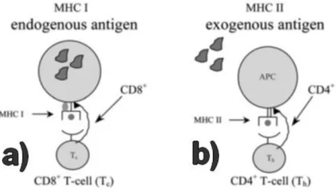
antigen codified in the DNA-based vector. As depicted in figure 1.(a), transfected cells may present the antigen, through Major Histocompatibility Complex (MHC) class I, to CD8+ lymphocytes [15, 16]. The target antigen production by non-APCs results in its exogenous take up by APCs, which will present the antigen to CD4+ lymphocytes via MHC class II, as can be seen in figure 1.(b) [15-17]. Such antigen presentation leads to the rise of a solid immune response [18].
Figure 1. Schematic representation of APC pathways. A) Antigen presentation by MHC I. B) Antigen
presentation by MHC II (adapted from [16]).
Beyond the capacity of DNA vaccines to generate all types of desired immunity, other advantages are related with DNA manufacturing, which can be easily obtained in large quantities with great purity, minimizing the risk of vaccine contamination with potential pathogens, being safety for a broad population administration [12]. In addition, the multivalent vaccination by administration of several antigenic genes reduces the total number of vaccinations that must be administered [19]. Furthermore, DNA vaccines also provide the potential for longer-lived antigen exposure in vivo, which could lead to increased immunogenicity, and could be easily transported and stored, because DNA provides more stability than other biological molecules [20].
Vaccination progress revealed that DNA vaccines can be used not only for preventive immunization but also as a versatile method to control and treat cancer [21-23]. It is estimated that, in 2012, 8.2 million people worldwide died of cancer, according to World Health Organization, and part of this problematic is caused by bacteria and viruses infection. Such numbers indicate that it is urgent to focus in alternative treatment strategies and recent studies suggest that DNA vaccines might be a partial answer for this problem.
4
1.1.3 Biological vs non-biological DNA therapy approaches
Nucleic acids, viruses or genetically engineered microorganisms, according to Food and Drug Administration (FDA), are an example of different vectors used for DNA therapy. Each vector present different advantages and drawbacks and can be divided in two separate categories, the biological and non-biological vectors [24].
1.1.3.1 Biological approaches
Viruses and bacteria are two different types of vectors that fit in this group. While viruses have been extensively studied and described in DNA therapy, bacteria have not yet been able to draw much attention in this area. Viruses represent 67% of the currently vectors in use in DNA therapy trials, as represented in figure 2. The strong application of this sort of vector might be related to the profound study of viruses in gene therapy, being referred to as the first-generation therapy vectors [9].
Figure 2. Vectors used in DNA-based therapy trials (adapted from [10]).
In general, the ability to easily infect the host cells is the main benefit of viruses in DNA therapy, contributing to increased gene expression levels and representing the key factor for viruses popularity. Different viruses are known to be used as vectors, however, adenoviruses and retroviruses stand out for being the most used. Adenoviruses are characterised by high efficiency transduction rates, capacity to transduce non-dividing and dividing cells and oncolytic power [9, 25, 26]. These appealing characteristics are probably the main reason behind the extensive study of this virus. However there are some drawbacks that cannot be overlooked.
5
In 1999, gene therapy faced the first death of a patient undergoing gene therapy treatment for nitrogen metabolism disorder ornithine transcarbamylase deficiency with an adenovirus which carried the gene for ornithine transcarbamylase [27]. Adenoviruses, being human pathogens, are highly immunogenic and its administration may be seen by the host defences as an infectious agent, triggering an immune response and, thereby, limiting its effectiveness and following administration, as well as compromising the patients health [9, 27, 28]. On the other hand, retroviruses present very low immunogenicity and are easily manipulated [25]. Nevertheless, their ability to randomly integrate the host cells in a permanent way raises several biosafety concerns [25]. The insertion of foreign nucleic acids into the host genome may disrupt important genetic information or be placed near a strong promoter, leading to its overexpression, perhaps contributing to the development of major health issues [26]. Another drawback in the use of viruses as DNA therapy vectors is the very small amount of DNA that they can carry, limiting the number and the size of genes to be inserted, and the high cost in its production [4].
Bacteria are microorganisms which present several properties useful for their application as DNA therapy vectors. Since 1980, when it was first reported the transfer of DNA into mammalian cells by bacteria [29], the study of bacteria in DNA therapy has come a long way. Nowadays, bacteria might be used in DNA therapy through two different approaches, namely, bactofection and tumour-specific bacterial replication [24]. Bactofection consists in the transfer of DNA into mammalian cells through the use of bacteria [24, 30, 31]. This procedure usually relies on using invasive type bacteria, which are capable of fully entering the target cells, and so deliver the genetic material [24, 30]. The tumour overgrowth can lead to the development of hypoxia, a condition which consists in low levels of oxygen due to insufficient blood supply [24, 30]. Under these circumstances, the use of anaerobic bacteria is useful in targeting tumour tissues, providing a specific delivery system of genetic material. Therefore, different non-invasive bacterial strains may be genetically engineered in order to secrete therapeutic proteins within the tumour cells [24, 30]. Invasive bacterial strains may also be used with the purpose of improving bactofection targeting [24]. However, safety issues associated with immunogenicity, bacterial sepsis and reversion of pathogenicity, suggest that further improvements are necessary [31].
1.1.3.2 Non-biological approaches
In the past years, non-biological vectors and techniques have been widely used, and their popularity is expected to keep rising. In 2004, 14% of DNA-based therapy clinical trials were developed with non-viral techniques, increasing to 18.3% in 2012 [32]. Although the general transfection efficiency of non-biological approaches is usually lower than biological vectors, non-biological techniques present certain characteristics that definitely surpass the apparent drawbacks. The main advantage of using such methodology relies on the biosafety of the
6
transfection procedure, besides allowing to transfect larger amounts of DNA at a reduced cost [33]. Plasmid DNA (pDNA) is currently very popular as a non-biological vector and has been widely studied in the biotechnology field. This double-stranded biomolecule has a simple low cost manufacture and is able to maintain acceptable expression levels, without presenting signs of pathogenicity [8, 32]. This vector is minutely described below, in section 1.3.
In general, the cellular entry of naked DNA is quite difficult, resulting in poor transfection and gene expression [5, 9, 34]. In order to overcome this limitation, naked DNA has been coupled with several physical and chemical methods which are able to increase the naked DNA cellular uptake.
1.1.3.2.1 Physical methods
Needle injection is a physical method with clinical interest for direct injection of naked DNA into tissues, organs or blood streams. The use of this methodology has been specially directed for DNA vaccines, due to the triggering of the immune system through the rupture of the skin by the needle [35]. Although needle injection has its advantages, like its simplicity and lack of toxicity, the poor level of gene expression has lead researchers to search for other alternatives [5].
Gene gun transfer is also a physical method often used when it is necessary to transfect skin, mucosa, surgically exposed tissues or tumour cells [5, 8]. This methodology consists in using gold particles coated with the target DNA [8]. Afterwards, the particles shall be accelerated through pressurized gas and expelled onto the target cells or tissue, delivering the DNA directly to the cytoplasm [8, 36]. Gene gun transfer is an appealing technique, especially for its simplicity and relative safety [8]. However, there are some drawbacks concerning the transient gene expression, which may imply several applications in order to achieve the therapeutic effect [5, 37].
On the other hand, electroporation is another physical method capable of successfully increase transgene expression in several types of tissue [5, 8]. Through the use of electrodes, a high-voltage electrical current is applied to the target cells, leading to the appearing of nanometric pores by which the naked DNA can enter into the cell [5, 38]. This technique allows delivering large DNA molecules, besides sustaining transgene expression for over a year [5]. Nevertheless, several disadvantages arise when using electroporation, namely the limited transfection area, tissue damage due to high voltage applied, and obligatory surgical procedure for treatment of internal organs [5, 8, 39].
7
1.1.3.2.2 Chemical methods
Cationic lipids represent one of the most commonly used complex agents for non-biological gene delivery, due to its lack of pathogenicity, low price and relatively simple production [5, 39, 40]. Cationic lipids include liposomes, closed spherical constructs which contain one or more structures of concentric phospholipid bilayers surrounded by an aqueous phase [40]. Plasmid DNA, when combined with liposomes, condenses into small particles named lipoplexes, which will protect the nucleic acids from enzymatic degradation and improve transfection efficiency [8]. Even though cationic lipids have been the subject of profound study in the past years, the successful mechanism of gene delivery and expression is still not fully understood. It is certain that factors such as chemical structure, size, surface charge, colloidal stability and others, contribute of transfection efficiency improvement [5, 8, 39]. After binding to cell surface due to opposite charges between lipoplexes and the cell-surface, the lipoplexes internalization occurs mainly by endocytosis [5, 34, 39]. In order to evade the lysosomal degradation, the endosomal release is necessary. Lipoplexes are known to destabilize the endosomal membrane, particularly with the help of co-lipids, which are able to relocate DNA contained in lipoplexes into cytoplasm, allowing the nuclear entry of DNA, since lipoplexes are too large to pass the nuclear pores [5, 8, 34, 39]. While this DNA therapy approach has been target of major improvements throughout the years, there are still some concerns regarding lipoplexes toxicity and the rapid plasma clearance [7].
Cationic polymers popularity has been growing in the past years due to its ability to condense nucleic acids through mild electrostatic interactions [41]. Nonetheless, different polymers result in different transfection activity and underlying toxicity. For instance, chitosan is one of the most studied non-biological gene carrier, due to its biosafety, even in high concentrations, and ability to effectively bind and compact DNA. However, it has low delivery efficiency in most cell lines, a drawback that has been trying to be surpassed by production of chitosan derivatives in order to improve its transfection ability [4, 41]. On the other hand, Poly-L-Lysine (PLL) was one of the first polymers to be utilized in gene delivery. Its charged amine groups interact with negative charged DNA, resulting in a good packaging of the DNA into nanosized particles. Although the uptake of PLL-DNA complexes is effective, the endosomal escape is still an obstacle when using such cationic polymer, leading to poor transfection activity. Polyethylenimine (PEI) is another polymer often used as a gene delivery carrier. Although its transfection rates in vivo are quite satisfactory, this polymer is nonbiodegradable, which may lead to an increase of toxicity in the cellular environment [8].
1.1.4 Ongoing clinical trials
Nowadays there are 1996 ongoing clinical trials focused on DNA therapy. The majority, representing 63.8% as depicted in table 1, are directed towards cancer diseases. Most of the 1274 cancer diseases targeting DNA-based therapies rely on the use of non-viral techniques.
8
As mentioned before, DNA vaccines have great potential in the treatment of such pathologies, due to the creation of an immune response towards to a target antigen.
For instance, Koen Oosterhuis and colleagues, have an ongoing trial with a DNA vaccine, consisting in the administration of naked pDNA which encodes a fusion protein domain1 of tetanus toxin fragment C and the shuffled version of the HPV E7 oncoprotein in order to produce E7 specific T cell immunity for the treatment of stage IV squamous cell cancer patients [42, 43]. Another example is the work developed by Cornelia Trimble and colleagues, which has an ongoing trial for the treatment of patients with stage III or IV HPV 16-positive head and neck squamous cell carcinoma, based on the development of a DNA vaccine, which also targets an immune response towards E7 oncoprotein through the administration of pDNA pNGVL4a-Sig/E7(detox)/HSP70 [44]. The E7 oncoprotein is produced when an infection by Human Papillomavirus (HPV) occurs and is responsible for transforming the host’s cellular cycle, coupled with E6 protein, as it shall be further described. This problematic issue has drawn attention from several researchers, whom have been working in the development of different DNA-based therapies toward such oncoproteins [23, 45, 46]. With this in mind, our research group has focused in the development of a DNA vaccine able to encode both oncoproteins E6 and E7 with the purpose of potentiating an immune response towards both antigens, preventing the emergence of a future infection and helping in the treatment of an ongoing infection.
9
1.2 Human papillomavirus
The link between the development of several types of cancer with the infection by HPV has been extensively described in the past years. Cervical cancer is the second most common cancer amongst women, according to the World Health Organization, and a recent study made in Brazil found HPV DNA in 99% of 172 cases of invasive cervical cancer [47].
Besides cervical cancer, HPV has been also associated with anal, vaginal, vulval, penile, head and neck cancers [49]. There are several types of HPV, which are identified by DNA sequence similarity and reflect on the tissue tropism of the virus. HPV can be divided into two different phylogenetic groups, the alpha genus and the beta genus. While alpha HPVs infect mucosal tissues, beta HPVs infect cutaneous tissue, leading to different clinical outcomes [50]. HPV can also be categorized by risk of inducing cancer, being thus classified as high, intermediate and low risks. Table 2 serves as an example of classification for different HPVs.
Table 2. Classification of several HPVs (adapted from [48]).
Group Prototypes Site of infection
Acute
consequences
Chronic
consequences Other features
Cutaneous (alpha)
HPV1, HPV2 Skin Warts None
Synchronous regression, lasting immunity Mucosal (beta) HPV6, HPV11, Genital
mucosa Warts None
Slow resolution in immunosuppressed individuals Mucosal High risk (beta) HPV16, HPV18, HPV31, HPV33, HPV45 Anogenital mucosa (other mucosa surfaces) Flat lesion (CIN 1) ~2% persist, ~1% progress to invasive cancer Slow resolution in immunosuppressed individuals, variable malignant potential Cutaneous High risk (alpha)
HPV5, HPV8 Skin Flat lesion or
none warts Promotes SCC
SCC more common in
immunosuppressed individuals
10
HPV 16 and HPV18 are high risk HPVs, which are usually associated with development of cervical cancers. Therefore, the development of an efficient therapeutic against these HPV types gains a significant importance, in order to prevent or eliminate an infection by HPV.
1.2.1 Human papillomavirus molecular biology
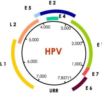
Human Papillomavirus is one of the most common viruses responsible for sexually transmitted infections, despite not being only sexually transmitted [51]. Around 40 different types of HPVs can be found in the genital tract, from over 100 HPV types molecularly characterized [52]. This virus is strongly associated with the transformation of infected cells, what might lead to solid replication of abnormal cells and, consequently, to the tumour outgrowth [53]. The HPV genome, represented in figure 3, apart from being responsible for encoding 8 proteins, 6 of them at an early stage (E1, E2, E4-E7) and 2 of them at a late stage (L1 and L2), has a non-protein coding upstream regulatory region (URR) which contains cis-elements and is able of regulating the gene expression, genome replication and viral packaging [52]. While the early proteins are associated with the infection and transformation of the infected cells, the late proteins are accountable for the spread of the infection in the host system.
Figure 3. HPV genome structure. HPV genome is represented as a 7857 base pair circular DNA molecule.
The open reading frames (ORFs) for the 8 proteins encoded are represented in different colours. The blank space between L1 and E6 ORFs represents the upstream regulatory region [52].
In figure 4, it is represented the evolution of HPV infection. Through microabrasions, the virus is able to penetrate the epithelium [48]. The expression of proteins E1 and E2, upon infection of undifferentiated basal cells, is responsible for the regulation of viral replication and expression of the other early stage proteins [54]. Hereafter, the expression of oncoproteins E5, E6 and E7 leads to the uncontrolled cell proliferation, cell survival and keratinocyte cell
11
differentiation. In cancers associated to HPV, E6 and E7 are responsible for the cancer phenotype [53, 55]. L1 and L2 are components of the viral capsid, allowing the assembling of the virus particles, which will be released within the superficial epithelial cells [48].
Figure 4. Evolution of infection by HPV [48].
1.2.2 Papillomavirus E6 oncoprotein
Papillomavirus E6 oncoprotein is a 151 amino acid protein characterized by two zinc finger domains, with a molecular weight of 18 kDa [56, 57]. The main action of this oncoprotein in the cellular cycle of the infected cells involves the ability of E6 protein to interfere with tumour suppressor p53, hindering the p53 protein capacity to trigger signalization pathways for cell repair and apoptosis [58]. The disabling of p53 contributes to continuous replication of damaged DNA and survival of abnormal cells, which would usually be repaired when p53 is expressed in normal levels. There are several mechanisms by which E6 protein is able to incapacitate p53 protein role [55, 59]. E6 is known to bind to E3 ubiquitin ligase, commonly known as E6 Association Protein (E6AP), through LXXLL motifs. E6AP is unable to bind to p53 without E6 oncoprotein. The docking of such proteins leads to the recognition of p53, preventing the binding of p53 to specific sequences of DNA [56, 60].
In addition, the association between E6AP-E6 complex to p53, ubiquitinate p53 leading to its degradation by the proteasome, as shown on figure 5 [61]. On the other hand, E6 oncoprotein is able to affect p53 functions through hindering of transactivation of p53 responsive genes. Such mechanism may be seen, for instance, through the p300 interaction with E6. The
12
promotor p300 is capable of acetylation of p53 when cellular damage exists, leading to higher recognition of specific DNA sequences by p53. The binding of E6 to p300 obstructs p53 acetylation, assisting in the expression decrease of responsive p53 genes [59, 60]. High risk HPV E6 proteins show increased affinity to p53, resulting in greater levels of inhibition, therefore contributing to a tumorous phenotype unlike low risk HPVs [59].
Figure 5. Degradation of p53 in the presence of HPV 16 E6 protein (adapted from [61]).
1.2.3 Papillomavirus E7 oncoprotein
Papillomavirus E7 oncoprotein is a 98 amino acid protein with a molecular weight of 16-17 kDa, which comprises three functional conserved regions (CR) [57, 62]. CR1 is associated with transformation of cell cycle independent of the gene product of retinoblastoma tumour suppressor (pRB), while CR2 is linked to the transformation of cell cycle by binding to pRB through its LXCXE motif [57]. The main transforming pathway of E7 oncoprotein relies on the binding to pRB, disrupting its binding to transcription factors such as E2F. Usually, pRB has the ability to phosphorylate E2F, forming a pRB-E2F complex which is capable of interfering with the progression of the cellular cycle. When the binding of E7 to pRB occurs, E2F disengagement results in stimulation of E2F responsive genes, which are responsible of cellular cycle transition to S phase [54, 57]. CR1 and CR3 domains of E7 protein are involved with destabilization of p107 and p130, two proteins which are also able of forming complexes with E2F. The association of E7 protein to pRB, p107 and p130 leads to uncontrolled cell proliferation and progression to malignant transformation [57, 62]. Similarly to E6 and p53 binding, high risk HPV E7 proteins show increased affinity for pRB binding [54].
13
1.2.4 Preventive and therapeutic vaccination
The rising of awareness for prevention of HPV infection has been increasing in the past years. Screening tests, such as pap smears, have been implemented as a routine test, contributing to an early diagnose and a decrease in the cancer mortality [51]. Prophylactic vaccines have been developed for prevention of infection. Its functionality resides in the administration of viral-like proteins (VLPs) that shall mimic a natural HPV infection. These VLPs consist in HPV late protein L1, a constituent of the viral capsid [63]. So far, there are only two available vaccines commercialized for the prevention of HPV infection: Gardasil and Cervarix [51]. Up to now, efficacy of prophylactic vaccines has been point out to be high [63]. However, it is estimated that the sexual behaviour of patients undergoing vaccination has a major influence in the outcome of the vaccines efficacy [51, 54]. The risk of infection increases with the number of lifetime sexual partners of the patient. However, the main failure of the VLPs therapy is related with the vaccination of a person already infected with HPV, which shall not produce any kind of effect within the individual [54].
Therefore the investment in a therapeutic alternative is extremely necessary, which might be able to prevent and, at the same time, treat infection by HPV. Nowadays there are several ongoing investigation and clinical trials regarding therapeutic vaccination treatments for HPV infection, taking mostly advantage of the previously described DNA vaccine technology. Each research has focused on different antigens and different antigen modifications in order to produce the ideal therapeutic HPV vaccine. Nonetheless, it is undeniable the preference for choosing E6 and E7 as target antigens for immunization and treatment responses [18, 45, 46, 64-66]
14
1.3 Plasmid DNA technology
Plasmid DNA has been widely studied throughout the years for its use as a successful cloning tool and for its ability in the eukaryotic cell transfection and subsequent induction of proteins expression [67]. Its capacity for carrying DNA information and lack of toxicity led this biomolecule to be strongly exploited as a DNA-based therapy vector, currently resulting in the most popular non-viral technique used in DNA-based therapy clinical trials, as previously depicted in figure 2 [68]. This circular double-stranded DNA molecule has bacterial origin and its size varies until 120 kilo base pairs, allowing this vector to carry larger DNA amounts than viral systems [28, 67, 69, 70].
The pDNA can be presented in different isoforms, the supercoiled (sc), open circular (oc), linear (ln), oligomeric or denatured isoforms which can occur depending on DNA sequence and different environment conditions. However, the sc conformation is the most produced isoform by Escherichia coli (E. Coli) host. The increasing of temperature, for instance, may lead to the uncoiling of the double DNA helix, promoting the conversion of the sc pDNA isoform to other pDNA isoforms. Also, undamaged sc pDNA can be converted to other isoforms through random enzymatic cleavage [71]. The isoform with target biological function is the sc pDNA, which has been proven to provide greater transfection rates than other isoforms [72, 73]. Thus, the sc pDNA isolation from the non-effective pDNA topologies and from the host components, that may be unveiled as toxic, is extremely important.
1.3.1 Construction, production and primary isolation of pDNA
The construction of a recombinant plasmid was firstly reported by Stanley Cohen and colleagues in 1973, making use of restriction enzymes, which are able to recognize specific DNA sequences and consequently cutting it, and DNA ligase in order to re-attach such DNA sequences [2]. A pDNA vector shall comprise an origin of replication for efficient cloning of the molecule in the target bacterial host, a strong promoter for expression in eukaryotic cells, the therapeutic target gene, an antibiotic resistance gene and finally, a polyadenylation termination sequence. The origin of replication is necessary for the amplification of the pDNA molecule within the bacterial host. The introduction of a gene capable of inducing a specific-antibiotic resistance is necessary to guarantee the solely growth of transformed bacteria with the target pDNA. The polyadenylation termination sequence is important for the translated RNA protection from degradation[74]. In figure 6 it is schematically represented the plasmid which was chosen to be manipulated throughout the present work.
15
Figure 6. Schematic representation of HPV-16 E6/E7 pDNA. (plasmid 8641, adapted from [75]).
Fermentation allows the production of pDNA in transformed bacteria, by adjusting the growth conditions according to the chosen bacterial host, which is usually E. coli [67, 76]. Once the target optical density of transformed bacteria is achieved, it is necessary to proceed to the downstream processing of the cell lysate in order to obtain the biopharmaceutical pDNA. Cell lysis should be performed through mechanical or chemical methods. The most used method is the alkaline lysis, which relies on the use of basic solutions capable of disrupting the cells membrane, releasing the pDNA and all cell components. Concentration and washing steps of the lysate sample can be performed to recover a clarified lysate to be further purified [77]. In order to achieve the suitable purity of the target biomolecule, the chromatography field has very much evolved in the past years, providing us a variety of techniques for such purpose. In figure 7 it is represented the main steps by which a target pDNA shall pass in order to accomplish a high pharmaceutical grade.
16
Figure 7. Production, recovery and purification of pDNA in order to achieve high pharmaceutical grade
for application [69].
1.3.2 Plasmid DNA purification
Chromatography is an interesting area in biotechnology, providing methodologies and processes able to eliminate contaminants from a target molecule and purifying it. Impurities such as genomic DNA (gDNA), oc plasmid and RNA represent a hurdle to the sc pDNA purification that shall be surpassed through the aid of chromatography techniques. This methodology is very important to guarantee the compound safety for application. In matter of fact, purification of DNA vaccines has major significance and contributes to most of the expenses associated with vaccine development. In this way, the constant development of suitable chromatographic strategies to isolate the sc pDNA from a lysate sample in a single step is required, in order to reduce the costs associated to the DNA vaccine manufacturing. Table 3 represents the several specifications which pDNA must fulfil in order to be safely administrated. To achieve such specifications, several chromatographic techniques have been explored, taking advantage of biomolecule characteristics, namely size, charge, hydrophobicity or affinity between the different molecules present in the extract to be purified and the chromatographic ligands used [78].
17
Size exclusion chromatography relies in the separation of different biomolecules according to their molecular size [79]. The greater the biomolecules are (as for instance the gDNA and pDNA) the less time it will take to pass through the support, since they do not enter to the pore particles, while smaller molecules like RNA will need more time [80, 81]. Nonetheless, such technique is unable to separate different isoforms of pDNA, showing poor selectivity and capacity for this molecule [76, 78].
On the other hand, anion exchange chromatography is known to fractionate the sample elements by charge differences. The principle behind this chromatographic process is based on the attraction of the target molecule to its opposite charge, immobilized into the stationary phase [81, 82]. Polymeric amines are usually the ligands used in anion exchanger chromatographic supports for pDNA purification [69]. After the sample binding, the salt concentration should gradually vary in order to elute the different molecules bound to the support. Nonetheless, some hurdles arise with the use of such technique due to its inability to distinguish different molecules with similar chemical and physical properties [81].
Hydrophobic interaction chromatography, also a very popular chromatography procedure, relies on the hydrophobic interaction between the target molecule and the chromatographic support. High concentrations of salt shall be applied for the sample binding, followed by a gradual decrease of ionic strength in order to separate the molecules with an increasing Table 3. Specifications for pDNA to be considered as safe for administration (adapted from [78]).
Characteristics Specifications Appearance Clear Colourless solution Plasmid Homogeneity >97% sc
Proteins Not detectable, via BCA
RNA Not detectable, via 0.8% agarose gel
gDNA <2 µg/mg plasmid, via PCR
18
hydrophobicity degree [76, 81]. Even so, the use of high salt concentrations represents a downside due to the high costs and the environmental impact associated [81].
Affinity interactions between target molecules and certain ligands have been object of intense study in the past years, leading to the development of affinity chromatography. Such technique is based on the specific interactions, as for instance molecular recognition, in order to selectively purify the target biomolecule [76]. The type of ligand used in this procedure should be chosen according to the biomolecule function or chemical structure [81]. The main drawback pointed to this chromatographic principle is the strong binding of the target molecule to the support, sometimes making it difficult to elute [83]. Within affinity chromatography, different strategies may be distinguished for the purification of pDNA, namely, immobilized metals affinity chromatography, triple-helix affinity chromatography, protein-DNA and amino acid-DNA affinity chromatography.
Immobilized metals affinity chromatography (IMAC) is based on the fractionation of molecules according to their affinity with metallic ligands of the stationary phase [76, 81]. RNA and single-stranded nucleotides show high affinity for the IMAC matrices, while gDNA, pDNA and other double-stranded nucleic acids have very low binding affinity [76, 81, 84]. This technique in particular can be used for polishing purposes, eliminating RNA and endotoxin impurities, but it has no significant importance in pDNA isoforms separation [76, 81].
On the other hand, triple-helix affinity chromatography (THAC) is based on the recognition of specific sequences of DNA by immobilized oligonucleotides, forming a triple-helix at acidic pH [76, 81, 85]. In comparison to IMAC, THAC has the advantage of being able to successfully fractionate different DNA isoforms, isolating the sc pDNA in one single chromatographic step. However, long chromatographic runs and low yields, in part due to the stronger retention of sc pDNA, suggest this might not be the ideal procedure for sc pDNA purification [81].
On the other side, amino acid-DNA affinity chromatography allows multiple interaction between the target DNA and the immobilized amino acid ligands, natural compounds whose chromatographic use provides an efficient single step chromatographic purification of sc pDNA and successful elimination of RNA, gDNA and other possible contaminants [76, 81]. Our research group has focused the purification of pharmaceutical-grade pDNA to be therapeutically applied, through the use of histidine, lysine and arginine amino acids as affinity ligands.
For instance, the sc pDNA purification with histidine and arginine agarose matrices suggested the involvement of different interactions between each amino acid and the different nucleic acids, revealing a selective retention according to the oligonucleotide bases sequence in the target biomolecule [86, 87]. Moreover, the isolation of sc pVAX1-LacZ from clarified cell lysate was successfully achieved, through the use of a histidine-agarose matrix, with an overall yield of 40% [88]. Likewise, through the use of lysine-agarose matrix, the sc
19
purity attained with this two amino acids, the recovery yields were relatively low. Meanwhile, the use of arginine-agarose to purify the sc pVAX1-LacZ revealed a recovery yield of 79%, with the recommended purity degree [90].
Overall, the arginine agarose chromatography seems to be a good strategy to purify sc pDNA, owing to the selectivity achieved with this ligand under mild elution conditions and the satisfactory recovery yield. However, conventional stationary phases present some limitations related with work at high flow rates and low binding capacity for large biomolecules, such as pDNA, therefore leading to the exploration of alternative supports.
1.3.3.1 Monoliths: a new chromatographic support technology
Monoliths are chromatographic supports that have been gaining attention in the past years due to attractive characteristics, which are responsible for the outstanding behaviour of this new technology in comparison with conventional stationary phases. Such innovative stationary phases consist in continuous beds, resembling several stacked membrane sheets [69]. These supports are highly porous, whose sizes are dependent of the polymerization temperature, and comprise an interconnected three-dimensional network, allowing that all channel surface to be convectively accessible [91]. Such structural characteristics result in faster mass transfer, higher access of target molecule to the total monolithic structure and higher working flow rates [69]. The greatest advantage of monolith utilization relies on the combination of the convective flow with its high interconnectivity, allowing a swift separation in short beds [92]. The use of monoliths in sc pDNA purification has great value, considering conventional supports limitations [69].
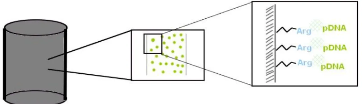
Taking into account the high specificity and selectivity of the arginine ligand by the sc pDNA, as well as the high versatility and capacity of the monolithic supports, the coupling of both strategies, by the immobilization of arginine ligand into the monolith, emerged as a promising solution for the pDNA vaccine purification [93]. Thus, the arginine monolith was able to purify sc HPV-16 E6/E7 pDNA through amino acid-DNA affinity chromatography (as it is schematized in figure 8). Although 100% of purity was achieved, only 39% of sc HPV-16 E6/E7 pDNA was recovered [94]. To improve such recovery yield, maintaining the high purity degree, the experimental design tool could be used.
20
Figure 8. Schematic representation of arginine modified monolithic support.
1.3.4 Design of experiments
To attain a high pDNA yield and purity, some tests of screening, optimization and robustness should be performed. Such achievement might be difficult by the usual ‘one by one’ method, in which factors are separately tested, revealing to be time-consuming and exhaustive. Design of Experiments, a statistical technique which allows to plan, conduct, analyse and interpret data from experiments, arises as a solution to such demanded procedure, permitting to evaluate different factors through the use of a small amount of experiments [95, 96]. The user usually chooses the different factors to take in consideration, as well as the range in which values should vary. This technique has different designs available that can be chosen accordingly to the final purpose of the experimental design, which will suggest different experiments that must be performed. This technique emerges as a useful tool to help investigators to achieve better results with few experiments and can be used in miscellaneous areas.
In the chromatography field, the application of experimental design is usually associated with analytical chromatography, particularly in high performance liquid chromatography and gas chromatography [95]. Nonetheless, our research group recently published the use of DoE in preparative chromatography, accomplishing the purification of pre-miR-29 with high purity and considerable yield through an O-phospho-l-tyrosine affinity chromatographic support [97]. For optimization purposes, there are several designs which can be chosen in order to achieve the target result. For instance, Central Composite Face (CCF), Box–Behnken or Doehlert are experimental design tools used with the intent of optimizing a given experiment. Central composite designs are usually the preferred strategy when choosing an optimization design, even though it requires more experiences than other optimization designs such as Box-Behnken or Doehlert designs [95]. CCF design can be a good choice to optimize a purification strategy, because this method provides a wider perspective of the optimum gap, when
21
compared for example to Box-Behnken, which excludes experiments that meet every higher or every lower level for all factors [96].
In figure 9 it is schematically represented the CCF design. Each dot represents a combination of different values of the different factors for one experiment. The black dots in the cube vertices represent the factorial design points in evaluation, while the centre black dots represent the star points, which in this design lie within the faces of the factorial design points [95]. In red is represented the centre point, which has to be replicated in order to provide a measure of pure error and stabilize the variance of the predicted response [96]. .
Figure 9. Schematic representation of CCF design (adapted from [98]).
CCF design is preferably used for optimization of experiments, which uses a full quadratic model that can be generally described as (n) = 2k–p + 2k + cp, where ‘k’ is the number of factors to be considered, ‘p’ the fractionalization number and ‘cp’ the number of centre points required for curvature estimation [99].
1.3.5 Plasmid application
Once the pDNA purity is guaranteed, further testing is necessary in order to assess the DNA vaccines success in transfecting the host cells, expressing the target gene and developing an immune response. With this in mind, several in vitro and in vivo procedures might be useful in assessing such information. Western blot and immunocytochemistry, for instance, are in
vitro procedures which allow the evaluation of the transfection efficiency and target gene
expression. Western blot technique allows to stain and quantify target proteins from a whole protein extract, while immunocytochemistry can be used to guarantee the pDNA entrance in the host cells by tracking the expression of the target gene, as well as quantifying it [100,
22
101]. Immunocytochemistry can also be used to monitor the nuclear structures, in order to verify if the transfection materials used are damaging the cells. On the other hand, in vivo studies are presented as the following step for pDNA application studies, permitting to verify the DNA vaccine ability to induce an immune response directed to the target antigens. Procedures such as flow cytometry for targeting of specific cytokines, serve as an example of studies that might be used to assess such application in in vivo experiments [102].
In this work, western blot and immunocytochemistry procedures were performed in order to assess the sc pDNA HPV-16 E6/E7 ability to transfect and produce the E6 and E7 target genes, also comparing its biological activity with other plasmid isoforms or plasmids prepared by using different purification methodologies.
25
CHAPTER 2 – Global Aims
The main objective of this work is to obtain a successful DNA vaccine for prevention and treatment of HPV infections. Such goal can only be reached if purification of the therapeutic molecule is achieved, considering the safety concerns regarding its administration. In order to accomplish this aim, the optimization of the implemented purification strategy for sc pDNA HPV16 E6/E7 has extreme importance, maximizing the performance of the purification process. In addition, the assessment of the purified target molecule ability for transfection is important, to evaluate if the DNA vaccine is able to perform its expected purpose. Hence, the goals of the present thesis are the optimization of sc pDNA HPV-16 E6/E7 purification by arginine monolith and evaluation of the transfection efficiency and gene expression ability of the purified molecule.
Therefore, the present thesis can be divided in two different steps:
- Establishment of an optimized purification strategy for sc HPV-16 E6/E7 by arginine monolith, in order to increase the recovery percentage of the target molecule, making use of design of experiments statistical tool.
- Transfection studies with purified sc HPV-16 E6/E7 pDNA molecule, as well as with the commercially obtained pDNA and oc pDNA, with the intent of assessing the transfection efficiency and gene expression ability of the different molecules.
![Figure 2. Vectors used in DNA-based therapy trials (adapted from [10]).](https://thumb-eu.123doks.com/thumbv2/123dok_br/18854274.929823/34.892.119.777.512.844/figure-vectors-used-dna-based-therapy-trials-adapted.webp)
![Table 2. Classification of several HPVs (adapted from [48]).](https://thumb-eu.123doks.com/thumbv2/123dok_br/18854274.929823/39.892.145.805.550.1108/table-classification-hpvs-adapted.webp)
![Figure 4. Evolution of infection by HPV [48].](https://thumb-eu.123doks.com/thumbv2/123dok_br/18854274.929823/41.892.145.756.244.635/figure-evolution-infection-hpv.webp)
![Figure 5. Degradation of p53 in the presence of HPV 16 E6 protein (adapted from [61])](https://thumb-eu.123doks.com/thumbv2/123dok_br/18854274.929823/42.892.290.568.315.561/figure-degradation-p-presence-hpv-e-protein-adapted.webp)
![Figure 7. Production, recovery and purification of pDNA in order to achieve high pharmaceutical grade for application [69]](https://thumb-eu.123doks.com/thumbv2/123dok_br/18854274.929823/46.892.253.600.51.435/figure-production-recovery-purification-order-achieve-pharmaceutical-application.webp)
![Figure 9. Schematic representation of CCF design (adapted from [98]).](https://thumb-eu.123doks.com/thumbv2/123dok_br/18854274.929823/51.892.324.564.387.654/figure-schematic-representation-ccf-design-adapted.webp)