O
R
I
G
I
N
A
L
A
R
T
I
C
L
E
In vitro
and
in vivo
cell tracking of PKH26-labeled
osteoblasts cultured on PLDLA scaffolds
Alice Rezende Duek
1, Gabriel Ciambelli Dias da Costa
1, Bruna Antunes Más
1,2, Maria Lourdes Peris Barbo
1,
Adriana Cristina Motta
2* and Eliana Aparecida de Rezende Duek
1,21
Laboratório de Biomateriais, Pontifícia Universidade Católica de São Paulo – PUCSP,
Sorocaba, SP, Brazil
2
Faculdade de Engenharia Mecânica, Universidade Estadual de Campinas – UNICAMP,
Campinas, SP, Brazil
Abstract
The importance of monitoring in vivo interaction that occurs between cells /bio/tissue recipient in the understanding of tissue regeneration processes becomes ever greater. This study aims to monitor and evaluate the inluence of scaffold implants of poly (L-co-D, L lactic acid) - PLDLA synthesized in the laboratory, previously cultured with primary osteoblastic cells heterologously stained with the luorescent vital dye, PKH26, on the tissue regeneration process in 8 mm central critical defects of the Wistar rat calvaria. The results obtained by MTT assay and monitoring of cells stained with PKH26 dye over 14 days of culture showed that the dye was cytocompatible with osteoblastic cells and did not exert a negative inluence on the growth of unstained cells. In the in vivo study, macroscopic observations made during deployment times corroborate the results in vitro, as no apparent signs of toxicity were observed in the implanted bone defect area. The use of mobile monitoring with the dye, PKH26 in vivo is an effective strategy for the understanding of cell behaviour in the presence of PLDLA polymer.
Keywords: PKH26, PLDLA, scaffolds, tissue engineering.
1. Introduction
The growth of tissue engineering and regenerative medicine in recent years reflects the efforts of researchers in the search for new strategies in an attempt to compensate for, as one example, the issue of limited availability of autologous tissue in procedures involving bone grafts. It is estimated that over 2.2 million grafts are performed annually worldwide for the repair of bone defects[1,2], and the growth forecast that surrounds this market reflects the significant volume of this industry, which was US $2.1 billion in 2013 and is projected to increase to $2.7 billion in 2020, according to Global Data.
Among the materials with the greatest potential to be used in tissue engineering, bioresorbable polymers belonging to the family of poly (α-hydroxy acids), such as poly (L-co-D, L lactic acid) (PLDLA), stand out for their properties such as biocompatibility and versatility in terms of degradation over time. Regarding the most efficient way to promote the organization, growth and differentiation of cells in the tissue formation process in injured tissue, the most appropriate type of devices are the scaffolds, which must meet a series of requirements, such as excellent biocompatibility, good mechanical properties and adequate porosity[3,4]. The degradation rate of the copolymer is intermediate between poly (L- lactic acid) and poly (DL- lactic acid), thus proving very interesting for most applications[5]. Furthermore the lactic DL- acid units in the copolymer chain sequence hinder the crystallization of lactic L- acid, while the strength of the material is maintained for the period necessary for recovering the treated tissue[6].
Understanding the wide range of cellular and molecular responses involved in an in vivo system is quite complex; this has often led to misunderstanding and erroneous or contradictory interpretation of the actual biological events that guide a biomaterial implantation success and therefore, which properties and features should be developed in the biomedical implantable devices[7].
The use of so-called “constructs”, which are scaffolds grown onto autogenous or allogenic bone cells employed in tissue engineering and aimed at providing a higher rate of regeneration in various tissues, but little is known about the biological function and behaviour of the transplanted cells in the in vivo environment, and unsatisfactory results are reported frequently, for example, low viability of the transplanted cells and immune response of the organ / tissue implant[8,9].
The monitoring of live biological interactions has been shown extremely important and the use of fluorescence microscopy techniques stands out in this sense, due to the ease of handling and low cost of application, both in relation to previous procedures for cell labelling, as does the cost of acquisition and maintenance of the equipment itself.
The use of PKH26 is extending to stem cell transplants for applications including diseases of the retina, myocardium and brain and bone regeneration[12,13].
In this work, the application of PKH26 dye, subjected to the method of evaluation and monitoring of polymeric constructs containing primary osteoblastic halogen cells, aims to contribute to the knowledge needed to develop new biomaterials and the preparation of strategies that provide a greater clinical success of the application of biomaterials focused on orthopaedics and bone regeneration processes.
2. Materials and Methods
2.1 Porous scaffolds
PLDLA 70/30 Solutions Lab synthesized in Biomaterials - PUC/SP for Motta and Duek[14] were prepared by dilution of the copolymer in methylene chloride (Merck) (10% w/v) at room temperature. To obtain scaffolds, PLDLA was added to the sucrose solution (Synth) (75% w/v) with controlled particle size between 200 and 500 μm. The solution was poured into a cylindrical mold of silicone with a diameter of 8 mm. After evaporation of the solvent and sucrose removal in a 1% solution of polyvinyl alcohol (PVA), scaffolds were dried under vacuum and stored in a desiccator.
2.2 Isolation and collection of osteoblastic cells
Osteoblastic cells were obtained by explantation of the calvarial bone fragments of three Wistar rats (250-300 g) at 20 days of age, according to the method of Declercq et al.[15].
2.3 Cell labelling
Distribution and cellular behaviour of cultured osteoblasts in the scaffolds in in vitro assays and in vivo were assessed by fluorescence microscopy. Prior to cell culture in the scaffolds, the cells were labelled with the fluorescent vital dye, PKH26 (General PKH26-GL cell linker kit, Sigma
) according to the manufacturer’s instructions. The cell
membrane of 2 x 10⁵ cells per mL were stained with the fluorescent marker and cultured in 48-well polystyrene culture plates containing pre-sterilized glass coverslips. After 24 and 14 days of culture, the coverslips were washed in PBS and fixed in 4% paraformaldehyde (PFA-Merck)[16]. Image acquisition and evaluation was performed using a NIKON ECLIPSE E800 fluorescence microscope (NIKON Instruments) with an excitation wavelength of 543 nm and an emission wavelength.
2.4 Growing cell scaffolds
The scaffolds of cylindrical PLDLA were cut into 0.5 mm thick slices, disinfected in 70% ethanol by 1 hour and rinsed in ultrapure water. Following cell staining with PKH26 dye, the stained cells were reseeded onto 96-well plates at a density of 2 x 10⁵ cells per mL and cultured on scaffolds for either 24 h or 14 days.
2.5 In vitro cell viability
2.5.1 MTT assay
Cell viability of osteoblasts marked by PKH26 dye was investigated by the metabolic MTT assay[17]. A concentration of 2 x 10⁵ cells per mL was seeded on supports and controls in standard DMEM, supplemented with 10% fetal bovine serum (FBS), and further incubated for 24 h in an incubator (5% CO2 at 37 °C). Then the medium was removed, the wells rinsed with 0.1 M PBS buffer and to each well was added 100 ul of DMEM medium containing 10 uL of 3-(4,5-dimethylthiazol-2-yl) -2,5 diphenyl tetrazolium bromide, MTT (5 mg/mL), followed by an incubation period of 4 h at 37 °C in the dark. After this time the solution containing MTT was replaced by 200 μL of dimethylsulfoxide (DMSO) solution, and 25 ul Glycine/Sorensen buffer. Afterwards, 100 ul of the solution contained in the wells were transferred to a new plate and the absorbance of MTT was determined at 570 nm by microplate reader Elx-800-UV (Bio-Tek Instruments, USA).
2.6 Preparation of implants
Cell viability and distribution of osteoblastic cells labelled by fluorescent dye, PKH26 was also investigated by fluorescence microscopy. After 24 h or 14 days of cultivation, the cured scaffolds were fixed in 13% gelatin solution, diluted with distilled water, plus 30% sucrose. The material was immersed in a solution containing 30% sucrose for a 2-3 day period and then frozen at -70 °C for 24 h and subjected to the criotomia technique in a cryostat (Shandon Cryotome E). Cryosections were obtained from
the series surface of the scaffold, allowing the monitoring of the distribution of cells marked with PKH26 for the process of proliferation and cell migration into the scaffold. The slides were photographed on a fluorescence inverted optical microscope (NIKON - E 800).
2.7 In vivo experiments
Thirty-four Wistar rats of both sexes (250-300 g) were used. Rats were divided into groups according to implant time (4, 8 and 12 weeks) and subdivided by treatment used, and only the 4-week time point (n = 7) was monitored for cells labelled by fluorescent dye, PKH26. The treatment groups were as follows: A control group was given pure PLDLA implants. The PLDLA group was given implants cultured with osteoblast cells marked by PKH26.
2.7.1 Implantation
extracted bone fragment (Figure 1C). Figure 1D illustrates the defect area and intact blood vessels, suggesting that there was no damage to the animal’s brain. The skin on the defect region was repositioned and sutured followed by local antisepsis (Figure 1F). All experimental procedures involving the use of animals were approved by the Ethics Committee on the use of animals in the Centre of Medical and Health Sciences at PUC/SP Sorocaba Campus (n.2013/13).
2.8 Processing of material for histological analysis
After fixation, the material was subjected to decalcification in EDTA solution (4.13%) for 21 days. Samples were prepared for histological analysis in accordance with techniques used for light microscopy, using wax as a means of inclusion. Bone fragments containing the implants were subjected to the following processes: dehydration in a sequence of three tanks containing ethanol solutions of increasing concentration (80, 90 and 100%) where they remained for 30 min; diafanization I and II in xylene for 30 min followed by soaking in liquid paraffin I and II at 60 °C for 30 min.
The material was embedded in cubic shapes measuring approximately 2.5 cm and filled with paraffin at 60 °C forming blocks that remained at rest for 24 h at room temperature to cure. We then trimmed the blocks for the following histological cutting process.
For in vivo monitoring of fluorescent cells, samples were fixed in a 13% hard gelatin solution diluted with distilled water plus 30% sucrose. The material was then immersed in a solution containing 30% sucrose for 2-3 days. The material was then frozen at -70 °C for 24 h and immediately subjected to freezing in a cryostat (Shandon Cryotome E).
Histological sections of 3 μm thickness were obtained using a Leica type RM2245 microtome, and stained with
hematoxylin and eosin (HE). The slides were photographed with a fluorescence optical microscope (NIKON - E 800).
The central region of each critical defect was subjected to histological analysis. To this end, the caps were divided into six sections following the coronal plane and covering the length of the defective region. With the aid of a calibrated examiner using an optical microscope fitted with incandescent and fluorescent light filters, observations and analysis of new bone formation, ACS waste, waste clot elements and tissue inflammation were carried out.
2.9 Fluorescent cell monitoring and analysis
The monitoring of fluorescent cells in vivo was carried out with the help of NIS -Elements Advanced Research software image analysis installed on an optical microscope (NIKON E800). Areas and fluorescent dots (corresponding
to labelled cells in culture) of the implant and new bone formation that exhibited fluorescence were detected and evaluated for distribution and cell migration inside the implants.
3. Results
The manufacture of polymeric scaffolds obtained by the porogeno leaching method is widely used for both in vitro
assays and in vivo, in order to assess the cytocompatibility, biocompatibility and influence of new materials on the biological behaviour of cells and tissues[18]. Some practical advantages of this method lie in its low manufacturing cost and pore size control, which are compatible with the biological specificity of each cell and tissue type[19].
3.1 In vitro study
3.1.1 Fluorescent labelling
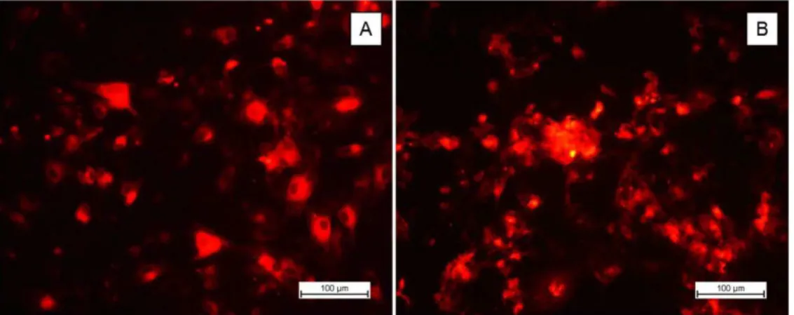
In order to monitor the manner of operation and colouring behaviour of PKH 26, primary osteoblast cells previously stained with PKH26 were grown on glass coverslips and monitored by fluorescence microscopy after 24 h and 14 days in culture (Figure 2). Cells were positive for the dye, PKH26, showing a fluorescence signal intensity sufficient for monitoring, which indicated PKH26 was suitable for use in this study.
After 24 h of cultivation (Figure 2A), PKH26-labelled cells were isolated to show the projections of well-defined cytoplasmic membrane and slightly rounded morphology or Splay typical of cellular adhesion to the substrate in the early stages of cultivation. In addition, there were no apparent signs of dye cytotoxicity, such as the appearance of cellular debris or altered cellular morphology.
After 14 days in culture (Figure 2B), the surfaces of all glass coverslips analysed (n = 3) were almost entirely covered with a confluent layer of cells exhibiting fluorescent signals with very similar intensity to that found after 24 h of cultivation. Due to the high density of overlapping cells adhered on the coverslip and the affinity of the dye for extracellular matrix lipophilic groups synthesized by the cells during the cultivation period, it was not possible to observe the division of the cytoplasmic membrane of osteoblasts or their morphology[20]. However, the presence of juxtaposed
Figure 1. Steps of the surgical procedure performed for the implantation of scaffolds: (A) incision to expose the parietal bone; (B) critical defect; (C) extraction of bone fragment; (D) defect area;
cell groups inserted into the extracellular matrix were clearly distinguishable in that they exhibited a sharp and strong bright red fluorescence intensity relative to the array[21].
3.2 Cell viability
According to the results presented in Figure 3 comparing the growth curves of stained and unstained osteoblast cells cultured in culture wells (n = 6), cell labelling with the dye did not cause any cytotoxic effects on osteoblast cells. From the growth curves of unstained (control) cells and osteoblasts stained with PKH26, it can be seen that the adhesion rate and cell proliferation displayed no significant difference over any of the cultivation times analysed (p > 0.01), confirming thus the cytocompatibility of the dye.
Because it is a cytocompatible, nonspecific cell membrane dye, PKH 26 is widely used in monitoring studies applying the practices of tissue engineering and cell therapy[8,22,23]. However, several researchers have shown that the long functional life of the dye, or fluorescence emission in vitro
and in vivo, and the degree of dye cytocompatibility may
be related to the relationship between the dye concentration and cell concentration used in the experiment as well as the greater or lesser affinity of the dye for a particular cell type or tissue[24,25].
Thus, the results obtained by in vitro monitoring analyses and cell viability assay of osteoblast cells in culture stained with PKH 26 allow us to conclude that the concentrations and procedures used in this experimental protocol have proven effective for cell labelling of osteoblasts, suggesting that the deployment of cultivated PLDLA scaffolds with PKH26-stained osteoblasts, proposed in the second stage of this project, will not present risks or negative influences on the process of bone repair in vivo.
3.3 In vivo experiments
After the implantation step, animals were observed according to the conditions resulting from the surgical procedure, as the clinical evolution of the neurological point of view, motor response, sensitivity, and power and water consumption. Such analysis showed that the process of implantation of scaffolds seeded with osteoblastic cells
and the surgical procedure did not exert negative influences on the recovery of the animals. After an implantation period of 4, 8 or 12 weeks, the rats were sacrificed by an overdose of halothane[26]. The calvaria containing the implant region were removed and immediately placed in Bouin’s fixative solution 10% for a period of 24 h.
3.3.1 Extraction and material processing
Macroscopic evaluation of the implanted material and appearance of the implantation site was performed throughout all of the bone regeneration times studied. An improvement in the appearance of the implant site with the gradual absorption of the material over 4, 8, and 12 weeks (Figures 4A-C) as well as improved biological interaction and integration of bulk tissue and scaffold PLDLA suggested that the implanted device served its function of temporarily synthetic matrix . No acute inflammatory response signal or rejection of the implanted material was found, proving the biocompatibility of PLDLA synthesized in the laboratory, which is in agreement with a study by Más et al.[27] on osteoblast interaction with a copolymer surface.
Figure 2. Confirmation of cell labelling with the fluorescent dye, PKH26. (A) Osteoblastic cells grown on glass coverslips after 24 h;
(B) osteoblast cell culture after 14 days.
Figure 3. Comparison of the growth of unstained osteoblast cells
(no fluorescent labelling) and osteoblast cells stained with PKH26
3.3.2 Histological analysis and fluorescent labelling
One of the points discussed in relation to the implantation of previously cultured cells is their identification; on way to distinguish newly formed tissue and to monitor the way it behaves in the new environment is by the labelling cells with specific dyes. Another issue is whether the defect is filled with surrounding cells or with implanted cells. To evaluate the effect of implantation of scaffolds seeded with osteoblast cells marked with PKH26, the devices were grown for 14 days and implanted into the central bony defects in the
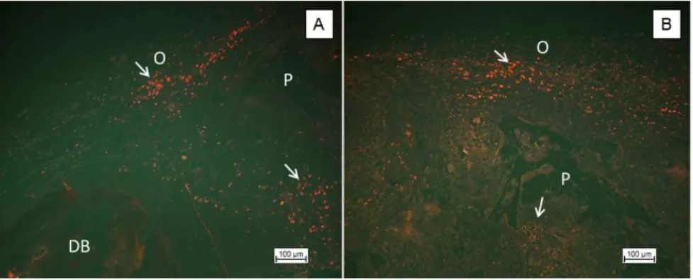
skull in Wistar rats. The first group of cells was labelled with fluorescent scaffolds for a duration of 4 weeks. The dye was clearly seen, which allowed monitoring of the cells. Figure 5 shows the micrographs of histological sections of the middle region of the implanted scaffold PLDLA area cultivated with osteoblast cells stained by PKH26.
The population of cells located at the edge of the bone defect (Figure 6A) in contact with the original bone suggests that, during cell culture, there was a preference for the region as a result of further diffusion of nutrients from the culture medium in peripheral regions of the scaffolds of PLDLA[28].
Figure 4. Calvarias removed from animals at days 4 (A), 8 (B) and 12 (C) weeks, showing the evolution of regeneration in the defect area.
Figure 5. Cell monitoring PLDLA scaffolds cultured with osteoblast cells after 4 weeks of implantation. (A) Photomicrograph illustrating the population of osteoblastic cells (O) displaying red fluorescence, located on the edge of the bone defect (DB - “bone defect”); (B) Population of cells located in the central region of the implant with visible presence of the polymer scaffold (P).
Figure 6. Histological and fluorescent PLDLA of scaffolds cultured with osteoblast cells after 12 weeks of implantation. BD - bone
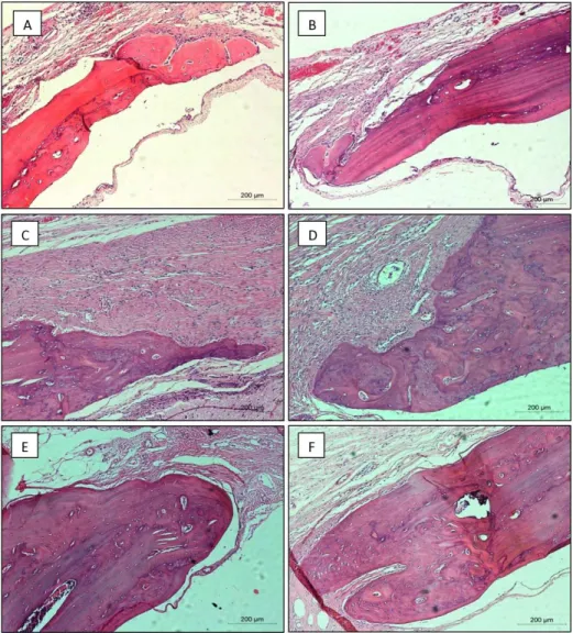
Figure 5B illustrates the central area of the critical defects implanted with PLDLA scaffold where, although some regions show small areas colonized by pre-cultured osteoblasts and labelled with PKH26, the physical structure of the scaffold was shown to promote migration/cellular distribution within the implanted material. Figure 7 shows histological images of the bone defect area after 4 weeks (A and B) 8 weeks (C and D) and 12 weeks (E and F).
On all edges illustrated in Figure 7, irregular formation and growth of bone tissue was observed. The resulting defects at 4 weeks (Figures 7A and 7B), appear to be smaller than the primary defect and, especially in areas where the external diploe is located in proximity to skeletal muscle growth. At the 4-week time point assessed, no cases of bone formation were observed without direct contact with the defect edges. At 8 weeks post implantation, the defect was apparently less severe than at the previous time point, showing continuous and asymmetric bone formation. Moreover, sharp bone growth can be noted in the closing trend from the edges, as evidenced by the greater amount of newly formed bone (Figures 7C and 7D). In no case were
Figure 7. Histological analysis of PLDLA scaffolds cultured with osteoblast cells after 4 weeks (A and B) 8 weeks (C and D) and 12 weeks (E and F) of implantation.
bone formation focuses forming calluses for the internal diploe observed, similar to that within the limits of the brain itself. At 12 weeks, the reduction of the defect was greater with irregular growth, but throughout the surrounding area. Secondary bone formation occurs preferentially in parallel beams between them (Figures 7E and 7F), simulating the original aspect diploe. At that time, it became a clear case of bone formation without direct contact with the edges (Figure 6).
Figure 8 shows the histological analysis of fluorescent and cultivated PLDLA scaffolds with osteoblast cells after 4 days (A and B), 8 weeks (C and D) and 12 weeks (E and F) of implantation.
as shown in the highlighted areas of Figure 8B. At 8 weeks, similarly, the image suggests the migration of dye-labelled cells, evidenced both in the defect area and at the edges of the newly formed bone. Already at 12 weeks, the image reveals the formation of bone spurs, irregular, primary and surrounded by osteoblast cells. Figure 6 shows, independent of defect edge (BD), an area of newly formed bone (TON) significant after 12 weeks of implantation.
In Figure 6B, the arrows point to cells precultured and implanted with fluorescent dye, both in the bone defect area linked to the edge and the unrelated surrounding area, suggesting a contribution of these active cells to the formation of this fragment.
Among the important requirements of a dye of choice stands out the following: the ability to remain detectable after a long time, ease of handling and non-interference in immunocytochemical responses of cells implanted in the
tissue. In this work, we verified the cytocompatibility the PKH26 dye in the cultivation of osteoblast cells for 14 days and also by MTT assay.
4. Conclusions
In this work, the cytocompatibility the PKH26 dye was verified in osteoblasts in cell culture for 14 days on PLDLA scaffolds by MTT assay. The dye did not exert negative influences on cell growth of osteoblasts in relation to unstained cells. In the in vivo study, macroscopic observations made during deployment times corroborated the in vitro results, as no apparent signs of toxicity were observed in the implanted bone defect area. The use of mobile monitoring with the dye, PKH26 in vivo is an effective strategy for the understanding of cell behaviour in the presence of PLDLA polymer.
5. References
1. Dantas, T. S., Lelis, E. R., Naves, L. Z., Fernandes-Neto, A. J., & Magalhães, D. (2011). Bone graft materials and their application in dentistry. UNOPAR Científica Ciências Biológicas e da Saúde, 13(2), 131-135. Retrieved in 9 November 2015, from pgsskroton.com.br/seer/index.php/biologicas/article/ viewFile/1248/1198
2. Kunz, F., Bergemann, C., Klinkenberg, E. D., Weidmann, A., Lange, R., Beck, U., & Nebe, J. B. (2010). A novel modular device for 3-D bone cell culture and nondestructive cell analysis.
Acta Biomaterialia, 6(9), 3798-3807. PMid:20227531. http://
dx.doi.org/10.1016/j.actbio.2010.03.015.
3. Ikeda, T., Ikeda, K., Yamamoto, K., Ishizaki, H., Yoshizawa, Y., Yanagiguchi, K., Yamada, S., & Hayashi, Y. (2014). Fabrication and characteristics of chitosan sponge as a tissue engineering scaffold. BioMed Research International, 6, 1-8.
PMid:24804246.http://dx.doi.org/10.1155/2014/786892. 4. Kroeze, R. J., Helder, M. N., Govaert, L. E., & Smit, T. H.
(2009). Biodegradable Polymers in Bone, Tissue Engineering.
Materials (Basel), 2(3), 833-856. http://dx.doi.org/10.3390/ ma2030833.
5. Alexander, J. T., Branch, C. L., Jr, Subach, B. R., & Haid, R. W., Jr. (2002). Applications of a resorbable interbody spacer via a posterior lumbar interbody fusion technique. Orthopedics,
25(10), S1185-S1189. PMid:12401030.
6. Moser, R. C., Mcmanus, A., Riley, S., & Thomas, K. (2005). Strength retention of 70:30 Poly(Llactide-co- D, L lactide) following real-time aging. Journal of Biomedical Materials Research. Part B, Applied Biomaterials, 75(1), 56-63.
PMid:16001395. http://dx.doi.org/10.1002/jbm.b.30238. 7. Brazelton, T. R., & Blau, H. M. (2005). Optimizing techniques
for tracking transplanted stem cells in vivo. Stem Cells, 23(9), 1251-1265. PMid:16109764.http://dx.doi.org/10.1634/ stemcells.2005-0149.
8. Yoo, J. J., Bichara, D. A., Zhao, X., Randolph, M. A., & Gill, T. J. (2011). Implant-assisted meniscal repair in vivo using a chondrocyte-seeded flexible PLGA scaffold. Journal of Biomedical Materials Research. Part A, 99(1), 102-108. PMid:21800420. http://dx.doi.org/10.1002/jbm.a.33168. 9. Polzer, H., Volkmer, E., Saller, M. M., Prall, W. C., Haasters,
F., Drosse, I., Anz, D., Mutschler, W., & Schieker, M. (2012). Long-term detection of fluorescently labeled human mesenchymal stem cell in vitro and in vivo by semi-automated microscopy. Tissue Engineering. Part C, Methods, 18(2),
156-165. PMid:21951128. http://dx.doi.org/10.1089/ten. tec.2011.0275.
10. Matz, R. L., Erickson, B., Vaidyanathan, S., Kukowska-Latallo, J. F., Baker, J. R., Jr., Orr, B. G., & Banaszak Holl, M. M. (2013). Polyplex exposure inhibits cell cycle, increase inflammatory response, and can cause protein expression without cell division. Molecular Pharmaceutics, 10(4), 1306 -1317. PMid:23458572. http://dx.doi.org/10.1021/mp300470d. 11. Wang, W.-J., Wu, S.-P., Liu, J.-B., Shi, Y.-S., Huang, X., Zhang, Q.-B., & Yao, K.-T. (2013). MYC regulation of CHK1 and
CHK2 promotes radioresistance in a stem cell-like population
of nasopharyngeal carcinoma cells. Cancer Research, 73(3), 1219-1231. PMid:23269272. http://dx.doi.org/10.1158/0008-5472.CAN-12-1408.
12. Canola, K., Angenieux, B., Tekaya, M., Quiambao, A., Naash,
M. I., Munier, F. L., Schorderet, D. F., & Arsenijevic, Y. (2007). Retinal stem cells transplanted into models of late stages of retinitis pigmentosa preferentially adopt a glial or a retinal ganglion cell fate. Investigative Ophthalmology & Visual Science, 48(1), 446-454. PMid:17197566. http://dx.doi.
org/10.1167/iovs.06-0190.
13. Boomsma, R. A., Swaminathan, P. D., & Geenen, D. L. (2007).
Intravenously injected mesenchymal stem cells home to viable
myocardium after coronary occlusion and preserve systolic function without altering infarct size. International Journal of Cardiology, 122(1), 17-28. PMid:17187879. http://dx.doi.
org/10.1016/j.ijcard.2006.11.022.
14. Motta, A. C., & Duek, E. A. R. (2007). Synthesis and characterization of poly (L-co-D,L acid lactic). Polímeros: Ciência e Tecnologia, 17(2), 123-129. http://dx.doi.org/10.1590/ S0104-14282007000200011.
15. Declercq, H. A., Verbeeck, R. M. H., Ridder, L. I. F. J. M., Schacht,
E. H., & Cornelissen, M. J. (2005). Calcification as an indicator of osteocoindutive capacity of biomaterials in osteoblastic cells cultures. Biomaterials, 26(24), 4964-4974. PMid:15769532.
http://dx.doi.org/10.1016/j.biomaterials.2005.01.025.
16. Duffy, G. P., Mcfadden, T. M., Byrne, E. M., Gill, S. L., Farrell,
E., & O’Brien, F. J. (2011). Towards in vitro vascularisation of collagen-GAG scaffolds. European Cells & Materials,
21(12), 15-30. PMid:21225592. http://dx.doi.org/10.22203/ eCM.v021a02.
17. Ignatius, A. A., & Claes, L. E. (1996). In vitro biocompatibility of bioresorbable polymers: poly (L, DL-lactide) and poly(L-lactide-co-glycolide). Biomaterials, 17(8), 831-839. PMid:8730968.
http://dx.doi.org/10.1016/0142-9612(96)81421-9.
18. Thadavirul, N., Pavasant, P., & Supaphol, P. (2014). Development of polycaprolactone porous scaffolds by combining solvent casting, particulate leaching, and polymer leaching techniques for bone tissue engineering. Journal of Biomedical Materials Research. Part A, 102(10), 3379-3392. PMid:24132871. http:// dx.doi.org/10.1002/jbm.a.35010.
19. Sabir, M. I., Xu, E. X., & Li, L. (2009). A review on biodegradable polymeric materials for bone tissue engineering applications.
Journal of Materials Science, 44(21), 5713-5724. http://dx.doi. org/10.1007/s10853-009-3770-7.
20. Chen, J., Wang, C., Lu, S., Wu, J., Guo, X., Duan, C., Dong, L., Song, Y., Zhang, J., Jing, D., Wu, L., Ding, J., & Li, D. (2005). In vivo chondrogenesis of adult bone-marrow-derived autologous mesenchymal stem cells. Cell and Tissue Research,
319(3), 429-438. PMid:15672263. http://dx.doi.org/10.1007/ s00441-004-1025-0.
21. Christian, W., Johnson, T. S., & Gill, T. J. (2008). In vitro and in vivo cell tracking of chondrocytes of different origin by
fluorescent PKH 26 and CMFDA.Journal of Biomedical Science and Engineering, 1(3), 163-169. http://dx.doi.org/10.4236/ jbise.2008.13027.
22. Kang, E. J., Byun, J. H., Choi, Y. J., Maeng, G. H., Lee, S. L., Kang, D. H., Lee, J. S., Rho, G. J., & Park, B. W. (2010).
In vitro and in vivo osteogenesis of porcine skin-derived
mesenchymal stem cell–like cells with a demineralized bone and fibrin glue scaffold. Tissue Engineering. Part A, 16(3), 815-827. PMid:19778183. http://dx.doi.org/10.1089/ten. tea.2009.0439.
23. Lee, J. Y., Choi, M. H., Shin, E. Y., & Kang, Y. K. (2011). Autologous mesenchymal stem cells loaded in Gelfoam for structural bone allograft healing in rabbits. Cell and Tissue Banking, 12(4), 299-309. PMid:20652421. http://dx.doi.
org/10.1007/s10561-010-9194-4.
24. Li, P., Zhang, R., Sun, H., Chen, L., Liu, F., Yao, C., Du, M., & Jiang, X. (2013). PKH26 can transfer to host cells in vitro and vivo. Stem Cells and Development, 22(2), 340-344.
PMid:22913652. http://dx.doi.org/10.1089/scd.2012.0357. 25. Park, B. W., Kang, E. J., Byun, J. H., Son, M. G., Kim, H.
J., Hah, Y. S., Kim, T. H., Mohana Kumar, B., Ock, S. A., & Rho, G. J. (2012). In vitro and in vivo osteogenesis of human mesenchymal stem cells derived from skin, bone marrow and dental follicle tissues. Differentiation, 83(5), 249-259.
26. Lemos, M. M. (2006). Experimental study on the effect of induced tendinitis in the gastrocnemius muscle: histopathology and raman spectroscopy. Franca: Universidade de Franca. 27. Más, B. A. (2011). Imobilização de colágeno em arcabouços de
poli (L-co-D,L ácido lático) (Master’s dissertation). Faculdade
de Engenharia Mecânica, Universidade Estadual de Campinas, Campinas.
28. Tavassol, F., Schumann, P., Lindhorst, D., Sinikovic, B., Voss, A., von See, C., Kampmann, A., Bormann, K. H., Carvalho, C., Mülhaupt, R., Harder, Y., Laschke, M. W., Menger, M. D.,
Gellrich, N. C., & Rücker, M. (2010). Accelerated angiogenic host tissue response to poly (L-lactide-co-glycolide) scaffolds by vitalization with osteoblast-like cells. Tissue Engineering. Part A, 16(7), 2265-2279. PMid:20184434. http://dx.doi. org/10.1089/ten.tea.2008.0457.