Ciências da Saúde
New therapeutic approaches for bone
regeneration
Ricardo Gil Fradique
Dissertação para obtenção do Grau de Mestre em
Ciências Biomédicas
(2º ciclo de estudos)
Orientador: Professor Doutor Ilídio Correia
Ciências da Saúde
Novas abordagens terapêuticas para regeneração
óssea
Ricardo Gil Fradique
Dissertação para obtenção do Grau de Mestre em
Ciências Biomédicas
(2º ciclo de estudos)
Orientador: Professor Doutor Ilídio Correia
List of Publications
• Articles in peer reviewed international journals:
Diogo G. S., Gaspar V. M., Serra I.R., Fradique R., Correia I.J.; Manufacture of β-TCP/alginate scaffolds through a Fab@home model for application in bone tissue engineering. Biofabri-cation, 2014. 6(2):025001. doi:10.1088/1758-5082/6/2/025001
Torres,A. L., Gaspar, V. M., Serra, I. R., Diogo, G. S., Fradique, R., Silva, A. P., Correia, I. J.; Bioactive Polymeric-Ceramic Hybrid 3D Scaffold for Application in Bone Tissue Regen-eration. Materials Science and Engineering: C, Materials for Biological Applications, 2013. 33(7):4460–9. doi:10.1016/j.msec.2013.07.003
• Poster communications:
Diogo G.S., Gaspar V.M., Serra I.R., Fradique R., Correia I.J.; Bone scaffolds produced by rapid prototyping, IX Annual CICS Symposium, 30 June - 1 July, 2014, Covilhã, Portugal. Diogo G.S., Gaspar V.M., Serra I.R., Fradique R., Correia I.J.; Rapid prototyping of com-posite bone scaffolds for tissue engineering, Biannual encounter of the Technical Divisions of Portuguese Materials Society (SPM), 4th of May, 2014, Covilhã, Portugal.
• Oral presentations:
Fradique R., Correia I.J.; Design and production of 3D Scaffolds by Rapid prototyping for bone tissue engineering, III National Bioengineering Conferences, 28-29th of May, Covilhã, Portugal
The most exciting phrase to hear in science, the one that heralds new discoveries, is not ’Eureka!’ but ’That’s funny...’ Isaac Asimov
Acknowledgments
I would like to thank my supervisor professor Ilídio Correia, for the opportunity to develop my master’s thesis with him. For all the dedication, time, and patience spent in getting here. To Sónia for all the daily help, and for the acquisition of the scanning electron microscopy im-ages.
To professor Abílio Silva, for the availability and help with the mechanical characterization of the scaffolds.
To all my group colleagues. Thank you for all the help and support. A special thanks to Mariana, with whom I divided the long hours spent in the laboratory. Your help was invaluable throughout this year.
A very special “thank you” to my girlfriend, Cristiana. Thank you for always being by my side, for all your love, and for the infinite patience in my busy lab days.
To my parents and my sister. Without a doubt the most important people in my life, who always helped me unconditionally. I would not be here without them.
Resumo
O envelhecimento da população mundial tem associado o aumento do número de casos de doenças ou defeitos ósseos. Estas situações patológicas, que têm diferentes causas, afetam a mobilidade e a qualidade de vida do indivíduo. Os tratamentos actuais contemplam a utiliza-ção de enxertos ósseos, que na sua maioria são autoenxertos. No entanto, estes apresentam algumas limitações, como seja o estado de saúde do paciente que limita a obtenção do enxerto, a extensão da lesão óssea, e ainda a possibilidade de inflingir um maior sofrimento no doente. Este tipo de enxertos nem sempre permitem uma completa recuperação. Neste contexto, surge a Engenharia de Tecidos como área interdisciplinar que utiliza biomateriais e moléculas bioacti-vas para desenvolver dispositivos que permitam efectuar a reparação ou substituição do tecido ósseo. Uma das abordagens mais utilizadas envolve a produção de “scaffolds”, que são matrizes tridimensionais, que tomam papel de molde temporário para a adesão e proliferação celulares, fornecendo suporte mecânico durante a regeneração óssea. Uma abordagem particularmente promissora utiliza a tecnologia de prototipagem rápida para produzir “scaffolds” com estru-tura detalhada, utilizado modelos computacionais ou dados provenientes de exames médicos de rotina. O presente estudo descreve a caracterização fisicoquímica, mecânica e biológica de “scaffolds” tridimensionais produzidos com β-Tricalciofosfato/Alginato, utilizando prototi-pagem rápida. A Espectroscopia de Infravermelho por Transformada de Fourier, Difracção de Raio-X, Ângulo de Contacto e Microscopia Electrónica de Varrimento foram usadas na caracter-ização da estabilidade mecânica, superfície e porosidade dos “scaffolds” produzidos. O perfil citotóxico dos “scaffolds” foi estudado através de ensaios de MTS in vitro, utilizando osteoblas-tos humanos como células modelo. Os resultados obtidos demonstraram que os “scaffolds” produzidos apresentam óptimas propriedades biológicas e fisicoquímicas, suportando a adesão e proliferação celulares. Os materiais produzidos demonstraram possuir excelente resistência mecânica, igualando ou excedendo as propriedades do osso trabecular, demonstrando o seu potêncial para serem usados na regeneração de tecido ósseo.
Palavras-Chave
“Scaffolds” β-Tricalciofosfato/Alginato, Desenho Assistido por Computador, PVA, Prototipagem Rápida, Engenharia de Tecidos
Resumo alargado
O osso é um tecido altamente dinânimo e vascularizado, e é o principal componente do es-queleto humano. Este tecido é capaz de se auto-regenerar, e é também responsável por muitas outras funções no organismo. A sua estrutura complexa pode ser dividida em matrix orgânica, constituida na sua maioria por colagénio, e matriz inorgânica, maioritariamente hidroxiapatite, assim como os vários tipos de células ósseas, osteoblastos, osteoclastos e osteócitos. Apesar da sua capacidade de regeneração, vários factores como a idade, doenças, ou traumas têm levado a um aumento no número de defeitos ósseos a nível mundial.
As terapias mais utilizadas para a reparação do tecido ósseo envolvem o uso de enxertos ósseos, na sua maioria autoenxertos. No entanto, factores como a disponibilidade limitada, ou a possi-bilidade de dor crónica, têm limitado a sua utilização.
A Engenharia de Tecidos trata-se de uma área interdisciplinar, que combina o uso de biomateri-ais com moléculas bioactivas, de forma a estimular a reparação e regeneração do tecido ósseo. Neste campo, as matrizes tridimensionais, ou “scaffolds”, têm demonstrado grande potencial para aplicação na regeneração óssea. Estes devem possuir várias propriedades essenciais para permitirem a reparação do tecido: biocompatibilidade, biodegradabilidade, porosidade e re-sistência mecânica adequadas. Também devem mimetizar a matriz extracelular do local de implantação, de forma a favorecer a adesão e proliferação celulares.
Na produção destas estruturas têm sido utilizados diferentes materiais, nomeadamente metais, polímeros, e cerâmicas, assim como vários métodos de produção. As tecnologias de prototi-pagem rápida têm facilitado o desenvolvimento de estruturas tridimensionais com arquitectura controlada, através de modelos CAD e de dados provenientes de exames médicos de rotina. O presente estudo descreve a produção e caracterização das propriedades químicas, mecânicas, e biológicas, de scaffolds 3D produzidos com β-TCP/Alginato. Foram testadas seis formulações diferentes, utilizando três rácios de β-TCP/Alginato (60/40 % (p/p), 70/30 % (p/p), 80/20 % (p/p)), com, e sem PVA na sua composição. Os scaffolds foram produzidos por prototipagem rápida através de uma 3D plotter (Fab@Home).
As propriedades fisicoquímicas dos scaffolds produzidos foram caracterizadas por Espectroscopia de Infravermelho por Transformada de Fourier, Difracção de Raio-X, Ângulo de Contacto, e Mi-croscopia Electrónica de Varrimento. Os resultados mostraram que não existiram alterações na estrutura cristalina dos materiais utilizados, sendo que os “scaffolds” produzidos apresen-taram um caracter hidrofílico, o que permitiu a adesão celular. Por outro lado, também se verificou que a superfície dos materiais é irregular e rugosa, o que favorece a adesão celular. Foram ainda avaliadas a porosidade e resistência mecânica dos scaffolds, sendo que a formu-lação 60/40 demonstrou as melhores propriedades mecânicas, similares às propriedades nativas do osso trabecular. O perfil citotóxico foi avaliado através do ensaio de MTS in vitro, utilizando osteoblastos humanos. Os resultados obtidos apresentaram excelentes propriedades biológicas para todas as formulações, suportando e favorecendo a proliferação celular. De forma a avaliar a adesão celular na superfície dos materiais, imagens de microscopia electrónica de varrimento foram adquiridas após incubação com as células, onde foi possível observar as células aderidas e esticadas à superfície dos materiais.
Os resultados obtidos demonstraram que os scaffolds produzidos têm boas propriedades quími-cas, biológicas e mecâniquími-cas, que poderão permitir a aplicação destas estruturas na regeneração de tecido ósseo.
Abstract
The aging of the worldwide population has led to an increase in bone tissue diseases. Wether due to trauma or pathology, if the bone tissue is compromised it constitutes a serious threat to the well being of the individual. Current solutions for bone defects involve the use of bone grafts, particularly autografts. However, these present serious limitations for their use, such as limited availability, or the possibility of chronic pain, and may not allow the patient’s full recovery. In this context, Tissue Engineering emerged as a potential solution, since it uses bio-materials and bioactive molecules to develop devices that allow the repair or regeneration of bone tissue. One of the most researched approaches are scaffolds, that are three dimensional matrices, aimed to act as temporary templates for cell adhesion and proliferation, offering mechanical support while the bone tissue regenerates. A particularly promising approach uses rapid prototyping technology to produce 3D scaffolds with highly controlled structure, either from CAD models, or routine exam medical data. The present study describes the physicochem-ical, mechanical and biological characterization of 3D β-TCP/Alginate scaffold, produced by rapid prototyping. Fourier Transform Infrared Spectroscopy, X-Ray Diffraction, Contact Angle, and Scanning Electron Microscopy were used to characterize the porosity, surface hydrophilic character and chemical composition of the produced scaffolds. In addition, the mechanical stability (Compressive Strength and Young’s Modulus) was also evaluated. The biocompatibil-ity of the scaffolds was evaluated by in vitro MTS assays, using human osteoblasts as model cells. The results obtained showed that the produced scaffolds present excellent biological and physicochemical properties, allowing for the adhesion and proliferation of human osteoblasts. Moreover, the produced scaffolds presented remarkable mechanical strength, matching or sur-passing the properties of native trabecular bone, which is fundamental for their potential use in the regeneration of bone tissue.
Keywords
β-Tricalcium Phosphate/Alginate scaffolds, Computer-Aided Design, PVA, Rapid prototyping,
Contents
1 Introduction 1 1.1 Bone tissue . . . 1 1.2 Bone anatomy . . . 1 1.3 Bone histology . . . 3 1.3.1 Bone matrix . . . 3 1.3.2 Bone cells . . . 3 1.3.3 Bone remodelling . . . 4 1.4 Bone disorders . . . 6 1.5 Bone grafts . . . 7 1.6 Tissue engineering . . . 8 1.6.1 3D scaffolds . . . 81.6.1.1 Biomaterials for scaffold production . . . 9
1.6.2 Rapid prototyping techniques for 3D scaffold production . . . 12
1.6.2.1 Fab@Home 3D plotter for scaffold production . . . 13
1.7 Aims . . . 14
2 Materials and Methods 15 2.1 Materials . . . 15
2.2 Methods . . . 15
2.2.1 Rapid Prototyping of beta tricalcium phosphate/Alginate composite scaffolds 15 2.2.2 Physicochemical and morphological characterization of the scaffolds . . . 16
2.2.2.1 Scanning Electron Microscopy analysis . . . 16
2.2.2.2 Fourier Transform Infrared Spectroscopy analysis . . . 16
2.2.2.3 X-Ray Diffraction analysis . . . 16
2.2.2.4 Energy Dispersive Spectroscopic analysis . . . 16
2.2.3 Mechanical characterization of the scaffolds . . . 17
2.2.4 Swelling studies . . . 17
2.2.5 Porosity evaluation . . . 17
2.2.6 Characterization of the degradation profile of the scaffolds . . . 18
2.2.7 Contact Angle Measurements . . . 18
2.2.8 Biological characterization of the scaffolds . . . 18
2.2.8.1 Cell culture in the presence of the scaffolds . . . 18
2.2.8.2 Evaluation of cell viability and proliferation in the presence of the scaffolds . . . 18
2.2.8.3 Scanning Electron Microscopy analysis . . . 19
2.2.9 Statistical Analysis . . . 19
3 Results and Discussion 21 3.1 Morphological characterization of the scaffolds . . . 21
3.2 Physicochemical characterization of the scaffolds . . . 24
3.2.1 Fourier Transform Infrared Spectroscopic analysis . . . 24
3.2.2 X-ray diffraction analysis . . . 25
3.2.3 Energy dispersive spectroscopic analysis . . . 25
3.2.5 Swelling studies . . . 27 3.2.6 Porosity evaluation . . . 28 3.2.7 Characterization of the degradation profile of the produced scaffolds . . . 29 3.2.8 Contact angle measurements . . . 29 3.2.9 Analysis of the scaffolds biological properties . . . 30 3.2.9.1 Characterization of the cytotoxic profile of the scaffolds . . . . 30
4 Conclusions and Future Perspectives 35
Bibliography 37
List of Figures
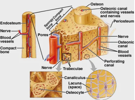
1.1 Schematic view of bone tissue structure and organization. . . 2
1.2 Schematic view of bone tissue cells and bone remodelling. . . 4
1.3 Sodium alginate chemical structure. . . 11
1.4 Image of a Fab@Home plotter used to produce the scaffolds used in this study . . 12
3.1 Images of the CAD model used (left) and of the final printed model (right) . . . . 21
3.2 Macroscopic images of the different produced scaffolds . . . 22
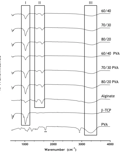
3.3 SEM images showing the morphology of the scaffolds at different magnifications . 23 3.4 FTIR analysis of the powders and 3D scaffolds. . . 24
3.5 XRD spectra of β-TCP and of the produced scaffolds . . . 25
3.6 EDS analysis of the produced scaffolds . . . 26
3.7 Mechanical characterisation of the scaffolds . . . 26
3.8 Swelling profile of the scaffolds . . . 27
3.9 Porosity evaluation of the scaffolds . . . 28
3.10 Degradation profile of the scaffolds . . . 29
3.11 Contact angle of the produced scaffolds . . . 30
3.12 Macroscopic images of human osteoblasts in the presence of the scaffolds . . . . 31
3.13 SEM images of osteoblasts morphology in the presence of the scaffolds . . . 32
3.14 Evaluation of human osteoblast cell viability cultured in contact with the different scaffolds . . . 33
List of Tables
Acronyms
3D 3 dimensional. 7, 8, 10, 12–15, 35 ANK progressive ankylosis protein. 5 BMP bone morphogenic protein. 5 BMU basic multicellular unit. 4 BSA bovine serum albumin. 15 CAD computer assisted design. 12, 13
DMEM-F12 Dulbecco’s modified Eagle medium: nutrient mixture F12. 15, 18 ECM extracellular matrix. 3, 8, 10, 21, 23
EDS Energy Dispersive Spectroscopy. 16, 26 EDTA ethylenediaminetetraacetic acid. 15, 18 EtOH ethanol. 15, 17–19
FBS Fetal bovine serum. 15, 18 FDM fused deposition modelling. 12 FGF fibroblast growth factor. 5
FTIR Fourier Transform Infrared Spectroscopy. 16, 24 HA hidroxyapatite. 1, 3, 5, 10
IGF-I insulin like growth factor 1. 4 IL-6 interleukin 6. 4
MCP-1 monocyte chemoattractant protein 1. 4 M-CSF macrophage colony-stimulating factor. 4 MMP matrix metallopeptidase. 5
MTS 3-(4,5-dimethylthiazol-2-yl)-5-(3-carboxymethoxyphenyl)-2-(4-sulfophenyl)-2H-tetrazolium. 15, 18, 32
PBS phosphate buffer saline. 15
PCL polycaprolactone. 10
PGA polyglicolic acid. 10 PLA polylactic acid. 10
PMS phenazine methosulfate. 18 PTH pituitary hormone. 4
PVA poly(vinyl) alcohol. 15, 16, 24, 25, 27, 28, 30 RANK receptor activator of nuclear factor κ-B. 4 RANKL receptor activator of nuclear factor κ-B ligand. 4 RGD Arginyl-glycyl-aspartic acid. 5
RT room temperature. 13, 16, 18, 19
SEM Scanning Electron Microscopy. 16, 19, 22, 31 STL Standard Tessellation Language. 13
β-TCP beta tricalcium phosphate. 10, 14–16, 21, 22, 24–28, 30, 33, 35
TGF-β transforming growth factor β. 4, 5 TNF-α tumour necrosis factor α. 4 UV ultraviolet. 18
Chapter 1
Introduction
1.1 Bone tissue
Bone is a highly dynamic and vascularised tissue, and it is the main component of the human skeleton1,2. The principal function of the skeleton is to provide structural support to the body3,
allowing motion by opposing muscular contractions and withstanding functional loads1,4, while
protecting the internal organs, especially the heart, lungs, and spinal cord5. It is also involved
in different biological functions, namely the production of blood cells through hematopoiesis3,
and ion storage, mainly phosphorous and calcium2,3.
In spite of a complex structure, bone tissue can be divided in cells and bone matrix. Different types of bone cells are found in bone tissue, each presenting a specific function: osteoblasts, that are known as bone forming cells; osteocytes, involved in bone maintenance; and osteo-clasts, that perform bone remodelling5. With distinct origins and functions, each cell type is
essential for the production and maintenance of the bone matrix.
Bone matrix is composed by an inorganic and organic phase, in a 65/35% ratio6. The organic
phase is mainly composed of collagen and proteoglicans, while the inorganic phase is composed by calcium phosphate crystals, in the form of HA (hidroxyapatite)2,3,6.
1.2 Bone anatomy
Bones are exceptionally well suited for the structural demands of the human body, providing high strength and durability while minimizing weight. Moreover, during the lifetime of each individual, the shape, anatomy and mechanical properties may be changed in response to ex-ternal stimulus4.
Bones can be classified according to their shape into long, short, flat or irregular3,6. Long bones
present a cylinder like shape, representing most of the bones of the upper and lower limbs. As hollow tubes, they provide great strength and durability against axial compression forces, while minimizing weight4. Short bones resemble a cubic or spheric shape, such as the carpal bones
from the wrist, or the tarsal bones, from the ankle6. Flat bones, on the other hand, present
thin, curved or flat shapes, being the ribs and scapulae examples of this type of bone. Irregular bones have complex shapes that are neither long, short, nor flat, such as the vertebrae and facial bones3,6. Bones can also be classified by structure in trabecular, also called cancellous
or spongy bone (about 20% of the total skeleton), and cortical or compact bone (about 80% of the total skeleton)2,3,6.
In addition, the outer surface of the bone is covered by a bi-layered connective tissue mem-brane, the periosteum. The outer, fibrous layer, is made of irregular collagenous tissue, which contains blood vessels and nerves, while the inner layer is composed by a single layer of bone cells. These facilitate the fixation of tendons or ligaments to the bone.
The endosteum is a connective tissue membrane that covers the internal surfaces of all bone cavities, and it is composed by a single layer of cells6,7.
Each growing long bone can be further characterized by three different sections: the diaphysis that composes the bulk of the bone, the epiphysis, located at the ends of the bone, and the epiphyseal plate, located where new bone is formed during growing6–8.
An important structure within the long bones is present in the diaphysis, that can contain the medullary cavity. This cavity, together with the cavities in cancellous bone, is filled with mar-row. Blood cell formation occurs in the red marrow, while yellow marrow is mainly adipose tissue, with adults presenting mostly the former, except for a few particular bones6,7. This
yel-low marrow can act as a localized energy reservoir in emergency situations, like bone fractures9.
Flat, short and irregular bones usually present a cancellous interior surrounded by two layers of compact bone, with the cancellous centre filled with marrow6,8.
On a smaller scale, bone can be also classified as woven or lamellar, according to the fibres organization3,4,6. Woven bone presents randomly oriented collagen fibres, and usually occurs
during growing stages, such as foetal development or fracture repair. It is typically remodelled to form lamellar bone3,4.
Lamellar bone is a layered type of bone, composed of 3-7 µm lamellae, with collagen fibres lying parallel to each other4,6.
A brief schematic of the internal structure and organization of bone tissue is presented on figure 1.1.
1.3 Bone histology
1.3.1 Bone matrix
As previously described, bone matrix is a composite of organic and inorganic materials, in a 35/65% ratio6. The organic phase is mainly constituted by collagen and proteoglycans, and the
inorganic by calcium phosphate crystals. These components are in a delicate balance, if the mineral is diminished, the bone becomes more flexible, due to increased collagen. If, on the other hand, the collagen is removed, the bone becomes mainly composed of mineral, and thus very brittle6,11.
The bone matrix composition also changes with age, with the decreasing bone turnover causing an increase in the mineralization degree, thus leading to a decrease in bone collagen con-tent12,13.
1.3.2 Bone cells
Osteoblasts are mononuclear and basophilic cells with a large spherical nucleus, and highly de-veloped rough endoplasmic reticulum and Golgi apparatus14. In addition, these cells present
secretory behaviour, producing collagen and proteoglycans and releasing them to the surround-ing environment6,15. They also release matrix vesicles, on which Ca2+and PO
43–are concentrated
to form HA crystals. These will then act like seeds for mineralization of the matrix. In this way, osteoblasts create new ECM (extracellular matrix) and regulate its mineralization. These cuboid shaped cells can live up to 8 weeks, depositing 0.5-1.5 µm of osteoid per day4,6. On the other
hand, osteoblasts also act as bone lining cells during the quiescence phase of bone16.
Osteocytes are osteoblasts that have become surrounded by bone matrix, but are morphologi-cally and functionally different16,17. These types of cells account for 90/95% of all cells in the
bone. They are relatively inactive, but can still produce the components needed to maintain the bone matrix6,17.
Currently, it is believed that these cells may actually modulate the spatial and temporal forma-tion and resorpforma-tion of bone tissue, through the high number of dendritic processes that inter-connect the osteocytes and the bone lining cells4,6,17. It was also proposed that these cells can
act like mechanosensor cells, transducing mechanical stress and guiding bone remodelling16,17.
Nitric oxide, Wnt and cadherin-mediated signalling have been suggested as transduncing mech-anisms, being activated following mechanical strain. However, the precise mechanical stimulus and response remain unclear17–19.
Osteoclasts are responsible for bone reabsorption15,20, mobilizing Ca2+and phosphate ions from
the bone matrix, resorbing the bone in a multiple stage process4,21. These are highly
special-ized giant cells, multi-nucleated and highly migratory4,15,20, derived from the red bone marrow
monocyte/macrophage lineage6,14. Capable of absorbing up to 200 000 µm3/day of bone
ma-trix4, they also present numerous mitochondria, a well developed Golgi apparatus around the
nuclei, endoplasmic reticulum, vacuoles and lysosomes. This cellular organization is the result of their great involvement in protein synthesis, in particular lysosomal enzymes15.
1.3.3 Bone remodelling
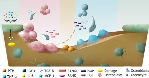
Bone is a dynamic tissue, constantly being remodelled. In this process, represented in figure 1.2, old bone matrix is replaced by new matrix, whether due to bone growth or repair, adjustments to stress, or calcium regulation22,23. Osteoblasts and osteoclasts gather in a temporary assembly
called BMU (basic multicellular unit)17,22–24, regulated by mechanical forces, bone cell turnover,
hormones (PTH (pituitary hormone), growth hormones, etc), cytokines and local factors17.
The bone remodelling process can be divided into four main phases: activation, resorption, reversal, and formation4,20,22,23,25and the full process can take 3-6 months to be completed4.
Figure 1.2: Schematic view of bone tissue cells and bone remodelling.
Activation phase
This stage involves the detection of an initiation signal, that can be a mechanical strain, dam-age, or factors released on the bone microenvironment20,22,25. In response to either the direct
endocrine signals, or signals produced by osteocytes, osteoblasts react by recruiting osteoclast precursors to the remodelling site, through the release of a chemoattractant, MCP-1 (monocyte chemoattractant protein 1)20.
In particular, IGF-I (insulin like growth factor 1), TNF-α (tumour necrosis factor α), IL-6 (inter-leukin 6), and PTH, activate bone lining osteoblasts, causing an increase in the surface expres-sion of the RANKL (receptor activator of nuclear factor κ-B ligand) by these cells. On the other hand, responding to osteoclastogenic stimulus, osteoblastic cells present in the bone marrow secrete M-CSF (macrophage colony-stimulating factor). This induces the expression of RANK (re-ceptor activator of nuclear factor κ-B) in pre-osteoclastic cells. The RANKL/RANK interaction leads to pre-osteoclast differentiation into multinucleated osteoclasts22,23.
In addition, damage to the bone or immobilization can result in osteocyte apoptosis, leading to increased osteoclastogenesis. This is due to a decrease in the levels of TGF-β (transforming growth factor β), which is an osteoclastogenesis inhibitor secreted by osteocytes in basal con-ditions20.
Another osteoclast activation signal can be induced by hormones. PTH is an endocrine remod-elling signal used to maintain calcium homoeostasis. This hormone is secreted by the parathyroid
glands in response to low calcium levels in the serum20, and can stimulate both bone formation
when secreted intermittently, and bone resorption when secreted continuously24.
In the bone, it activates a transmembrane G-protein-coupled receptor on the surface of os-teoblasts, that activates protein kinase A, protein kinase C and intracellular calcium signalling pathways. This osteoblast activation causes a wave of transcriptional responses that modulate the secretion of molecules responsible for the recruitment, differentiation, and activation of osteoclasts, establishing bone resorption20.
Resorption phase
During this stage, osteoclasts adhere to the bone surface and start to dissolve the bone20,22.
Cell anchorage is done via αvβ3 integrin molecules, creating an isolated microenvironment
un-derneath the cell20,25.
The organic and inorganic components of the bone are degraded through different processes. The inorganic component is mostly dissolved by decreasing the pH22, which is performed by
pumping hydrogen ions into the sealed zone20,25. The organic components are degraded by
lyso-somal enzimes, MMP (matrix metallopeptidase) and cathepsin K, released by osteoclasts, that degrade the unmineralized osteoid lining at bone surface20,22. This exposes RGD
(Arginyl-glycyl-aspartic acid) adhesion sites that facilitate further osteoclast attachment, enhancing bone re-sorption20. To avoid excessive bone resorption, osteoclasts suffer apoptosis after this stage.
Reversal phase
This stage marks the transition from osteoclast to osteoblast activity, with osteoblast precur-sors being recruited and differentiating25,26. In this phase, bone lining cells prepare the bone
surface for the subsequent bone deposition by osteoblasts, cleaning it from bone matrix left-overs20,24,25.They are also believed to be responsible for the signals that allow the transition
from bone resorption to bone formation20,24.
Formation phase
Throughout the formation phase osteoblasts lay down bone until they have replaced the re-sorbed bone completely24. The resorption phase releases a variety of growth factors stored in
the bone matrix, such as BMP (bone morphogenic protein), FGF (fibroblast growth factor), and TGF-β, that are responsible for the recruitment of osteoblasts into the absorbed area22,25.
Once osteoblasts are on site, and fully differentiated, both the organic and inorganic compo-nents are deposited, like collagen type I, which is the main organic component of the bone matrix, proteoglycans, osteonectin, bone sialoprotein and osteocalcin20,25. The precise
mech-anism of bone mineralization is still unclear, although it is believed that alkaline phosphatase, nucleotide pyrophosphatase phosphodiesterase, and ANK (progressive ankylosis protein) take part in creating the optimum concentration of extracellular inorganic phosphate to allow for HA formation20.
The remodelling cycle is concluded when all reabsorbed bone is replaced. At the end of the cycle, osteoblasts either enter apoptosis, revert back to bone lining phenotype, or differentiate into osteocytes within the matrix20. Even though the bone remodelling process is one of the most
in bone disorders are almost always related to this cycle, influencing either bone formation or resorption4.
1.4 Bone disorders
Bone disorders can be due to a variety of factors. Abnormal growth can cause gigantism or dwarfism, while abnormal collagen contents can lead to osteogenesis imperfecta. Mineral and vitamin deficiencies cause rickets, and bacterial infections can cause bone destruction, as is the case of osteomyelitis. Osteomalacia and osteoporosis are also responsible for bone damage, through decalcification6.
As already described, bone disorders arise primarily from derails in the bone remodelling pro-cess. Many of these metabolic diseases, with the exception of osteoporosis, present increased bone turnover, and are often associated with increased bone resorption due to high levels of serum PTH27. The most common metabolic bone diseases are acquired25, that is, derived from
a disease or endocrine dysfunction, and include osteoporosis, renal osteodystrophy (the skeletal changes of hyperparathyroidism and chronic renal failure), as well as Paget’s disease25,28.
Osteoporosis
Osteoporosis is the most common metabolic bone disease28, with an estimated 27.5 million
peo-ple affected in the European Union29. Osteoporosis is characterized by low bone mass and
struc-tural bone deterioration, causing increased bone fragility and vulnerability to fractures25,27,28.
It is most commonly due to an imbalance between bone resorption and bone formation. While both are heightened, they are altered with different intensities, resulting in higher bone resorp-tion than bone formaresorp-tion, and leading to a loss in bone mass28. This disorder is more common in
older populations, especially in women, due to the hormonal changes that they suffer with age. Nevertheless, excluding the hormonal effects of menopause, the disease has the same rate of progress in both genders28.
Renal osteodystrophy
Renal osteodystrophy includes a heterogeneous group of metabolic bone diseases derived from chronic kidney disease25,28. Among these are included osteomalacia25, deriving from vitamin D
deficiency with profound defects in mineralization27; osteitis fibrosa25, a loss of bone mass
de-rived from overproduction of PTH; osteopenia25, when the bone density is lower than normal, a
pathological situation that can precede osteoporosis; and osteosclerosis25, a localized increase
in bone density, that is associated with osteopetrosis. This type of disorders clearly reflect the importance of PTH and vitamin D on bone metabolism25,28, since most of bone related diseases
derive from abnormalities in the metabolism of either or both of these components30.
Paget’s disease
Paget’s disease is a disorder of the bone remodelling process, involving the abnormal destruction of needed bone, and construction of unneeded bone25, leading to the production of structurally
abnormal tissue27,28. Although the causes are not entirely understood, it is believed that it can
be caused by a viral infection during childhood, and several genetic mutations have also been identified on patients with the disease25,28.
It is clear that there is a need for new therapies to treat bone defects, either caused by disease or trauma.
1.5 Bone grafts
As already described, bone presents self-regeneration and remodelling. However, this is a lim-ited ability. There are certain traumas that the organism is unable to heal without surgical intervention, called critically sized osseous defects31. In such cases, bone grafts are usually
used for enhancing the formation of new bone. The capacity of bone grafts to regenerate tissue is measured according to their osteogenic, osteoconductive and osteoinductive potential32. An
osteogenic graft is capable of inducing the maturation of osteoprogenitor cells into osteoblasts, facilitating the formation of bone tissue. Osteoconduction is related with the formation of bone on a surface, in particular, the ability of the graft to support the bone growth in a 3D (3 dimensional) defect. Finally, osteoinduction refers to the recruitment and maturation of osteo-progenitor cells into the defect site31.
These bone grafts are divided into three main categories: autografts, allografts and xenografts32.
Autografts
Autografts are considered the gold-standard of bone grafting33,34, and are the basis to which
all of the other methods are compared. They consist of transplanting bone tissue from a donor to the lesion site, within the same patient31,32. They usually present high success rates31, with
excellent biological properties: the collected bone is rich in osteogenic cells, osteoconductive bone matrix and growth factors32,33,35. In addition, there is no risk of rejection or disease
trans-mission, since only tissue from the patient’s own body is used35.
However, the harvesting of bone from a different site can cause pain32,33, infection, scarring32,
blood loss, and donor site morbidity32–34. Since the graft is being taken from the patient, there
is also limited tissue availability33. In addition, there is no guaranty that the cellular
compo-nents survive transplantation33, and questions have been raised about the osteoinductivity of
these grafts, since the uncertain quantities of certain growth factors on autografts can cause insufficient tissue regeneration35.
Allografts
Allografts are used as an alternative to autografts, and are obtained from human donors or ca-davers31,34. Even though these types of bone grafts eliminate the need for a donor site, and the
supply limitations, they lack the osteoinductive capacity of autografts31,33. They also present
the risk of viral disease transmission and immunogenicity31–34. The processing techniques used
to lower these risks also decrease the mechanical resistance of the grafts, and usually eliminate the cellular phase of the bone tissue33.
Xenografts
Also worth mentioning are xenografts, bone grafts obtained from other species. Since the poten-tial for immune rejection and disease transmission is much higher from animal bone than from human bone, these types of bone grafts are processed through more intensive treatments,34,
which further limits the biologic and mechanical properties of these materials.
Due to all the aforementioned limitations, bone graft substitutes have been the focus of intense research in the area of Bone Tissue Engineering, with has as main objective to create bone scaffolds that mimick the microenvironment of the bone32.
1.6 Tissue engineering
Tissue engineering emerges as “an interdisciplinary field that applies the principles of engi-neering and the life sciences toward the development of biological substitutes that restore, maintain, or improve tissue function”2. In particular, bone tissue engineering tries to create
grafts with the capacity to induce the restoration of bone structure and functions34,36,
favour-ing regeneration over replacement2, and are usually composed of an ECM-mimicking structure
(scaffold), cells, and growth factors3,34,37.
The ideal tissue engineered construct should be able to completely replace autologous bone grafts3. However, even when the constructs cannot fully replace these grafts, they can be
added to the autologous graft to increase its volume, diminishing the amount of bone needed3.
Bone tissue engineering tries to recreate the appropriate environment for the regeneration through the extensive use of 3D scaffolds38. Nonetheless, the scaffolds currently generated
are limited by the current manufacturing techniques, and are based on a randomly distribu-tion of cells, matrix, and bioactive molecules37. As such, the mimicking of both functional and
biological complexity is seen as the current challenge to achieve full tissue regeneration37.
1.6.1 3D scaffolds
Fundamentally, scaffolds are templates for tissue regeneration38, capable of delivering cells and
growth factors to a damaged tissue, while providing mechanical support during the regeneration phase37. Scaffolds have been created through a variety of techniques using different
biomateri-als that present certain key qualities for a successful regeneration38: biocompatibility3,36,38,39,
biodegradability3,36,38–40, osteoconductivity3,36,39, osteoinductivity3,36,39, appropriate
mechani-cal properties3,36,38,39, adequate structure3,36,38,39, and ease of manufacture38,41.
• Biocompatibility
This is considered to be one of the most important properties of bone scaffolds3. Neither
the scaffold, nor any of its components and by-products, can be toxic or cause any adverse response in the host3. In addition, it must allow for normal cell adhesion, functioning, and
proliferation38.
• Biodegradability
A bone scaffold is designed to serve as a temporary matrix, and eventually be replaced with bone tissue. As such, it must degrade within the body at the same rate of new tissue formation40. To accomplish this, a controlled inflammatory response must occur, with an
not only to the local of implantation of the scaffold39, but also taking into account the
age of the patient who receives it38.
• Osteoconductivity
For a scaffold to be able to improve the regeneration of a damaged bone tissue, cells must be able to migrate, adhere, proliferate, and deposit new bone matrix3,39.
• Osteoinductivity
The scaffold should also be able to induce the formation of new bone, as well as cell differentiation36. While this characteristic is usually connected to growth factors, recent
studies have shown that bone induction can derive from the physical structure of the scaffold as well3.
• Appropriate mechanical properties
In order to provide adequate mechanical support, a bone scaffold must have mechanic properties similar to the host bone36,38,39. The Young’s Module of bone ranges from
0.1-0.2GPa of cancellous bone, to 15-20GPa in cortical bone. The compressive strength also presents very different ranges: 2-20MPa for cancellous bone, and 100-200MPa for cortical bone39. Since these properties vary largely according to the bone type and location,
scaf-folds must be tailored to their specific application, taking into account the loads they will have to bear once implanted.
• Adequate structure
The structure of the scaffold is closely connected to its mechanical properties, and struc-tural modifications can drastically affect the mechanical resistance. However, it also has profound effects on the biological properties. The porosity of a scaffold increases the available area for cell growth and attachment3,38. In addition, interconnected porosity
allows the diffusion of essential nutrients and oxygen3,38,39, as well as waste products38.
To facilitate bone ingrowth, scaffolds must possess pores with a minimum of 100 µm in diameter39, with the ideal range between 200 to 300 µm39. Recent studies have shown
that a combination of micro and macro porosities can perform better than macro-porous scaffolds39. The presence of micropores on a structure greatly increases the surface area
for cell adhesion, and can also contribute for a greater concentration of calcium and phos-phate ions, by providing a sheltered environment within the scaffolds, thus improving bone regeneration42.
• Acceptable manufacturing technology
Ultimately, for a scaffold to be able to have any clinical impact and viability, it must be cost effective and its production scalable38,41. Whether the scaffold needs in-vitro culture
before implantation, how easy it is to handle, its storage needs, as well as the sterilization methods, are all factors that can affect the clinical viability of a scaffold38.
1.6.1.1 Biomaterials for scaffold production
Biomaterial is currently defined by the European Society for Biomaterials as a “material in-tended to interact with biological systems to evaluate, treat, augment or replace any tissue, organ or function of the body”38. These types of materials have suffered a great evolution since
their inception, spanning already four generations43.
The first biomaterials were biologically inert, and the main goal was simply to replicate the physical properties of the native tissue with minimal toxic effects43,44. When the properties of
these materials proved to be insufficient, and in conjunction with a better understanding of the foreign body response, the research focus changed towards the creation of a biological response, with bioactive materials. This marked the rising of the second generation of biomaterials, that elicited not only a biological response, but also presented a controllable resorption within the body43,44. Osteoconductive implants became routine, with the use of synthetic HA and
bioac-tive glasses on porous implants and coatings. Also, the creation of structures that facilitated tissue ingrowth and 3D interlocking with the surrounding tissue contributed to the formation of a mechanically strong interface43. The advances in scientific knowledge, mainly in molecular
biology and proteomics, allowed the design of biomaterials capable of creating specific cellular responses, giving origin to the third generation of biomaterials43. Moreover, these materials are
being modified with biological components obtained from the ECM, that are specifically recog-nized by cells seeded on them44. Finally, the fourth generation of biomaterials combines the
properties of all previous generations, while trying to mimic the native tissue: biomimetic ma-terials. These materials try to recreate the complexity of the physiological environments43,44,
with some of them being already capable of modulating genetic activation and expression after implantation44, as well as respond to variations in temperature, pH and ionic force45–47.
It is thus clear that the choice of materials used in the production of scaffolds for bone re-generation is a key aspect in their performance. A variety of biomaterials is used for bone scaffold manufacturing, although they usually fall within four main categories: ceramic, poly-meric, metallic, or composite scaffolds32,39.
Ceramics
The ceramics used in bone tissue engineering are commonly composed of calcium and phos-phate3. Ceramic scaffolds can be divided into bioinert or bioactive scaffolds, with the latter
further divided into resorbable and non-resorbable scaffolds32. Bioactivity refers to the ability
of the scaffold to modify its surface structure and establish connections with the surrounding bone when implanted41.
The most used ceramics are HA and β-TCP (beta tricalcium phosphate), which closely resemble the mineral phase of the bone, and are therefore highly biocompatible, bioactive and osteocon-ductive38,41,48. However, these materials are different when it comes to their degradation rates.
While β-TCP presents an almost ideal biodegradability in vivo, HA shows almost no biodegra-dation3,41,49. In addition, the ability of β-TCP to bond directly to tissue, and regenerate bone
without intermediate tissue, as well as proper bio-resorption rate, have made it one of the most used ceramics in tissue engineering3,32,39,43,49.
Also, in spite of its great compressive strength, which is usually equal or greater than the native bone tissue31, ceramics tend to present a hard and brittle nature31,38,40,43. In addition, it can be
difficult to process them into highly porous structures, which further limits their use in scaffold production40. The mechanical properties of native bone results from the combination of both
its organic and inorganic phases. When ceramics are used alone they have limited properties for load-bearing applications41.
Polymers
Polymers can either have a natural origin, such as collagen36,50, starch34, silk34, or alginate34,
PGA (polyglicolic acid)36,38,39.
Since some natural polymers actually are structural constituents of tissues, these materials present greater biocompatibility and biodegradability50. However, they usually present lower
mechanical resistance and faster degradation rates, which limits their use in tissue engineer-ing50. Synthetic polymers are highly versatile31,50, with controlled properties, although they
have a lack of bioactivity. Furthermore, the degradation products of these polymers can some-times present negative effects on the regeneration process31.
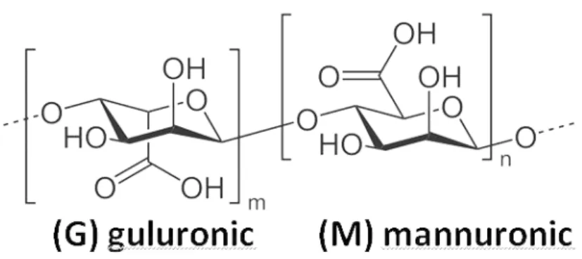
Alginate, which was the polymer used in this study for scaffolds production, is a natural polysac-charid derived from brown seaweeds43,52. Composed of 1,4-linked β-D-mannuronic acid (M) and α-L-guluronic acid (G) residues, this polymer can form stable hydrogels cross-linked with
diva-lent cations52,53. This cross-linking happens between two G blocks of adjacent polymer chains,
through interaction with the carboxylic groups and, as a consequence, the stiffness of the gel produced is directly related to the M/G ratio of the polymer and quantity of free divalent cations in solution53,54.
Figure 1.3: Sodium alginate chemical structure. (Adapted from Narayanan et al.55)
Alginate is extremely versatile, easily forming gels, fibers, foams, and nanoparticles, at physio-logical conditions53, while being biocompatible, and non-immunogenic54. Still, the dissolution
rates of ionically cross-linked alginates cannot be perfectly controlled, and it presents slow degradation kinetics in vivo.
Metals
Titanium and tantalum feature among the most common used metals for scaffold production. While these materials have great compressive strength and fatigue resistance, they are not biodegradable. In addition, they do not allow the incorporation of biomolecules39, requiring
coatings and surface treatments to immobilize biologically active peptides56. The release of
toxic metallic ions is also a concern32,39.
Composites
Composite materials have in their constitution two or more distinct materials, for example ce-ramics and polymers39,57. These combinations try to overcome the drawbacks of the brittleness
1.6.2 Rapid prototyping techniques for 3D scaffold production
The current challenge in tissue engineering is to mimic the natural structure of living tissue38.
Until now, the most used methods of scaffold production included solvent casting58,59, phase
inversion60,61, fiber bonding62,63, melting64,65and freeze drying66,67. However, these methods
present severe disadvantages, such as the use of toxic solvents, the inability to create large structures with suitable mechanical properties or lack of structure and porosity control1.
The advances in computer technology allowed the development of rapid prototyping techniques for tissue engineering applications. These enable the manufacturing of highly reproducible 3D scaffolds68, with increased complexity and control, from a CAD (computer assisted design)
model57. In addition, the model used for scaffold production can be derived from medical
data, and thus be tailored specifically for a particular damaged tissue with high anatomic accu-racy57,69,70.
The most common rapid prototyping techniques in use are 3D printing, FDM (fused deposition modelling) and 3D plotting1,71.
3D printing uses a liquid binder dispensed onto thin layers of powder, following the objects profile, creating a final structure in a layer-by-layer assembly1,72. This approach is limited by
the binder used, since many of the available binders are toxic, and the structures cannot have closed surfaces, otherwise unbounded powder would accumulate inside73.
FDM employs the deposition of a thermoplastic filament on a platform, by melting extrusion, also in a layer-by-layer fashion, the stacking of layers creating the final structure1. Given the
temperatures used for the polymer extrusion this technique does not allow for the incorporation of cells nor biomolecules on the interior of the material used74.
Finally, in 3D plotting a hydrogel is dispensed through a syringe onto a platform1. This deposition
can be achieved by pneumatic action, screw-driven, or piston action, with the latter providing the best flow control. This robotic deposition has resolutions in the order of 200 µm, with high fabrication speeds, and is one of the most promising technologies37.
In this work a Fab@Home 3D plotter was used for scaffold production (figure 1.4). This model was developed by Malone and Lipson75, and was previously used for tissue engineering
applica-tions76,77.
1.6.2.1 Fab@Home 3D plotter for scaffold production
As described above, the 3D plotting technology has great advantages over other methods, mainly the possibility of using viscous solutions for scaffold production at RT (room temperature). The viscosity of the solution used has a great effect on the accuracy and resolution of the equipment. In addition, several settings should be taken into account before a scaffold can be produced. The dispensing pressure, the nozzle diameter, the depositon rate, and the printspeed, depend on the solution used, as well as the CAD model, and must be adjusted accordingly78.
In brief, the CAD model is designed or scanned, and converted to STL (Standard Tessellation Language) format. This is then loaded onto the Fab@Home software, that slices the model into several layers. With the syringe loaded with the solution and the equipment properly configured, the scaffold is deposited layer-by-layer onto the plotter platform75,78.
1.7 Aims
Additive manufacturing of β-TCP/Alginate composite scaffolds for bone tissue engineering • Optimization of the design of the 3D structure
• Optimization of the viscosity of the solution
• Study the influence of β-TCP content on the scaffolds properties
• Study the influence of a β-TCP binding agent on the scaffolds properties
• Evaluate the mechanical, physicochemical and biological properties of the produced scaf-folds
Chapter 2
Materials and Methods
2.1 Materials
Amphotericin B, BSA (bovine serum albumin), cacodylate buffer (Mw=214.03 g/mol), calcium chloride (Mw=110.98 g/mol), DMEM-F12 (Dulbecco’s modified Eagle medium: nutrient mixture F12), EtOH (ethanol), EDTA (ethylenediaminetetraacetic acid), 25% (v/v) glutaraldehyde, L-glutamine, penicillin G, PBS (phosphate buffer saline), PVA (poly(vinyl) alcohol) (Mw=31 000 g/mol), sodium alginate (Mw=120 kDa to 190 kDa), streptomycin, trypan blue and trypsin were pur-chased from Sigma-Aldrich (Sintra, Portugal). β-TCP powder (Mw=310.20 g/mol) was obtained from Pancreac® (Barcelona, Spain). Sodium hydroxide was acquired from Pronalab (Barcelona,
Spain).
MTS (3-(4,5-dimethylthiazol-2-yl)-5-(3-carboxymethoxyphenyl)-2-(4-sulfophenyl)-2H-tetrazolium) reagent, inner salt was purchased from Promega (Madison, USA). FBS (Fetal bovine serum) was purchased from Biochrom AG (Berlin, Germany). Human osteoblast cells (CRL-11372) were ob-tained from American Type Culture Collection (VA, USA). 96-well plates were acquired from Orange Scientific (Braine LÁlleud, Belgium). Tris Base was obtained from Fischer Scientific (Portugal).
2.2 Methods
2.2.1 Rapid Prototyping of beta tricalcium phosphate/Alginate composite
scaf-folds
To produce the scaffolds herein described an additive manufacturing method was used. In par-ticular, a Fab@Home plotter was used, for the creation of well defined, highly reproducible 3D structures75. This type of system deposits a highly viscous solution in a controlled manner, in
this case a β-TCP/alginate composite.
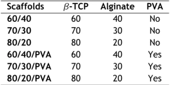
For this study, six types of scaffolds were produced, using different ratios of β-TCP/alginate, and PVA.
Briefly, a 1% (w/v) PVA solution was prepared by dissolving the polymer in double deionized and filtered water (obtained using a Milli-Q Advantage A10 ultrapure Water Purification System; resistivity=18.2MΩ/cm at 25 °C), with overnight agitation. Then, a 15% (w/v) alginate solution was prepared, dissolving the alginate in either water or in the PVA solution. This solution was then homogenized in a X10/25 ultraturrax (Ystral, Germany) for 30 minutes. Finally, β-TCP powder was added to the solution according to the specific ratios studied (table 2.1), and the solution was again homogenized.
Following, a 5% CaCl2 solution was added to the composite solution, following a 0.14:1 (% v/v) ratio of CaCl2to alginate. In this step, alginate goes through a ionic crosslinking process, which allows for the increased viscosity of the solution through the formation of a hydrogel53. This
Table 2.1: Chemical composition of the produced scaffolds
Scaffolds β-TCP Alginate PVA
60/40 60 40 No 70/30 70 30 No 80/20 80 20 No 60/40/PVA 60 40 Yes 70/30/PVA 70 30 Yes 80/20/PVA 80 20 Yes
increase in the viscosity is essential for the later deposition process.
The Fab@Home model reproduces a computer designed 3D model, in a layer-by-layer process. As such, the model of the scaffold was produced in OpenSCAD software (version 2014.3, ©2009-2014 Marius Kintel and Clifford Wolf), and then exported to STL format. Afterwards, the solution was filled into a 10 cm3 disposable syringe barrel, and the scaffolds plotted through a 22G
polypropylene tapered nozzle. Following, the scaffolds were maintained in a 5% CaCl2 bath for 24 hours, for full crosslinking, after which they were left to dry at RT for 24 hours. Finally, the scaffolds were freeze-dried for 24 hours.
2.2.2 Physicochemical and morphological characterization of the scaffolds
2.2.2.1 Scanning Electron Microscopy analysis
SEM (Scanning Electron Microscopy) micrographs were acquired in order to characterize the morphology, porosity and surface of the scaffolds. The samples were mounted onto aluminium stubs with Araldite glue, and sputter-coated with gold using a Quorum Q150R ES sputter coater. The SEM images where then captured with variable magnifications, at an acceleration voltage of 20 kV, using a Hitachi S-3400N Scanning Electron Microscope.
2.2.2.2 Fourier Transform Infrared Spectroscopy analysis
To measure the physicochemical characteristics of the scaffolds, FTIR (Fourier Transform In-frared Spectroscopy) was used. The FTIR spectra obtained for the samples are the average of 128 scans, between 400 and 4000 cm−1, with a spectral resolution of 4 cm−1. All the samples
were crushed to a powder, mounted on a diamond window, and the spectra recorded with a Nicolet iS10 FTIR spectrophotometer (Thermo Scientific, Waltham, MA, USA). All the compo-nents of the scaffold were also analysed in pure state for comparison with the samples79.
2.2.2.3 X-Ray Diffraction analysis
XRD (X-Ray diffraction) measurements were performed in order to evaluate the phases and crys-tallinity of the ceramic component of the scaffolds after their production.
All samples were mounted in silica supports, and the data recorded over a range of 5°to 90°2θ degrees, with continuous scans of 1 °/min, using a copper ray tube operated at 30 kV and 20mA70,77.
2.2.2.4 Energy Dispersive Spectroscopic analysis
EDS (Energy Dispersive Spectroscopy) was used for elemental characterization of the various scaffolds. The samples were placed on aluminium stubs, air-dried at RT, and analysed in a
XFlash Detector 5010(Bruker Nano).
2.2.3 Mechanical characterization of the scaffolds
Compression assays were performed in order to evaluate the mechanical behaviour of the scaf-folds. The dimensions of the final scaffolds were measured and introduced into a Zwick® 1435 Material Püfung (Ulm, Germany). The assays were performed using a crosshead speed of 3 mm/min and a load cell of 5 kN. Five specimens of each sample were used for each assay. The compressive strength of each scaffold was calculated according to equation (2.1)80.
C s = F
w ∗ l (2.1)
Where F is the load at the time of fracture, and w and l represent the width and length of the scaffold, respectively.
The Young Modulus was calculated using the values from the equation (2.1), and applying equa-tion 2.2.
Y M = C s Hd (2.2)
Where Hd stands for the height deformation at maximum load, and Cs is the scaffold tensile strength. Average values and standard deviations (s.d.) were determined for each sample (n=5).
2.2.4 Swelling studies
The swelling capacity of the scaffolds was determined following a method adapted from Valente et al.52. Samples from each scaffold were placed in eppendorfs containing 5 mL of Tris buffer
(pH=7,4), at 37 °C. The samples were then retrieved from the solution at predetermined inter-vals, and weighted, after removing the excess of Tris with filter paper. After this process, the samples were re-immersed in buffer solution. Three samples of each scaffold were used and the swelling ratio was evaluated using equation (2.3).
Swelling ratio (%) =Wt− W0
W0 ∗ 100
(2.3) Where Wt is the final weight of the scaffolds, and W0 their initial weight.
2.2.5 Porosity evaluation
To determine the microporosity of the different scaffolds a liquid displacement method was adapted from Torres et al.80. In brief, scaffolds were weighted, immersed in absolute EtOH
for 48 hours, and weighted again. EtOH was chosen for its ability to penetrate throughout the scaffolds without shrinking nor swelling the matrix81. The porosity was then calculated by the
Porosity (%) = Ww− Wd
Dethanol∗ Vscaf f old ∗ 100
(2.4)
Where Wwand Wdare the wet and dry weights of the scaffolds, respectively, Dethanolrepresents the density of EtOH at RT and Vscaffoldthe volume of the wet scaffold.
2.2.6 Characterization of the degradation profile of the scaffolds
The degradation of the composite scaffolds was investigated through a method adapted from Jeong et al.82 and Freed et al.83. In brief, scaffolds were placed in 24 well plates, fully
im-mersed in DMEM-F12, at 37 °C. At predetermined intervals the samples were removed, com-pletely dried and weighted. The degradation percentage at each point was calculated through equation (2.5):
Weight Loss (%) = (1 −Wi− Wt
Wi ) ∗ 100 (2.5)
Where Wicorresponds to the initial weight of the sample and Wtto the weight of the sample at time t.
2.2.7 Contact Angle Measurements
Contact angle measurements were performed using the sessile drop technique, adapted from Diogo et al.77, using water as reference fluid. Drops were placed at different points on the
surface of the scaffolds, and the data was acquired usind a Data Physics Contact Angle System
OCAH 200, operated in static mode at RT.
2.2.8 Biological characterization of the scaffolds
2.2.8.1 Cell culture in the presence of the scaffolds
Human osteoblasts (CRL-11372) were cultured in DMEM-F12, supplemented with 10% heat inacti-vated FBS, streptomycin (100 µg/mL) and gentamicin (100 µg/mL) in 75 cm2T-flasks. Cells were
maintained in a humidified environment at 37 °C, with 5% CO2, until confluence was attained.
Subsequently, cells were trypsinized with 0.18% trypsin (1:250), and 5 mM EDTA, centrifuged at 300 RCF, for 5 min, and the pellet resuspended in 5 mL of complete culture medium. Cellular density was then determined using a Neubauer chamber and trypan blue.
Prior to cell seeding, the scaffolds were cut into pieces with appropriate sizes and placed into 96-well plates for sterilization (n=5). This was achieved by subjecting the scaffolds to UV (ultra-violet) light for 30 min. Subsequently, cells were seeded at a density of 10x103cells per well,
for cell viability and proliferation evaluation. The culture medium was replaced every two days until the end of the assay.
2.2.8.2 Evaluation of cell viability and proliferation in the presence of the scaffolds Cell viability was determined using an MTS assay, at 4 and 7 days after seeding. The metabolic activity of the cells was assessed by quantifying the metabolic conversion of MTS to formazan. Briefly, the medium in each well was replaced with a mixture of 100 µL of fresh culture medium
containing 20 µL of MTS/PMS (phenazine methosulfate) reagent solution. The cells were then incubated for 4 hours at 37 °C. Following the incubation period, 80 µL of the supernatant were transfered into a 96-well microplate and the fluorescence intensity measured at 492 nm using a microplate reader (Anthos 2020, Biochrom). Cells cultured without materials were used as negative control (K–) and cells cultured with EtOH (96%) were used as positive control (K+). 2.2.8.3 Scanning Electron Microscopy analysis
In order to evaluate the morphology and topography of the scaffolds as well as the cellular be-haviour in their presence, SEM was performed according to the method adapted from Lee and Chow84. Briefly, the samples were washed at RT with cacodylate buffer solution, then fixed for
30 minutes in 2.5%(v/v) glutaraldehyde in 0.1M sodium cacodylate solution. The samples were then frozen in liquid nitrogen for 2 min and then freeze-dried for 2 hours.
For SEM analysis, the samples were mounted onto aluminium stubs with Araldite glue, and sputter-coated with gold using a Quorum Q150R ES sputter coater. The SEM images were cap-tured at variable magnifications, with an acceleration voltage of 20 kV, using a Hitachi S-3400N Scanning Electron Microscope.
2.2.9 Statistical Analysis
Statistical analysis of the obtained results for the different groups of scaffolds, and the various conditions, was performed by using one-way analysis of variance (ANOVA), with the Newman-Keuls post hoc test. The statistical test was used for comparison of the mean and the differences between groups. A p value less than 0.05 (p<0.05) was considered statistically significant.
Chapter 3
Results and Discussion
3.1 Morphological characterization of the scaffolds
As previously described, biodegradable scaffolds for bone tissue regeneration must fulfill cer-tain requirements, such as good biocompatibility and suitable structure. They must also provide mechanical support during the regeneration phase, as well as adequate physicochemical envi-ronment for cell proliferation.
Since most types of materials present certain disadvantages when used alone, in this work com-posite materials were used for scaffold development. In particular, a polymer and ceramic composite was chosen, mimicking the natural bone matrix. Thus, this study describes the devel-opment, optimization and production of scaffolds aimed for bone tissue regeneration, composed of β-TCP and alginate. Figure 3.1 presents the CAD model used, as well as the printed scaffold by rapid prototyping. The designed model is a 13 mm x 13 mm x 13 mm cube, with porous structure.
Figure 3.1: Images of the CAD model used (left) and of the final printed model (right)
Herein, alginate was chosen for its biocompatibility, and ability to act as temporary ECM. In addition, the ability to control the degradation rate of this polymer is a great advantage for the tailoring of the properties of the scaffold53,54. On the other hand, β-TCP was chosen for
its resemblance with the natural ceramic component of bone tissue, and consequently, its in-creased biocompatibility and osteoconductivity38,41,48, allied to its enhanced mechanical
resis-tance. Furthermore, the combination of these materials has already been shown to improve cell adhesion and proliferation, with the potential to support cell growth and differentiation before implantation77,85.
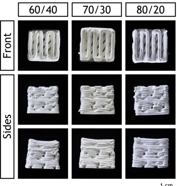
Macroscopic images of the produced scaffolds were acquired, to assess their morphology (figure 3.2).
Figure 3.2: Macroscopic images of the different produced scaffolds
It is possible to observe that the β-TCP content had a direct effect on the scaffolds structure, namely on the scaffolds dimensions. It was previously described that alginate gels and scaffolds present shrinkage during air drying86. Rassis et al.87 have related that the presence of solid
fillers in an alginate solution has a direct effect on the volume loss after drying. The compres-sion of the polymeric matrix leads to the comprescompres-sion of the β-TCP particles against each other. In effect, the scaffolds containing the highest percentage of β-TCP were capable of maintain-ing their dimensions showmaintain-ing a lower amount of shrinkage, since the amount of incompressible ceramic particles limits the shrinking that the scaffolds can suffer. Nonetheless, all the formu-lations maintained their shape, with a well defined structure and design.
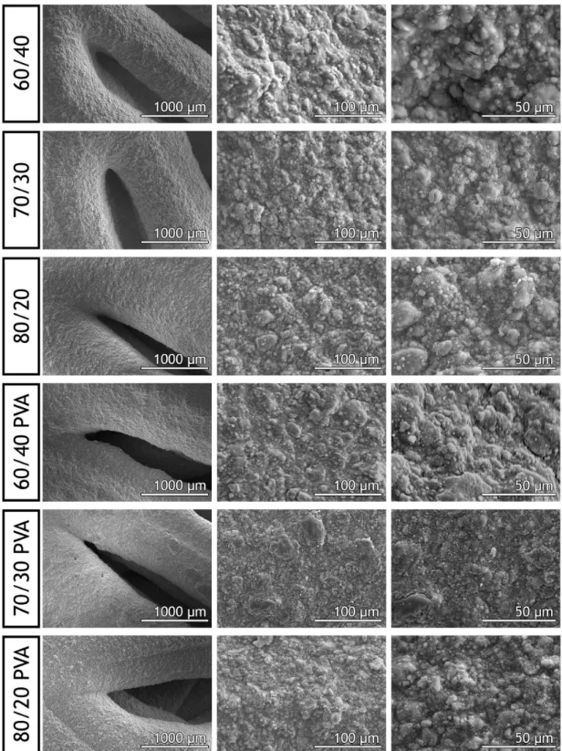
Following, SEM images were acquired, to characterize the scaffolds surface morphology (Figure 3.3).
Figure 3.3: SEM images showing the morphology of the scaffolds at different magnifications
In figure 3.3 it is possible to observe that all the scaffolds presented similar surface character-istics, with high roughness and irregularities. It has been previously described that the cellular morphology, adhesion and proliferation are directly affected by the scaffolds surface charac-teristics. In particular, human osteoblasts present increased metabolism, and ECM production, when in contact with rough surfaces. This effect is caused by an increased contact surface, directly related to the increased adhesion points on irregular surfaces88.