UNIVERSIDADE NOVA DE LISBOA
‘
Assessment of the genetic determinants involved in the expression of high
level of oxacillin resistance in contemporary clinical methicillin-resistant
Staphylococcus aureus (MRSA) strains’
MARIA GABRIELA GREGO RODRIGUES BENTO
Dissertation to obtain a Master Degree in Medical Microbiology
iii
Acknowledgments
To Dr. Catarina Milheiriço, my supervisor, for the tremendous effort in helping me writing this thesis, for the guidance and tutoring during the development of this work. To Professor Hermínia de Lencastre, Head of the Laboratory of Molecular Genetics at Instituto de Tecnologia Química e Biológica, it was an honor to be accepted and to have the opportunity to work in a laboratory with excellent working conditions, and for the support with the thesis.
I would like also to thank the great help of the high qualified team elements: Ana Tavares, Celine Coelho, Diana Espadinha, Joana Rolo, Inês Grilo, Maria Miragaia, Nuno Faria, Ons Bouchami, Teresa Conceição and Teresa Figueiredo who helped me in every way. To Ana Cristina Paulo, Carina Valente, Débora Tavares, Raquel Sá Leão, Sara Handem, Sofia Félix and Sónia Almeida.
To Mrs. Manuela Nogueira, for her kindness and assistance during this period.
To my family for being next to me during these past months of hard work and dedication. A special thanks to my friends Tamar Perloiro and Marta Nascimento.
iv
Abstract
Methicillin resistant Staphylococcus aureus (MRSA) is one of the leading causes of life threatening infections in hospital and community settings. In large part, this is due to the acquisition of resistance to ß-lactams, the most used class of antimicrobials. The majority of clinical MRSA isolates express β-lactam resistance in a heterogeneous way and several genetic factors are needed for expression of full resistance. One of the aims of our work was to identify genetic determinants responsible for the optimal expression of β-lactam resistance in strains belonging to the Brazilian clone (ST239-III). Whole genome sequencing (WGS) of two pairs of strains belonging to this clone was performed.
Our results demonstrated the presence of seven genes affected with non-synonymous mutations in both pair of strains. These genes were: rpoB, sasC, sdrD, coa, ebh, saK, int. Additionally we identified other genes mutated in one or the other pair of strains: fmt,
murA2, relA, valS, lysS, guaB, dfrB, gyr, tcaB, pbp2, pbp4, ccrA, blaZ, scn, cadA, mecA, merB, geh, lytA, nuc, tagB and pbp3. However more studies are needed in order to
confirm that the identified mutations have in fact a role in the phenotypic resistance level in the strains in study.
The mobility of the staphylococcal chromosomal cassette (SCCmec), which contains the
mecA gene, the central element of methicillin resistance, is due to recombinase genes,
namely ccrA, ccrB and ccrC. To assess the allelic variability of the ccrB locus (the most ubiquitous of the ccr genes) we sequenced this locus in a collection of representative MRSA strains selected in order to maximize temporal and geographical differences. The results obtained show very low mutation rate in ccrB locus among the clonal lineages studied. Further studies to confirm the functionality of these ccrB alleles and their role in the stabilization of the SCCmec cassettes should be performed.
v
Resumo
Staphylococcus aureus resistentes à meticilina (MRSA) são agentes frequentes de infeções bacterianas, a nível hospitalar e na comunidade. Uma das causas é a sua capacidade de aquisição de resistência aos β-lactâmicos, a classe de antimicrobianos mais usada em clínica. A maioria dos isolados de estafilococos expressa resistência aos β-lactâmicos de forma heterogénea e diversos fatores genéticos são necessários para a expressão total da resistência.
Um dos objetivos do trabalho apresentado nesta tese foi a identificação de determinantes genéticos responsáveis pela elevada expressão de resistência aos β-lactâmicos em estirpes do clone brasileiro (ST239-III). Com este objetivo, fizemos a sequenciação completa do genoma de dois pares de estirpes representativos deste clone.
Os resultados demonstraram a presença de sete genes afetados com mutações não-sinónimas nos dois pares de estirpes: rpoB, sasC, sdrD, coa, ebh, sak, e int. Adicionalmente, foram identificadas mutações nos genes fmt, murA2, relA, valS, lysS,
guaB, dfrB, gyr, tcaB, pbp2, pbp4, ccrA blaZ, scn, cadA, mecA, merB, geh, lyt A, nuc, tagB e pbp3 num ou no outro par de estirpes. Contudo, são necessários mais estudos para
confirmar que as mutações encontradas e identificadas como associadas com a resistência aos β-lactâmicos têm de facto um papel no nível de resistência
A mobilização do elemento SCCmec que contém o gene mecA, o elemento central da resistência à meticilina, é devida a três genes que codificam recombinases: ccrA, ccrB e
ccrC. De forma a estudarmos a variabilidade do gene ccrB (o mais comum de entre os
genes ccr), sequenciámos este locus numa coleção de estirpes MRSA selecionadas, com o objectivo de maximizar diferenças tanto a nível geográfico como temporal. Os resultados obtidos sugerem um baixo grau de mutação no locus ccrB entre as clones estudados. Contudo serão necessários estudos complementares para confirmar o papel destes alelos ccrB na estabilização das cassetes SCCmec.
vi
Table of contents
Acknowledgments ... iii Abstract ... iv Resumo ... v Table of contents ... vi Figures Index ... ix Tables Index ... x Abbreviations ... xi Chapter I - Introduction... 11. Staphylococcus aureus – General Features ... 1
1.1. S. aureus cell wall ... 1
1.2. Colonization and infection ... 2
1.3. S. aureus genome ... 2
2. Beta-lactam (β-lactam) resistance in S. aureus ... 3
2.1. Mechanism of action of β-lactam antibiotics ... 4
2.2. β – lactam resistance in S. aureus ... 4
2.2.1. Penicillin resistance ... 4
2.2.2. Methicillin resistance ... 5
3. Staphylococcal cassette chromosome mec - SCCmec ... 6
3.1. Integration and excision of SCCmec elements ... 9
3.2. Origin and evolution of the SCCmec ... 10
3.3. MRSA clones ... 10
4. Heterogeneous β-lactam resistance ... 13
5. Whole genome sequencing (WGS) ... 13
6. Aim………..15
vii Study A: Assessment of genetic determinants involved in the expression of high level of
beta-lactam resistance in contemporary clinical MRSA strains ... 16
1. Strain collection ... 16
2. Media and growth conditions ... 16
3. Phenotypic analysis of β-lactam resistance in S. aureus ... 16
3.1. Disk diffusion method ... 17
3.2. Etest ... 17
3.3. Population analysis profiles (PAPs) ... 18
4. DNA isolation ... 18
5. SCCmec typing ... 18
6. Whole genome sequencing (WGS) ... 19
6.1. DNA isolation ... 19
6.2. Genome sequencing ... 20
6.2.1. Detection of variations ... 20
Study B: Assessment of allelic variation in the ccrB locus in MRSA clones ... 21
7. Strain collection ... 21
8. Media and growth conditions ... 24
9. DNA isolation ... 24
10. ccrB sequencing ... 24
11. ccrB allelic variation ... 25
Chapter III – Results ... 26
Study A: Assessment of genetic determinants involved in the expression of high level of beta-lactam resistance in contemporary clinical MRSA strains ... 26
1. Strain selection ... 26
2. Population analysis profiles (PAPs) results ... 28
viii
4. Preparation of genomic DNAs for WGS ... 34
5. WGS results ... 35
Study B: Assessment of allelic variation in the ccrB locus in MRSA clones ... 39
6. ccrB allelic variation results ... 39
6.1. ccrB allelic variation among strains belonging to the same MRSA clone ... 42
Chapter IV – Discussion and conclusions ... 43
Study A: Assessment of genetic determinants involved in the expression of high level of beta-lactam resistance in contemporary clinical MRSA strains ... 44
Study B: Assessment of allelic variation in the ccrB locus in MRSA clones ... 46
Chapter V - References ... 48 Chapter VI – Annexes ... 58 Annex 1 ... 58 Annex 2 ... 60 Annex 3 ... 61 Annex 4 ... 62 Annex 5 ... 63 Annex 6 ... 64 Annex 7 ... 65 Annex 8 ... 66
ix
Figures Index
Figure 1. Regulation systems controlling the expression of β-lactamase and PBP2a ... 6
Figure 2. Types of SCCmec (I-XI). ... 8
Figure 3. Schematic illustration of the general arrangement of SCCmec. ... 10
Figure 4. Cefoxitin susceptibility determined by Etest for strains HU107 and HGSA145.. ... 27
Figure 5. Cefoxitin susceptibility determined by Etest for strains CPS68 and HDG2.. 28
Figure 6. Cefoxitin susceptibility determined by population analysis profile for the pair of strains A on TSA plates at 37°C. ... 28
Figure 7. Cefoxitin susceptibily determined by population analysis profile for the pair of strains B on TSA plates at 37°C. ... 29
Figure 8. Cefoxitin susceptibility determined by population analysis profile for the pair of strains B on TSA plates at 30°C. ... 30
Figure 9. Multiplex PCR results for SCCmec types identification ... 31
Figure 10. PCR results for the detection of mecA ... 31
Figure 11. PCR results for the detection of mecI ... 32
Figure 12. PCR results for the detection of RIF4 . ... 32
Figure 13. PCR results for the detection of RIF5 . ... 33
Figure 14. PCR results for the detection of pT181 e IS431. ... 33
x
Tables Index
Table 1. SCCmec types identified in S. aureus…. ... 9
Table 2. An overview of the major HA-MRSA clones.. ... 12
Table 3. An overview of the major PVL-positive CA-MRSA clones….. ... 12
Table 4. Strains used in study B. ... 21
Table 5. Oxacillin resistance profiles of a subset of strains in study. ... 26
Table 6. Cefoxitin resistance profiles of a subset of strains in study. ... 27
Table 7. Number of non-synonymous mutations in pair A (HU107/HGSA145) ... 36
Table 8. Number of non-synonymous mutations in pair B (CPS68/HDG2). ... 36
Table 9. Non-synonymous mutations shared by the two pairs of strains in study. ... 38
xi Abbreviations A – adenine AA – amino acid Arg - arginine Asn – asparagine
Asp – aspartic acid (aspartate)
ATCC – American Type Culture Collection bp - base pairs
C – cytosine
CA-MRSA – community-associated methicillin-resistant Staphylococcus aureus
ccr – cassette chromosome recombinase
CDC – Centers for Disease Control and Prevention CFU – colony forming units
CLSI – Clinical and Laboratory Standards Institute DNA – deoxyribonucleic acid
dNTP – deoxynucleoside triphosphate DR – direct repeat
EDTA – ethylenediamine tetraacetic acid
EMBL – European Molecular Biology Laboratory ENA – European Nucleotide Archive
FOX – cefoxitin fs – frameshift G – guanine
gDNA – genomic deoxyribonucleic acid Gln – glutamine
Glu – glutamic acid (glutamate) Gly – glycine
HA-MRSA – hospital-associated methicillin-resistant Staphylococcus aureus His – histidine
Ile – isoleucine IR – inverted repeat IS – insertion sequence
xii Abbreviations (continuation)
kb – kilobase kDa – kiloDalton LTA – lipoteichoic acid Lys – lysine
Mb – megabase
MGE – mobile genetic element MHA – Mueller Hinton agar MHB – Mueller Hinton broth
MIC – minimal inhibitory concentration ml – milliliter
MLST – multilocus sequence typing MNV – multiple nucleotide variation
MRSA – methicillin-resistant Staphylococcus aureus MSSA – methicillin-susceptible Staphylococcus aureus ND – not determined
ng – nanogram NT – non typeable OD – optical density ORF – open reading frame OX – oxacillin
PAP – population analysis profile PBP – penicillin-binding protein PCR – polymerase chain reaction PFGE – pulsed-field gel electrophoresis PG – peptidoglycan
pmol – picomole
PVL – Panton-Valentine leukocidin SAP – Shrimp Alkaline Phosphatase
SCC – staphylococcal cassette chromosome Ser – serine
xiii Abbreviations (continuation)
SNV – single nucleotide variation
Spa – Staphylococcus aureus protein A
ST – sequence type T – thymine TAE – Tris-acetate-EDTA TBE – Tris-borate-EDTA Thr – threonine Tn – transposon TSA – tryptic soy agar TSB – tryptic soy broth Tyr – tyrosine
V – volt Val – valine
WGS – whole genome sequencing WTA – wall teichoic acid
1
Chapter I - Introduction
1. Staphylococcus aureus – General Features
The genus Staphylococcus currently has more than 40 species and several subspecies (http://www.bacterio.cict.fr/s/staphylococcus.html).
The name Staphylococcus aureus has its origin from the Greek word staphylé, that means cluster of grapes and the latin word aureus, that means gold (1). S. aureus are gram-positive cocci with approximately 1µm in diameter that divides sequentially in three orthogonal planes over three consecutive division cycles (2, 3).
S. aureus is a non-motile, non-flagellate, coagulase and catalase positive,
non-spore-forming bacteria that grows in aerobic and anaerobic conditions (1).
1.1. S. aureus cell wall
One of the bacterial most important cellular structures is the cell wall. The cell wall is the first and major line of defense, providing the bacteria their cell shape, the strength to resist the high internal osmotic pressure and acts also as a protective barrier against threats from the environment (4). Peptidoglycan (PG), also called murein, is the main constituent of the bacterial cell wall. The PG polymer is composed by series of glycan strands of alternating N-acetylglucosamine and N-acetylmuramic acid cross-linked by short peptide bridges (5). The rigid sugar chains perpendicularly cross-linked with flexible peptide bridges results in a strong but also elastic stress-bearing structure that protects the underlying protoplast from lysing due to the high internal osmotic pressure (4, 6).
In S. aureus the majority of chains have a length of 3 to 10 disaccharide units. Attached to the N-acetylmuramic acid residues are stem peptides that are synthesized as pentapeptide chains, composed of L-alanyl-D-isoglutaminyl-L-lysil-D-alanyl-D-alanine. Neighbor glycan chains are cross-linked via an amino acid bridge of five glycines by the action of a transpeptidase that links the D-alanine from one stem peptide to the L-lysine of an adjacent stem peptide (6).
2 1.2. Colonization and infection
S. aureus is a commensal organism that colonizes the skin and mucous membranes of
humans and animals (7). The anterior nares are the primary niche and the main carriage site of S. aureus in humans being 30% of healthy human intermittent carriers, while 20% are permanent carriers. 50% are non-carriers (8). Transmission of S. aureus can be accomplished by direct contact (e.g. via colonized hands) or indirect contact (contact with contaminated surfaces) (9). S. aureus is also considered an opportunistic organism because is capable of causing infection when skin and mucous membranes became disrupted (10).
S. aureus can infect almost every tissue of the human body. These infections can vary
from mild skin and soft tissue infections, such as impetigo, folliculitis, cellulitis, wound and surgical infections to life-threatening infections, such as endocarditis, pneumonia, meningitis, bacteraemia and toxic shock syndrome (11).
The ability to cause severe diseases can be related to the type of strains that cause the infection and with the presence or absence of virulence factors (12).
1.3. S. aureus genome
S. aureus has a single circular chromosome of approximately 2,800,000 bp in size with
a low DNA G+C content (average 33%) (13). The genome can be divided in core and accessory genes (14). Approximately 75% of the S. aureus core genome is composed by genes associated with central metabolism and housekeeping functions, such as carbohydrate metabolism, protein synthesis and DNA replication (14). Additionally to these, there are genes not essential for survival and growth, including surface binding proteins, exoenzymes, toxins, capsule proteins and virulence genes (14). The accessory genome accounts for approximately 25% of the S. aureus genome and encodes for nonessential functions ranging from antibiotic and metal resistance to virulence (15). The accessory genome primarily comprises mobile genetic elements (MGEs) horizontally transferred between strains such as staphylococcal cassette chromosomes (SCC), bacteriophages, genomic islands, pathogenicity islands, plasmids and transposons (14, 16). The relative ease of transfer of some MGEs between staphylococcal species, containing mainly antibiotic-resistant genes,
3 outstands the potential pandemic problem, not only in hospital but also in the community setting (17).
2. Beta-lactam (β-lactam) resistance in S. aureus
The first β-lactam antibiotic, penicillin, was discovery in 1928 by Alexander Fleming (18) but it was the work of Howard Florey and Ernest Chain with their efforts on purification and chemistry that turned penicillin into a live-saving drug (18).
The introduction of penicillin G (benzylpenicillin) into the clinical practice in the early 1940s has been recognized as one of the greatest advances in therapeutic medicine (18). Until then, the mortality rate of individuals with an S. aureus infection was around 80% (19).
However, soon after the introduction of penicillin, in 1942, penicillin-resistance staphylococci were observed, first in hospitals then in the community (20). These strains became resistant due to the production of β-lactamases, which are able to inactivate the penicillin.By the late 1960s, about 80% of both hospital-associated and community staphylococcal isolates were resistant to penicillin (21).
The first semi-synthetic penicillin, methicillin, originally called celbenine, was introduced into clinical practice in 1960 to avoid the β-lactamase-producing staphylococci activity (22). Methicillin has an altered β-lactam ring with two carboxyl groups (CH3).
One year following the introduction of this antibiotic into clinical practice, the first MRSA was reported in England (23).
Although β-lactam resistance is a matter of major concern, β-lactams are still the most widely class of antibiotics prescribed, because of their high effectiveness, low cost, ease of delivery and minimal side effects (24). β-lactams in clinical use today are penicillin, narrow and extended spectrum cephalosporins, monobactams and carbapenems (25).
4 2.1. Mechanism of action of β-lactam antibiotics
β-lactam antibiotics are bactericidal agents that act by inhibiting the synthesis of the bacteria cell wall (26). The synthesis of PG is catalyzed by penicillin-binding proteins (PBPs), which are the bacterial targets of β-lactams. These enzymes are involved in the final stages of PG synthesis. PBPs are a group of membrane anchored extracytoplasmatic proteins that have evolved from serine proteases (27). PBPs catalyze transglycosylation and transpeptidation reactions that are required to the formation of linear glycan chains and to their crosslink, respectively (28).
β-lactams irreversibly acylate the PBP active-site serine, by mimicking the D-alanyl-D-alanine terminus of the pentapeptide side chain,by acting as substrate analogs. As consequence, the transpeptidase activity of PBPs is blocked, resulting in weakening of the cell wall and eventual lysis of the cell.
S. aureus have four native PBPs: three high-molecular-weight PBPs (PBP 1-3), and
one low-molecular-weight PBP (PBP4). PBP2 is the only bi-functional PBP, with both transpeptidase and transglycosylase activities. PBP1, PBP3 and PBP4 have only transpeptidation activity.
In S. aureus, PBP1 and PBP2 are the minimal machinery required for PG synthesis (28). S. aureus also encodes other enzymes involved in PG synthesis such as MGT and SgtA, two monofunctional transglycosylases and two proteins with predicted transpeptidase activity, FmtA and FmtB (29).
2.2. β-lactam resistance in S. aureus
β-lactams resistance in S. aureus can be mediated by β-lactamase production, acquisition of an extra PBP (PBP2a) and changes in the active site of the native PBPs (30).
2.2.1. Penicillin resistance
Penicillin is inactivated by β-lactamases, encoded by blaZ gene, which is part of the
bla system. β-lactamases are extracellular enzymes that promote the hydrolysis of the
penicillin β-lactam ring (21). BlaR1 is a sensor/inducer transmembrane protein encoded by the blaR1 gene and BlaI is a repressor protein, encoded by the blaI gene
5 (31). Usually, the blaI-blaR1-blaZ genes are carried on a plasmid or located on a transposon into the chromosome (in S. aureus, transposon Tn552) (32).
2.2.2. Methicillin resistance
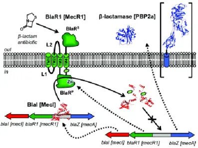
The central element conferring resistance to methicillin and others β-lactams antibiotics is the mecA gene (2.1 kb in length), that encodes for PBP2a, a transpeptidase protein with low affinity for β-lactam antibiotics, being intrinsically resistant to inhibition by these antibiotics (33). In the presence of β-lactams, the synthesis of PG is assured by the cooperation between the transglycosylase activity of PBP2 and the transpeptidase activity of the acquired PBP2a (33).
The mecA gene, is part of a mobile genetic element found in most MRSA strains, the so-called Staphylococcal Cassette Chromosome mec (SCCmec), and is highly conserved among different lineages (34, 35). The mecA regulatory locus is a three-component system. MecR1 is a sensor/inducer protein, encoded by mecR1 and MecR2 is an anti-repressor, encoded by mecR2, while MecI is a repressor protein, encoded by
mecI gene (36).
Although the MRSA characteristic phenotype is due to mecA gene, about 95% of MRSA strains have the blaZ gene, which means that besides the SCCmec element, most contemporary MRSA strains still carry the β-lactamase locus (37-39). The maintenance of a functional blaZ gene, even in the presence of mecA gene could be useful as a first-line of defense against β-lactams of the first generation (penicillins), which are still widely prescribed. The fitness cost associated to the expression of a secreted enzyme (BlaZ) is likely to be smaller than the fitness cost associated to the expression of a transpeptidase protein that have to be incorporated into the bacterial cell wall (PBP2a) (40).
The transcription of blaZ and mecA are controlled by homologous regulatory systems, BlaI-BlaR1 and MecR2-MecI-MecR1, respectively (37). These systems are similar in structure, showing sequence similarity with each other and function; both exhibit corepression but not coindution (41) (see figure 1).
6 Each one of these regulatory systems alone is able to control the transcription of blaZ and mecA (42). The induction of mecA gene expression when controlled by BlaI-BlaR1 is much faster than when controlled by MecR2-MecI-MecR1, taking from few minutes to several hours, respectively. (43).
Figure 1. Regulation systems controlling the expression of β-lactamase and PBP2a (shown in large brackets) in S. aureus. Adapted from reference (44).
3. Staphylococcal cassette chromosome mec - SCCmec
SCCmec is a mobile genetic element characterized by the presence of a mec gene complex, containing the mecA gene and its regulators and by the presence of a ccr gene complex, containing unique site-specific recombinases named cassette chromosome recombinases (Ccr) and finally by flanking direct and inverted repeats (45, 46). When a MSSA exogenously acquires the SCCmec, by horizontal transfer, a MRSA is generated (47, 48).
The mec gene complex is characterized by the mecA gene, intact or truncated copies of the mecA regulatory genes, namely mecI, mecR1 and mecR2 and a copy of the insertion sequence IS431.
In S. aureus there are three major mec classes. Class A has intact sequences for
mecI-mecR1 while classes B and C do not have mecI and have a partially deleted mecI-mecR1
7 The ccr gene complex is composed by the ccr genes and surrounding open reading frames (ORFs). Cassette chromosome recombinases (Ccr) are large serine recombinases of the resolvase/invertase family. Their main function is the excision and integration of the SCCmec element, i.e. the SCCmec mobilization.
Ccr recombinases are encoded by three phylogenetically different ccr genes, ccrA,
ccrB and ccrC, with nucleotide correspondences among them below 50%. Usually, ccr
genes with nucleotide identities more than 85% are assigned to the same allotype (42). To date, ccrA is classified into five allotypes, ccrB into seven allotypes and ccrC in two (49).
The gene ccrB is the most ubiquitous in SCCmec in MRSA and displays more sequence diversity than ccrA and mecA (50).
According to the guidelines proposed by the International Working Group on the Classification of SCC elements, SCCmec types are defined by the type of ccr gene complex and the class of the mec gene complex. Besides these two main components, SCCmec has also three regions called J, for joining (J1-3). These three hypervariable regions (J1-J2-J3), constitute nonessential components of the SCCmec, and include antibiotic resistance genes, pseudogenes, transposons, plasmids and insertion sequences. SCCmec subtypes are defined by the J regions. A SCCmec is considered non-typeable (NT) when it is not possible to ascertain a class of mec complex and/or a type of ccr (57).
8 Figure 2. Types of SCCmec (I-XI) identified in S. aureus. Adapted from reference(55).
To date, twelve different SCCmec types have been described (49) (see Table 1 and Figure 2).
9 Table 1.SCCmec types identified in S. aureus. Adapted from http://www.sccmec.org.
SCCmec type ccr gene complex ccr gene complex structure (allotypes) mec gene
complex mec gene complex structure
I Type 1 A1B1 B IS431-mecA-ΔmecR1-IS1272
II Type 2 A2B2 A IS431-mecA-mecR1-mecI
III Type 3 A3B3 A IS431-mecA-mecR1-mecI
IV Type 2 A2B2 B IS431-mecA-ΔmecR1-IS1272
V Type 5 C1 C2 IS431-mecA-ΔmecR1-IS431(the two IS431s
are arranged in the opposite direction)
VI Type 4 A4B4 B IS431-mecA-ΔmecR1-IS1272
VII Type 5 C1 C1 IS431-mecA-ΔmecR1-IS431(the two IS431s
are arranged in the same direction)
VIII Type 4 A4B4 A IS431-mecA-mecR1-mecI
IX Type 1 A1B1 C2 IS431-mecA-ΔmecR1-IS431(the two IS431s
are arranged in the opposite direction)
X Type 7 A1B6 C1 IS431-mecA-ΔmecR1-IS431(the two IS431s
are arranged in the same direction)
XI Type 8 A1B3 E blaZ-mecALGA251-mecR1LGA251-mecILGA251
XII Type 9 C2 C2 IS431-mecA-ΔmecR1-IS431(the two IS431s
are arranged in the opposite direction)
SCCmec types range in size from 21 to 67 kb and SCCmec III is considered to be the longest SCCmec element with 67 kb in length and it consists of two SCC elements, SCCmec type III and SCCmercury, harboring ccrC, integrated plasmid pl258 and tranposon Tn554. (46)
3.1. Integration and excision of SCCmec elements
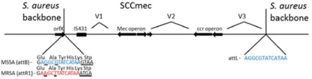
SCCmec elements are large pieces of DNA that are integrated at the 3´end of an open reading frame (orfX) in the S. aureus chromosome, at a specific site referred as attB (Figure 3), located near the replication origin, which is 66 kb – 89 kb upstream of the
spa gene and 10 kb downstream of purA, depending on the size of the integrated
10 OrfX is a staphylococcal ribosomal methyltransferase of the RlmH type. OrfX methylates 70S ribosomes and is conserved in all staphylococci (54).
After integration, SCCmec is flanked by direct repeats (DR) sequences, which serve as integration sites for the next SCC element (55).
Figure 3. Schematic illustration of the general arrangement of SCCmec. Adapted from
reference (54).
3.2. Origin and evolution of the SCCmec
The origin of SCCmec is still unclear. It was suggested that Staphylococcus sciuri harbored the ancestor of the gene mecA encoding PBP2a. Most S. sciuri are methicilin susceptible but in the presence of methicillin these bacteria could become resistant to the antibiotic due to an increase in the transcription of the mecA homologue caused by the appearance of mutations in the mecA promoter region (56). It has been proposed that the MRSA had been originated in vivo through the horizontal transfer of mecA gene from coagulase negative staphylococci (57).
3.3. MRSA clones
MRSA clones are defined by the combination of their multilocus sequence type (MLST) and by the SCCmec type they carry (e.g. ST239-SCCmec III, abbreviated as ST239-III) (58).
The first MRSA strain (NCTC10442) was isolated in 1961, in the United Kingdom and harbored SCCmec type I. This MRSA clone is known as the Archaic clone and was disseminated all over the world in the 1970s and 1980s. In 1982, in Japan, the
11 MRSA strain (N315) that harbored SCCmec type II was isolated. Strains with the characteristics of N315, were assigned to the New York/Japan clone that spread worldwide in the 1990s. Only three years later, in 1985, in New Zealand, another MRSA strain (85/2082) harboring SCCmec type III was isolated (48, 59). In the 1990s, some MRSA clones harboring SCCmec type IV have spread all over the world. In 2004, it was isolated in Australia, the MRSA strain WIS that harbored SCCmec type V. Other MRSA clones have been identified as described below (45, 60-64).
Some of the major MRSA clones included the Brazilian (ST239-III), New York/Japan (ST5-II), EMRSA 15 (ST22-IVh), EMRSA 16 (ST36-II), Berlin (ST45-II), Pediatric
(ST5-VI and ST5-IVa), USA 300 (ST8-IVa), European (ST80-IVNT and ST80-IVc),
Southwest/Pacific (ST30-IV) clone (65) (Table 2 and 3). The Brazilian clone (ST239) is one of the most successful and persistent clones, is multiantibiotic-resistant and accounts for 90% of hospital-associated MRSA (HA-MRSA) around the world. This clone spread throughout Asia, South America and is actually in Eastern Europe (66). The appearance and spread of different methicillin-resistant clones over time has been referred to as a wave-like phenomenon.
The first wave is associated with penicillin-resistant S. aureus, the second wave stands in for methicillin-resistant S. aureus, the third wave representing the hospital-associated MRSA (HA-MRSA) and the fourth wave is related to community-associated MRSA (CA-MRSA) in the late 1990s (67). Since then, CA-MRSA has emerged worldwide, not only in the community but also in healthcare facilities. Nowadays, the difference between HA-MRSA and CA-MRSA is beginning to fade (31, 68), it has been reported the presence of major HA-MRSA clones in the community and CA-MRSA clones as being the cause of nosocomial outbreaks. CA-MRSA strains (e.g strains usually carrying SCCmec type IV and type VI) when compared with HA-MRSA strains (e.g. strains usually carrying SCCmec type I-III) tend to be more virulent due to the presence of various virulence factors, faster growth, frequently susceptible to non-β-lactams classes of antibiotics and to have a lower degree of β-lactam resistance (69). The smaller size of some CA-MRSA strains such as type SCCmec type IV allow the rapid dissemination of MRSA clones with these SCCmec types, usually by horizontal gene transfer, presumably via bacteriophages (70).
12 Table 2. An overview of the major HA-MRSA clones. Adapted from reference (65).
Table 3. An overview of the major PVL-positive CA-MRSA clones. Adapted from
13
4. Heterogeneous β-lactam resistance
Several studies demonstrated that MRSA strains, highly resistant to β-lactam antibiotics and beta-lactamase producers, can have a nonfunctional MecI-MecR1 regulatory system. This inactivation can occur either through deletion of the mecI gene or by accumulation of point mutations within the mecI coding sequence or in the mecA promoter with subsequent increase in the production of PBP2a. Nevertheless, there are some cases reported in which strains phenotypically resistant to methicillin have no mutations in the mecI nor in the mecA promoter region (72). The amount of PBP2a does not fully correlate with the resistance level (73).
It has been reported the existence of genetic determinants that are responsible for the high β-lactam resistance levels in subpopulations of MRSA (72, 74). Mutations in genes that encode for products associated with distinct functional categories, such as transcription, translation, transport, cell division among others, have been associated with highly resistant MRSA strains; some examples are, rpoB, murE, murF, femB,
femX, hmrA, glmM, fmtA, dacA, sigB, spoVG, sarA, xdrA, ccpA, secDF, vraR, vraS, agrA, agrB (75).
Most contemporary MRSA strains present heterogeneous methicillin resistance. Such hetero-resistant MRSA strains have the majority of the cells resistant to relatively low or moderate concentrations of antibiotic, sometimes near the minimal inhibitory concentration (MIC) threshold of a susceptible strain, while subpopulations are able to grow at a much higher antibiotic MIC value (76). As a result, clinical isolates of MRSA can be improperly diagnosed and the use of β-lactams might lead to treatment failure (77).
5. Whole genome sequencing (WGS)
The first S. aureus genomes were sequenced in 2001 (51) and in the past fifteen years, the costs for performing a WGS analysis have drastically decrease, allowing this technique to became, in the near future, part of the routine typing methods (78). Several sequencing technologies are been used, from “sequencing by ligation” to “sequencing by synthesis” approaches. As each sequencing technology has strengths
14 and weaknesses, the choice of a specific platform to perform the WGS have to be selected in a way to obtain the desired experimental results.
One of the platforms commonly used is MySeq (Illumina Inc., San Diego, CA, USA), in which “sequencing by synthesis” consists in individual DNA molecules being attached to the surface of flow cells and isothermal ´bridging` amplification is used to amplify signals that are sequenced by using reversible fluorophore-labeled nucleotides, which are optically read from each flow cell. These techniques have low error rates, produce large amounts of raw data and have high accuracy, which provides sufficient discrimination between isolates of the same MRSA clone. One of the disadvantages is that individual read lengths tend to be short and this can be a problem for genomes with large repeats (78).
The applications of genome sequencing go from Clinical medicine, Bacterial evolution, Genomic archaeology to Metagenomics studies. Currently, full genome sequences of a large number of MRSA strains are available in the National Center for Biotechnology Information (http://www.ncbi.nlm.nih.gov).
The power of genome sequencing to identify multidrug-resistant pathogens is an invaluable tool to infection control (79).
Point mutations are the most frequent sequence variations in bacterial genomes. These point mutations result in single nucleotide polymorphisms (SNPs). There are SNP differences in housekeeping and other stable genes. Isolates belonging to the same lineage have a remarkably conserved core genome, even when separated by time and space (80).
The workflow of bacterial sequencing consists in sample preparation, library preparation, DNA sequencing, sequence assembly (de novo or against a reference) and bioinformatics analysis (78).
15
6. Aim
This thesis is composed by two different studies (study A and study B).
Study A was performed in order to identify the genetic determinants that could be on the basis of the different level of β-lactam resistance in strains belonging to the same genetic background and characterized by the same SCCmec type. Thus, two pairs of strains, belonging to the Brazilian clone, with the same genetic background (as ascertain by their MLST sequence type and/or spa type) and SCCmec types but with different β-lactam resistance profiles were selected. The two pairs of strains were compared by whole genome sequencing and the presence of non-synonymous mutations that could explain the phenotypic differences in the β-lactam resistance between the strains selected was analyzed.
Study B was developed to assess the allelic variability of the ccrB locus in successful clonal lineages for which the SCCmec stability remains to be determined (EMRSA15, EMRSA16, Berlin, Pediatric, USA300, European, SW/Pacific and Brazilian clones). Functionality assays will be performed in the future using the different alleles identified in this study in order to infer the role of the different ccrB alleles in the stabilization of the SCCmec elements in these particular clones.
16
Chapter II – Materials and Methods
Study A: Assessment of the genetic determinants involved in the expression of high level of beta-lactam resistance in contemporary clinical MRSA strains
1. Strain collection
The methicillin-resistant Staphylococcus aureus (MRSA) strains used in the present study are listed in Supplementary Table 1S (Annex 1). Seventy-five MRSA strains previously characterized as belonging to the Brazilian clone, with multilocus sequence type 239 (ST239), a spa type usually associated with this clone, and carrying SCCmec type III were selected from the collection of the Laboratory of Molecular Genetics, Microbiology of Human Pathogens Unit from Instituto de Tecnologia Química e Biológica, Oeiras, Portugal.
2. Media and growth conditions
S. aureus strains were grown overnight at 37°C on tryptic soy agar (TSA, Becton
Dickinson, Sparks, MD, USA) or on Mueller Hinton agar (MHA, Becton Dickinson, Sparks, MD, USA) or tryptic soy broth (TSB, Becton Dickinson, Sparks, MD, USA) or Mueller Hinton broth (MHB, Becton Dickinson, Sparks, MD, USA) under aerobic conditions.
3. Phenotypic analysis of β-lactam resistance in S. aureus
The susceptibilities of S. aureus strains to oxacillin and cefoxitin were determined by disk diffusion method, Etest (bioMérieux SA, Marcy-l`Étoile, France) and population analysis profiles (PAPs).
17 3.1. Disk diffusion method
The disk diffusion method (Kirby-Bauer) was performed according to the Clinical and Laboratory Standards Institute (CLSI) guidelines (81).
Briefly, a small aliquot of overnight cultures (grown in TSB at 37ºC) was diluted in MHB to an optical density at 620nm (OD620) of 0.08 (equivalent to 0.5 McFarland
turbidity) were spread on MHA plates, followed by placing oxacillin (OX) diffusion disks with 1 µg of antibiotic (Oxoid, Basingstoke, UK) and cefoxitin (FOX) diffusion disks with 30 µg of antibiotic (Oxoid, Basingstoke, UK) on the plates.
Minimum inhibitory concentration (MIC) values were evaluated after 18h incubation at 35ºC.
S. aureus American Type Culture Collection (ATCC) strain 25923, a mecA negative
isolate, was used as a quality control strain for the disk diffusion method. Results were only considered when the oxacillin and the cefoxitin disk halos for ATCC 25923 were 18-24 mm and 23-29 mm, respectively (81).
The following CLSI breakpoints for testing the susceptibility of S. aureus were used: oxacillin MIC susceptible (S) ≥ 13 mm, intermediate (I) = 11-12 mm, resistant (R) ≤ 10 mm; cefoxitin MIC susceptible (S) ≥ 22 mm and resistant (R) ≤ 21 mm.
3.2. Etest
Etest was performed according to the CLSI guidelines. Briefly, a small aliquot of overnight cultures (grown in TSB at 37ºC) was diluted in MHB to an optical density at 620nm (OD620) of 0.08 (equivalent to 0.5 McFarland turbidity) were spread on
Mueller Hinton agar plates (MHA, Becton Dickinson, Sparks, MD, USA), followed by placing the oxacillin (OX) or cefoxitin (FOX) Etest strips (bioMérieux SA, Marcy-l`Étoile, France) on MHA plates supplemented with 2% NaCl at 35°C for 24h. The following CLSI breakpoints for defining the susceptibility of S. aureus were used: oxacillin MIC susceptible (S) ≤ 2 µg/ml and resistant (R) ≥ 4 µg/ml; cefoxitin MIC susceptible (S) ≤ 4 µg/ml and resistant (R) ≥ 8 µg/ml.
18 3.3. Population analysis profiles (PAPs)
Population analysis profiles were performed for two pairs of strains (CPS68 vs HDG2 and HU107 vs HGSA145), by the agar plate method (76), in order to confirm their MICs.
Briefly, 25 µl of 100, 10-1, 10-3, 10-5 dilutions of an overnight culture (grown in TSB
at 37°C) were plated on TSA plates containing increasing concentrations of cefoxitin (Sigma-Aldrich). Colony forming units (CFU) were counted for every dilution with non-confluent growth after 48 h of incubation at 37°C.
4. DNA isolation
Chromosomal DNA from the two pairs of strains (CPS68 vs HDG2 and HU107 vs HGSA145) was prepared by using the Wizard Genomic DNA Purification Kit (Promega, Madison, WI, USA), according to the manufacturer`s recommendations, except for the lysis step with lysostaphin at 0.5 mg/ml and RNase at 0.3 mg/ml.
5. SCCmec typing
For the identification of the structural types/subtypes of the staphylococcal chromosomal cassette (SCCmec) and in order to confirm that the pairs of strains selected carried the same type/subtype of SCCmec, the Multiplex polymerase chain reaction (PCR) strategy described by Oliveira et al. in 2002 was performed as previously described (82).
The following prototype strains were used: COL as reference for SCCmec type I, N315 for SCCmec type II, ANS46 for SCCmec type III, HU25 for SCCmec IIIA, HDG2 for SCCmec IIIB and MW2 for SCCmec type IV.
PCR amplifications were performed in a T1 Thermocycler (Biometra, Alfagene). The PCR products (10 µl) were resolved in a 2% Seakem LE Agarose (Lonza, Rockland, ME USA), using the ladder 1-kb DNA Ladder Plus (Thermo Scientific, Fermentas) (Annex 2) in 0.5X Tris-borate-EDTA buffer (TBE) at 100 V for 45 min. Gels were
19 stained with ethidium bromide (0.15 µg/ml), then visualized and recorded under UV light in GelDoc XR+ System (Bio-Rad, Hercules, California, USA).
Uniplex PCR reactions were performed in dubious cases, with the same primers used in the multiplex methodology. Detection of mecA, mecI, RIF4, RIF5, IS431, pT181 and dcs was done in a final volume of 25 µl; containing 5 ng of the DNA template, 1X GoTaq Flexi buffer (Promega, Madison, WI, USA), 2.5 mM MgCl2 (Promega,
Madison, WI, USA), 160 µM of each dNTP (Bioron), 10 pmol of each primer, 0.625U of GoTaq FlexiDNA polymerase (Promega, Madison, WI, USA).
The cycling conditions were: predenaturation at 94°C for 4 min; 30 cycles of 94°C for 30 s, 53°C for 30 s, 72°C for 45 s; a final extension at 72°C for 4 min and soaking at 16°C. These assays were performed in a T1 Thermocycler (Biometra, Alfagene). The PCR products (10 µl) were detected by separation in a 1% Seakem LE Agarose (Lonza, Rockland, ME USA) using the ladder 1-kb DNA Ladder Plus (Thermo Scientific, Fermentas) in 1X Tris-acetate-EDTA buffer (TAE), at 80 V, for 40 min. Gels were stained with ethidium bromide (0.15µg/ml), then visualized and recorded under UV light in GelDoc XR+ System (Bio-Rad, Hercules, California, USA).
6. Whole genome sequencing (WGS)
6.1. DNA isolationGenomic DNA (gDNA) from strains HU107, HGSA145 and CPS68 was extracted using an optimization of the Qiagen DNeasy Blood & Tissue Kit (Qiagen) protocol (for details see Annex 3).
Samples were considered pure when the absorbance (A260/A280) ratio was between 1.7 and 1.9 and absorbance scans showed a symmetric peak at 260 nm. The DNA concentration was determined using the Nanodrop 1000 spectrophotometer (Thermo scientific, Wilmington, USA) and the Qubit 2.0 Fluorometer (Invitrogen, life technologies, Carlsbad, CA, USA).
20 6.2. Genome sequencing
Whole genome sequencing of MRSA strains CPS68, HU107 and HGSA145 was performed using the Illumina MiSeq platform at Instituto Gulbenkian de Ciência, Oeiras, Portugal, with a minimum coverage of 30X.
The coverage of the sequenced samples was confirmed using the following formula:
6.2.1. Detection of variations
Paired-end reads from each strain were mapped using CLC Genomics Workbench 8.0.1 (Qiagen Aarthus, Denmark) against the chromosome of the reference
Staphylococcus aureus ST239 strain TW20 (16) to identify single nucleotide variants
(SNVs), multi nucleotide variants (MNVs), deletions, insertions and replacements in the genome. WGS data from strain HDG2 were obtained from the European Nucleotide Archive (ENA) database (www.ebi.ac.uk/ena/data/view/ERS049891). The sequence and annotation of the TW20 genome were obtained from EMBL database with accession number FN433596 (16).
Variants analysis was performed with the tool “Quality-based variant detection”, using CLC Genomics Workbench 8.0.1.
21 Study B: Assessment of allelic variation in the ccrB locus in MRSA clones
7. Strain collection
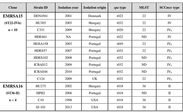
MRSA strains used in this study are listed in Table 4. Eighty MRSA strains belonging to the EMRSA15 (ST22-IVh), EMRSA16 (ST36-II), Berlin (ST45-IV and ST45-V), Pediatric (ST5-VI), USA300 (ST8-IVa), European (ST80-IVNT and ST80-IVc),
Southwest Pacific (ST30-IV) and Brazilian (ST239-III) clonal lineages and previously characterized in terms of their genetic background by spa typing and/or MLST and in terms of their SCCmec type, were selected from the MRSA culture collection of the Laboratory of Molecular Genetics at Instituto de Tecnologia Química e Biológica, Oeiras, Portugal. The selection of the strains was done with the objective to maximize temporal and geographical differences.
Table 4. Strains used in study B.
Clone Strain ID Isolation year Isolation origin spa type MLST SCCmec type
EMRSA15 DEN4561 2001 Denmark t022 22 IV
(ST22-IVh) HU303 2003 Hungary t032 22 IV
n = 10 C331 2009 Hungary t020 22 IVh HDE461 NA Portugal t022 ND IV HGSA158 2003 Portugal t849 22 IVh HDES57 2007 Portugal t032 22 IVh HDES102 2008 Portugal t032 ND IVh ICBAS12 2009 Portugal t032 ND IVh ICBAS46 2010 Portugal t032 ND IVh C124 2009 UK t032 22 IVh
EMRSA16 HU275 2002 Hungary t018 36 II
(ST36-II) HPH2 2006 Portugal t018 ND II
n = 4 C10 1998 USA t018 36 II
22 Table 4. Strains used in study B. (continuation).
Clone Strain ID Isolation year Isolation origin spa type MLST SCCmec type
Berlin 1150/93 NA NA t004 45 ND
(ST45-IV) DEN3050 2001 Denmark t015 45 IV
(ST45-V) HU281 2002 Hungary t038 45 IV
n = 7 PLN49 1997 Poland t015 45 IV
D17 1998 USA ND 45 IV
SJ-511 2013 USA t214 ND IVa
HDES79 2007 Portugal t004 45 IVa
Pediatric ARG164 1994-1996 Argentina t002 ND IV
(ST5-IVa) CLB1 1996 Colombia t002 ND IV n = 12 DEN698 2001 Denmark t002 5 IV HPV17 1992-1993 Portugal t311 5 VI HDE65 1993-1994 Portugal t311 ND VI HDE1 1992 Portugal t311 5 VI HDE383 1997 Portugal t311 ND VI HDES26 2007 Portugal t062 5 VI
HDES93 2007 Portugal t002 ND IVc
ICBAS43 2010 Portugal t002 5 IVa
C380 2005 Spain t311 5 IVa
STP46A 2012 STP t105 5 IVa
USA300 CR1 1996 CZ t024 8 IV
(ST8-IVa) CR43 1996 CZ t008 8 IV
n=11 DEN2988 2001 Denmark t008 8 IVa
HU288 2002 Hungary t008 8 IV
HU394 2005 Hungary t008 8 IVa
IPOP65 2001 Portugal t024 8 ND
ICBAS10 2009 Portugal t008 8 IVa
C368 2004 Spain t121 8 IVa
C377 2005 Spain t008 8 IVa
STP151 2012 STP t064 8 IVNT
USA300 1995-2003 USA ND 8 IVa
23 Table 4. Strains used in study B. (continuation).
Clone Strain ID Isolation year Isolation origin spa type MLST SCCmec type
European C006 2002 CZ t044 80 IVc
(ST80-IVNT) C014 2002 CZ t131 80 IVc
(ST80-IVc) DEN18851 1995 Denmark t044 80 IV (multiplex v1)
n=13 DEN4250 1996 Denmark t044 80 IV (multiplex v1)
DEN11819 1997 Denmark t044 80 IV (multiplex v1)
DEN2948 2001 Denmark t376 80 IV
E31 1997 Finland t044 80 IV (multiplex v1)
HT0401 2002 France t044 80 IV (multiplex v1)
C206 2005 Greece t044 80 IVNT
HU376 2002 Hungary t044 80 IVc
HFF189 2005 Portugal t044 80 IV
C273 2005 Romania t067 80 IVc
02-1418 2002 The Netherlands t044 80 IV (multiplex v1)
SW/Pacific C017 2004 CZ t019 30 IVc
(ST30-IV) SJ-031 2012 USA t021 30 IVa
n = 4 SJ123 2013 USA t019 30 IVc
DEN45 2001 Denmark t018 30 NT (multiplex v1)
Brazilian AGT120 1997 Argentina t037 ND ND
BZ48 1997 Brazil t037 239 IIIA
(ST239-III) CHL1 1997 Chile t037 ND ND
n = 19 CHL151 1998 Chile t037 ND ND
GRE18 1998 Greece t037 239 III
GRE317 1999 Greece t138 239 IIIA
HU125 1994 Hungary t037 239 III
HUR4 1997 Hungary t787 239 III
HU248 2001 Hungary t037 239 III
HU270 2002 Hungary t030 239 III
HU272 2002 Hungary t787 239 III
HU294 2003 Hungary t538 239 IIIA
HGSA15 1994 Portugal t037 239 IIIA
HGSA57 1995 Portugal t037 239 IIIA
HGSA142 2003 Portugal t037 239 IIIA
TAW97 1998 Taiwan t037 239 IIIA
TUR4 1996 Turkey t030 239 III
URU34 1997 Uruguay t037 ND ND
URU110 1998 Uruguay t037 ND ND
ST, sequence type; ID, identification; ND, not determined; NA, not available; NT, non typeable; SW, Southwest; var, variant UK, United Kingdom; USA, United States of America; CZ, Czech Republic; STP, São Tomé and Príncipe.
24
8. Media and growth conditions
Strains were grown overnight at 37°C on tryptic soy agar (TSA, Becton Dickinson, Sparks, MD, USA) or tryptic soy broth (TSB, Becton Dickinson, Sparks, MD, USA) under aerobic conditions.
9. DNA isolation
Chromosomal DNA was extracted using the boiling method (see Annex 4 for details) and in dubious cases, DNA was prepared by using the Wizard Genomic DNA Purification Kit (Promega, Madison, WI, USA), according to the manufacturer`s recommendations, except for the lysis step with lysostaphin at 0.5 mg/ml and RNase at 0.3 mg/ml.
10. ccrB sequencing
Seventy-one out of the 80 MRSA strains in study were analyzed using the sequencing approach described by Oliveira et al. in 2006 (83), in which the allelic variation in the
ccrB locus is evaluated by sequencing internal fragments of ccrB amplified by PCR,
using a pair of degenerated primers (ccrB F1 and ccrB R1). In each reaction (final volume of 50 µl), 5 ng of the DNA template, 1X PCR buffer with 1.5 mM MgCl2
(Applied Biosystems), 160 µM of each dNTP (Bioron), 100 pmol of each primer, and 1.25 U of AmpliTaq DNA polymerase (Applied Biosystems) was used. PCR amplifications were performed in a T1 Thermocycler (Biometra, Alfagene) with the following cycling conditions: predenaturation at 94°C for 4 min; 35 cycles of 94°C for 30 s, 42°C for 60 s, 72°C for 2 min; a final extension at 72°C for 4 min and soaking at 16°C.
The PCR products (5 µl) were detected by separation in a 1% Seakem LE Agarose (Lonza, Rockland, ME USA) using the ladder 1-kb DNA Ladder Plus (Thermo Scientific, Fermentas) in 1X Tris-acetate-EDTA buffer (TAE), at 80 V, for 40 min. Gels were stained with ethidium bromide (0.15 µg/ml) then visualized and recorded under UV light in GelDoc XR+ System (Bio-Rad, Hercules, California, USA).
25 After the visualization of the PCR products, the amplified fragments were purified using a mix of Exonuclease I and Shrimp Alkaline Phosphatase (SAP) enzymes, as described in the protocol provided by Dag Harmsen, Ridom Bioinformatics.
Briefly, to 30 µl of amplified PCR product, 6 µl of Exonuclease I (1U/µl) (New England Biolabs) and 6 µl of SAP (1U/µl) (USB Amersham) were added. The mixture was then subjected to a cycling program on the PCR machine with the following conditions: 30 min at 37°C followed by 20 min at 80°C.
The ccrB allele of nine strains was extrapolated from the available WGS data (strains highlighted in grey in Table 4), obtained from the European Nucleotide Archive (ENA) database (www.ebi.ac.uk/ena.).
11. ccrB allelic variation
DNA sequencing of both strands (forward and reverse) were performed at Macrogen (Amsterdam, The Netherlands)
The ccrB trace sequences were analyzed with DNA Star software (Lasergene, Madison, WI, USA) and alleles were assigned in accordance with the previously attributed ccrB alleles deposited in the ccrB database of the Laboratory of Molecular Genetics at Instituto de Tecnologia Química e Biológica, Oeiras, Portugal.
26
Chapter III – Results
Study A: Assessment of the genetic determinants involved in the expression of high level of beta-lactam resistance in contemporary clinical MRSA strains In order to analyze the genetic determinants responsible for the optimal expression of β-lactam resistance in strains belonging to the Brazilian clone, whole genome sequencing (WGS) for two pairs of strains belonging to the Brazilian clone (ST239-III) sharing identical genetic backgrounds but with different cefoxitin resistance profiles was performed.
1. Strain selection
Antimicrobial susceptibility tests to oxacillin and cefoxitin were performed in order to select two pairs of strains characterized by the same ST and/or spa types but with dissimilar oxacilin and/or cefoxitin resistance profiles (Supplementary Table 1S – in annexes section).
A subset of strains (HGSA339/HGSA145 and HU42/HU103) sharing identical genetic backgrounds and with discrepant values in the oxacillin disk halos were selected in order to determine their oxacillin MIC values by Etest.
Table 5. Oxacillin resistance profiles of a subset of strains in study.
Strain Isolation year Isolation origin SCCmec type SCCmec subtype spa type Sequence type (MLST) Oxacillin resistance Disk diffusion (mm) Etest (µg/ml)
HGSA339 2003 Portugal III IIIA t037 ND 6 256
HGSA145 2003 Portugal III IIIA t037 ND 9 256
HU42 1995 Hungary III ND t787 ND 6 2
HU103 1996 Hungary III ND t787 ND 15 8
Since no correlation could be established between the values of the oxacillin disk halos and the oxacillin MIC values determined by Etest (Table 5), oxacillin resistance profiles were replaced in favor of cefoxitin resistance profiles. Cefoxitin is used as a substitute for mecA-mediated oxacillin resistance according to CLSI guidelines (84).
27 When compared to oxacillin, cefoxitin is a better inducer of the mecA gene and tests using cefoxitin give more accurate and reproducible results than tests with oxacillin
(www.cdc.gov).
A correlation between the values of cefoxitin disk halos and MICs determined by Etest could be established for a new subset of strains (HU107/HGSA145 and CPS68/HDG2) sharing identical genetic backgrounds but with discrepant values in the cefoxitin disk halos (Table 6).
Table 6. Cefoxitin resistance profiles of a subset of strains in study.
Strain Isolation year Isolation origin SCCmec type SCCmec subtype spa type Sequence type (MLST) Cefoxitin resistance Disk diffusion (mm) Etest (µg/ml)
HU107 1996 Hungary III ND t037 ND 12 48
HGSA145 2003 Portugal III IIIA t037 ND 6 256
CPS68 1985 Portugal III ND t421 239 13 48
HDG2 1993 Portugal III IIIB t421 239 6 256
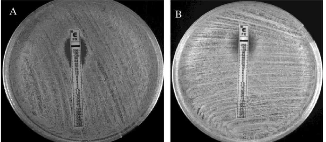
Two pairs of strains were selected for further study:Pair A composed by HU107 and HGSA145, both characterized by spa type t037 and with cefoxitin MICs of 48 and 256 µg/ml, respectively (Figure 4); and Pair B composed by CPS68 and HDG2, both characterized by spa type t421 and with cefoxitin MICs of 48 and 256 µg/ml, respectively (Figure 5).
Figure 4. Cefoxitin susceptibility determined by Etest for strains HU107 and HGSA145. A. HU107, MIC = 48 µg/ml and B. HGSA145, MIC = 256 µg/ml.
28
Figure 5. Cefoxitin susceptibility determined by Etest for strains CPS68 and HDG2.
A. CPS68, MIC = 48 µg/ml and B. HDG2, MIC = 256 µg/ml.
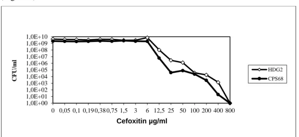
2. Population analysis profiles (PAPs) results
Population analysis profiles were performed for both pairs of strains in order to confirm their MICs and the differences in the MIC values of the strains belonging to the same pair. (Figures 6 and 7).
Pair A composed by strains HU107 and HGSA145, exhibited an MIC value of 12.5 µg/ml and 200 µg/ml as determined by PAP, respectively (Figure 6).
Figure 6. Cefoxitin susceptibility determined by population analysis profile for strains HU107 and HGSA145. A. HU107, MIC = 12.5 µg/ml and B. HGSA145, MIC = 200 µg/ml.
Colony-forming units (CFU) were determined by counting colonies after 48h of incubation on TSA plates at 37°C. 1,0E+00 1,0E+01 1,0E+02 1,0E+03 1,0E+04 1,0E+05 1,0E+06 1,0E+07 1,0E+08 1,0E+09 1,0E+10 0 0,05 0,1 0,19 0,38 0,75 1,5 3 6 12,5 25 50 100 200 400 800 C F U /ml CefoxitinPAP HU107 HGSA145 A B
29 Pair B composed by strains CPS68 and HDG2, showed an MIC value of 25 µg/ml for both strains as determined by PAP using TSA plates and 48h of incubation at 37°C (Figure 7).
Figure 7. Cefoxitin susceptibily determined by population analysis profile for strains CPS68 and HDG2. A. CPS68, MIC = 25 µg/ml and B. HDG2, MIC = 25 µg/ml.
Colony-forming units (CFU) were determined by counting colonies after 48h of incubation on TSA plates at 37°C.
Taking into account that the Etest results were significantly different from the results obtained with the PAPs, it was decided to determine the cefoxitin resistance for this pair of strains in different growth conditions, namely at a different temperature - 30°C instead of 37°C.
When the growth temperature of the PAPs was altered from 37°C to 30°C, using TSA as growth media, the MIC results were similar to the ones obtained by Etest, with CPS68 and HDG2, exhibiting MIC values of 50 and 200 µg/ml, respectively (Figure 8). 1,0E+00 1,0E+01 1,0E+02 1,0E+03 1,0E+04 1,0E+05 1,0E+06 1,0E+07 1,0E+08 1,0E+09 1,0E+10 0 0,05 0,1 0,19 0,38 0,75 1,5 3 6 12,5 25 50 100 200 400 800 C F U /ml Cefoxitin µg/ml HDG2 CPS68
30 Figure 8. Cefoxitin susceptibility determined by population analysis profile for strains CPS68 and HDG2. A. CPS68, MIC = 50 µg/ml and B. HDG2, MIC = 200 µg/ml.
Colony-forming units (CFU) were determined by counting colonies after 48h of incubation on TSA plates at 30°C.
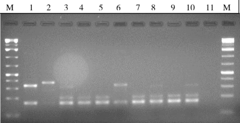
3. SCCmec results
In order to confirm that the pair of strains selected carried the same SCCmec, SCCmec typing was performed using the methodology described by Oliveira et al. in 2002 (82).
Strains COL, N315, ANS46 and MW2 were included as controls for SCCmec type I, II, III and IV, respectively; and strains HU25 and HDG2 were used as controls for subtypes IIIA and IIIB, respectively. Since, some of the patters were not clearly defined due to weak bands in the agarose gel, uniplex PCR reactions were additionally performed to confirm the dubious results.
1,0E+00 1,0E+01 1,0E+02 1,0E+03 1,0E+04 1,0E+05 1,0E+06 1,0E+07 1,0E+08 1,0E+09 1,0E+10 0 0,05 0,1 0,19 0,38 0,75 1,5 3 6 12,5 25 50 100 200 400 800 C F U /ml Cefoxitin µg/ml CefoxitinPAP CPS68 HDG2
31 Figure 9. Multiplex PCR results for SCCmec types identification. Lanes are as follows:
lane M – 1 kb DNA Ladder Plus; lane 1 – COL (SCCmec type I); lane 2 – N315 (SCCmec type II); lane 3 – ANS46 (SCCmec type III); lane 4 – HU25 (SCCmec type IIIA); lane 5 – HDG2 (SCCmec type IIIB); lane 6 – MW2 (SCCmec type IV); lane 7 – HGSA145; lane 8 – HU107; lane 9 -HDG2; lane 10 – CPS68; lane 11 – negative control
Uniplex PCR reactions were performed for the detection of mecA, mecI, RIF4, RIF5, IS431, pT181 (Figures 10-14).
Figure 10. PCR results for the detection of mecA (expected amplified fragment of 162 bp). Lanes are as follows: lane M – 1 kb DNA Ladder Plus; lane 1 - ANS46 (positive control);
lane 2 - HGSA145; lane 3 – HU107; lane 4 – HDG2; lane 5 – CPS68; lane 6 – negative control.
M 1 2 3 4 5 6 M M 1 2 3 4 5 6 7 8 9 10 11 M
32 Figure 11. PCR results for the detection of mecI (expected amplified fragment of 243 bp). Lanes are as follows: lane M – 1 kb DNA Ladder Plus; lane 1 – ANS46 (positive control);
lane 2 – HGSA145; lane 3 - HU107; lane 4 – HDG2; lane 5 – CPS68; lane 6 – negative control.
Figure 12. PCR results for the detection of RIF4 (expected amplified fragment of 243 bp). Lanes are as follows: lane M – 1 kb DNA Ladder Plus; lane 1 – ANS46 (positive control);
lane 2 – HGSA145; lane 3 - HU107; lane 4 – HDG2; lane 5 – CPS68; lane 6 – negative control.
M 1 2 3 4 5 6 M
33 Figure 13. PCR results for the detection of RIF5 (expected amplified fragment of 414 bp).
Lanes are as follows: lane M – 1 kb DNA Ladder Plus; lane 1 – ANS46 (positive control); lane 2 – HGSA145; lane 3 - HU107; lane 4 – HDG2; lane 5 – CPS68; lane 6 – negative control.
Figure 14. PCR results for the detection of pT181 and IS431 (expected amplified fragment of 303 bp). Lanes are as follows: lane M – 1 kb DNA Ladder Plus; lane 1 – ANS46
(positive control); lane 2 – HGSA145; lane 3 - HU107; lane 4 – HDG2; lane 5 – CPS68; lane 6 – negative control.
The combined results obtained in the SCCmec multiplex PCR and in the uniplex PCRs confirmed that the two pairs of strains selected were composed by strains belonging to the same SCCmec type (type III), but with different subtypes. Pair A is constituted by strains carrying SCCmec types III (HU107) and IIIA (HGSA145) and Pair B is pair B constituted by strains carrying SCCmec types III (CPS68) and IIIB (HDG2). Since the
M 1 2 3 4 5 6 M M 1 2 3 4 5 6 M
34 differences between the SCCmec III subtypes reside in the presence or absence of antibiotic resistance determinants for non-β-lactam antibiotics it was decided to pursue with the WGS analysis with the selected pair of strains.
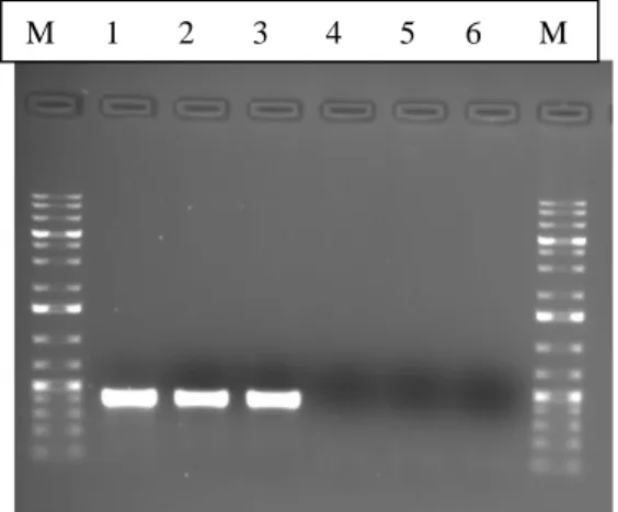
4. Preparation of genomic DNAs for WGS
To verify the chromosomal DNA integrity of the strains HGSA145, HU107 and CPS68 after the gDNA extraction using an optimization of the Qiagen DNeasy Blood & Tissue Kit (Qiagen) protocol, 2 µl of the extraction products were detected by separation in a 1% Seakem LE Agarose (Lonza, Rockland, ME USA) using the 1 kb DNA Ladder Plus (Thermo Scientific, Fermentas) in 1 X Tris-acetate-EDTA buffer (TAE), at 100 V, for 1h. Gels were stained with ethidium bromide (0.15 µg/ml), then visualized and recorded under UV light in GelDoc XR+ System (Bio-Rad, Hercules,
California, USA) (Figure 13).
With the gel electrophoresis results we could observe that the chromosomal DNAs were intact and do not present any state of degradation, since the bands were well defined and without smear.
Figure 15. Visualization of the genomic integrity. Lanes are as follows: lane M – 1 kb DNA
Ladder Plus; lane 1 – HGSA145; lane 2 – HU107; lane 3 – CPS68
The DNA concentration was determined using the Nanodrop 1000 spectrophotometer (Thermo scientific, Wilmington, USA) and the Qubit 2.0 Fluorometer (Invitrogen, life technologies, Carlsbad, CA, USA). The results obtained with Nanodrop and Qubit
35 respectively were as follow - HU107: 128 ng/µl and 192 ng/µl; HGSA145: 88.6 ng/µl and 154 ng/µl and for CPS68: 53.3 ng/µl and 65.6 ng/µl respectively.
5. WGS results
SNVs, MNVs, deletions, insertions and replacements detection
In order to identify differences at the genomic level between the two pairs of strains selected previously (HU107/HGSA145 and CPS68/HDG2), WGS was performed. The WGS samples coverage was confirmed to be 33X for HU107, 32X for HGSA145, 51X for CPS68 and 91X for HDG2. After the sequences trimming (the trimming percentage was around 99.9% for each strain and the number of reads after trim were for pair A HU107/HGSA145 376,291 and 331,989 respectively and for pair B CPS68/HDG2 594,861 and 3,653,828 respectively) (for details see Trim summary in annexes section, annex 5-8), pair-end reads were mapped to the reference
Staphylococcus aureus ST239 strain TW20 genome. In a first step the strains from
both pairs were compared with the reference strain Staphylococcus aureus TW20. Next, all the mutations shared between strains of the same pair, i. e., mutations of the same type in the same nucleotide position, were filtered out, making possible the comparison of mutations between the two strains of each pair (HU107 against HGSA145 and CPS68 against HDG2). Only non-synonymous mutations were considered and these mutations were divided in core and accessory regions.
SNVs at sites with heterogeneous mappings were filtered out if the SNV was present in less than 75% of the reads at that site.
The mutated gene, their product and the nature of the nucleotide and amino acid change are listed in Tables 2-7 deposited in the following link:
http://www.itqb.unl.pt/~m.gabrielabento/.
The number of non-synonymous mutations, in strains of pair A (HU107 vs HGSA145) demonstrated that SNVs (point mutations) are the most frequent mutation in accessory, and core regions (Table 7). This was also true for strains belonging to pair B (CPS68/HDG2) (Table 8).