UNIVERSIDADE DA BEIRA INTERIOR
Ciências da Saúde
The SCF/c-KIT system and imatinib actions in
prostate cancer: a cross-talk with RGN?
Henrique José Matos Morão Mingote Cardoso
Dissertação para obtenção do Grau de Mestre em
Ciências Biomédicas
(2º ciclo de estudos)
Orientador: Prof.a Doutora Sílvia Socorro
Co-orientador: Prof. Doutor Cláudio Maia
«Sei que tenho um cancro e que um dia me vai vencer. Mas esse dia não chegou e até lá
Tenciono continuar a aproveitar cada momento. Tive várias derrotas na vida,
Mas de todas as vezes caí de pé. É preciso nunca deixar de viver. A vida continua e continuarei a vivê-la,
Todos os dias, sem nunca me distrair Eu provavelmente morrerei da doença Mas o que nunca acontecerá é a doença matar-me»
Agradecimentos
O espaço que tenho nesta secção não é suficiente para agradecer a todas as pessoas que ao longo deste Mestrado e mais concretamente no último ano me ajudaram, direta ou indiretamente, a terminar esta fase da minha vida.
À professora Doutora Sílvia Socorro queria expressar o meu profundo agradecimento por ter acreditado nas minhas capacidades, e por me ter dado esta oportunidade. Queria também agradecer pelo apoio incondicional prestado, por toda a dedicação e pelos seus conselhos sábios. Agradeço também pela revisão crítica do texto e por todo o tempo que investiu em mim e no meu trabalho.
Ao Professor Doutor Cláudio Maia queria agradecer pela co-orientação neste projeto, pelas opiniões importantes que deu, pela disponibilidade demonstrada para ajudar e a motivação com que sempre o fez.
Agradeço ainda aos meus colegas de laboratório, Luís Rato, Inês Gomes, Margarida Grilo. E em especial agradeço à Sara Correia e à Cátia Vaz por toda a ajuda prestada e por todo o tempo que investiram em mim e no meu projeto. Sempre que eu precisei de bater à porta do vosso gabinete ela nunca esteve fechada. Jamais esquecerei alguns ensinamentos que me transmitiram. Quero também agradecer ao Carlos Gaspar pela ajuda na aquisição de imagens de microscopia de fluorescência e pela sua capacidade de me fazer sorrir mesmo quando era difícil. Ao Ricardo Marques agradeço por todo o apoio demonstrado ao longo deste ano.
Quero agradecer ainda a uma pessoa que durante este projeto foi quem mais me acompanhou. Marília Figueira, obrigado por tudo, obrigado por aqueles momentos em que tudo correu mal e me ajudaste a levantar, obrigado por teres celebrado as vitórias comigo e por acima de tudo por teres suportado o meu mau humor quando tudo parecia desabar. Obrigado pela tua amizade.
Agradeço também, às pessoas mais importantes da minha vida, a minha família. A vocês meus Pais agradeço-vos os bons valores que sempre me tentaram transmitir, todo o esforço que fizeram para me dar esta oportunidade e por toda a força que sempre me deram. Agradeço ainda à minha irmã porque sem ela não estaria aqui, demonstraste estar sempre disponível para mim. Longe ou perto estiveste sempre comigo, só te tenho a dizer Obrigado por tudo o que deste e darás. À minha avó queria pedir desculpa não ter tido a paciência contigo que merecias, mas é um orgulho saber que hoje ainda estás ao meu lado.
Agradeço ainda aos meus amigos por terem tido compreensão ao ouvirem: “não, hoje não posso” e continuarem ao meu lado.
Resumo
A progressão do cancro de próstata, de uma fase inicial com o tumor confinado à próstata para formas mais agressivas e invasivas, está associada à perda de resposta a androgénios. Neste estadio da patologia, as células malignas da próstata proliferaram independentemente da ação dos androgénios (o chamado cancro da próstata hormono-resistente), o que leva à falha das terapias clássicas de ablação de androgénios e restringe em muito as opções terapêuticas disponíveis para esta forma, normalmente letal, da doença. O Imatinib mesylate é uma droga quimioterapêutica que inibe a atividade de recetores tirosina cinase, como por exemplo o c-KIT, a qual tem vindo a ser usada com sucesso no tratamento de leucemias e tumores gastrointestinais. No entanto, a aplicação com imatinib no tratamento do cancro da próstata não tem sido totalmente eficaz, apresentando eficácia divergente em modelos pré-clínicos e testes in vivo. Por outro lado, a regucalcina (RGN), é uma proteína de ligação ao cálcio (Ca2+), que regula a homeostase de Ca2+ intracelular e a atividade de várias proteínas
envolvidas em vias de sinalização intracelular, como cinases e fosfatases, para a qual têm vindo a ser demonstrados os seus efeitos na supressão da proliferação celular na próstata. Isto levanta a questão de a RGN poder estar associada à regulação da expressão do c-KIT, e do seu ligando, o stem cell factor (SCF) em células da próstata. Assim, o objetivo principal da presente dissertação foi analisar os efeitos citotóxicos do imatinib em dois modelos celulares de cancro da próstata hormono-resistente, as linhas celulares DU145 e PC3. Para além disso, avaliou-se o efeito da RGN na expressão do sistema SCF/c-KIT na próstata de ratos através da utilização de modelo animal transgénico que sobre-expressa a RGN (Tg-RGN). Por último, foi estudada a localização sub-celular da RGN em células de cancro da próstata hormono-resistente, e a sua associação com a α-tubulina. As linhas celulares DU145 e PC3 foram incubadas com 20 µM imatinib durante 48 e 72 horas. O ensaio MTS foi utilizado para avaliar a viabilidade celular em resposta ao imatinib e a medição colorimétrica da atividade enzimática da caspase-3 foi incluída como um ponto final da apoptose. A expressão de reguladores do ciclo celular/apoptose em resposta ao imatinib, e a expressão do sistema SCF/c-KIT em ratos Tg-RGN vs. wild-type foi determinada através de PCR em tempo real e Western Blot. A expressão da RGN nas células DU145 e PC3 e a co-localização da RGN com α-tubulina foram avaliadas através de imunocitoquímica com marcação por fluorescência. O tratamento com imatinib diminuiu a viabilidade celular das células DU145 em ambos os tempos experimentais, 48 e 72 horas. No caso das células PC3, embora o imatinib tenha diminuído a sua viabilidade 6 horas após o tratamento, de seguida a viabilidade das células aumentou significativamente em relação ao controlo. O tratamento com imatinib aumentou a atividade enzimática da caspase-3 nas células DU145 enquanto nas células PC3 esta diminuiu significativamente, às 48 e 72 horas. Para além disso, as células DU145 exibiram um perfil de expressão reduzida da proteína anti-apoptótica Bcl-2 e níveis aumentados de proteínas
apoptóticas como a caspase-8 e caspase-9. Porém, não foram observadas diferenças nos níveis de expressão destas proteínas nas células PC3. A expressão do mRNA do p21, um reconhecido inibidor do ciclo celular, aumentou em ambas as linhas celulares em resposta ao tratamento com imatinib. Relativamente aos níveis de mRNA do fator de crescimento vascular endotelial (VEGF), estes encontraram-se diminuídos nas células DU145 em resposta ao imatinib, mas foi observado um efeito oposto nas células PC3. Com o intuito de tentar explicar a resposta diferencial das células DU145 e PC3 ao imatinib, caracterizou-se a expressão do c-KIT, e das suas isoformas, nestas linhas celulares. Análises por imunocitoquímica de fluorescência e Western blot mostraram que a expressão da forma membranar ativa do c-KIT se encontra diminuída nas células PC3 relativamente às células DU145, e que as células PC3 apresentam maior expressão das isoformas truncadas do c-KIT. Relativamente aos resultados da expressão do sistema SCF/c-KIT nas próstatas dos ratos Tg-RGN, verificou-se que esta se encontra diminuída. Confirmou-se ainda a expressão da RGN nas linhas celulares de cancro da próstata hormono-resistente e a sua co-localização com α-tubulina, um componente fundamental dos microtúbulos. Os resultados da presente tese demonstraram o efeito apoptótico do Imatinib nas células DU145, provavelmente através da inativação do c-KIT. Por outro lado, os efeitos paradoxais do imatinib observados nas células PC3 poderão estar associados à presença das isoformas truncadas do c-KIT, para as quais não está definitivamente estabelecido o seu papel. Estes resultados contribuíram igualmente para a compreensão da ineficácia do imatinib como opção terapêutica no cancro da próstata. Mais ainda, reforçam o papel da RGN como molécula antiproliferativa e reguladora do ciclo celular, o que é suportado pela diminuição da expressão do sistema SCF/c-KIT em resposta à sobre-expressão da RGN, assim como, pela associação da RGN com os componentes da maquinaria de divisão celular.
Palavras-chave
Resumo Alargado
A progressão do cancro de próstata, de uma fase inicial com o tumor confinado à próstata para formas mais agressivas e invasivas, está associada à perda de resposta aos androgénios. O cancro da próstata é uma doença cuja etiologia apresenta diversas causas. A presença de mutações em proteínas reguladoras da apoptose e proliferação é um acontecimento frequente nesta doença. Além disso, danos no epitélio prostático na sequência de inflamações recorrentes, infeções e/ou exposição a carcinogénios, pode despoletar o aparecimento do cancro. Nos estadios mais avançados da patologia, as células malignas da próstata proliferaram independentemente da ação dos androgénios (o chamado cancro da próstata hormono-resistente), sendo diversos os mecanismos que podem conduzir ao crescimento independente de androgénios. Entre muitos mecanismos, alguns dos quais ainda por esclarecer, destacam-se a amplificação da expressão do recetor de androgénios (AR), as mutações no gene do AR, as modificações em vias de sinalização celular que modulam a função do AR, as alterações na expressão de coreguladores do AR e ainda alterações no metabolismo. Todas estas alterações culminam no crescimento do tumor, mesmo com níveis reduzidos de androgénios, o que leva à falha das terapias clássicas de ablação de androgénios e restringe em muito as opções terapêuticas disponíveis para esta forma, normalmente letal, da doença. Portanto, revelar os mecanismos moleculares que desencadeiam o aparecimento de cancro da próstata resistente ao tratamento hormonal e o desenvolvimento de terapias eficazes para esta fase da doença é de crucial importância. O c-KIT é um recetor que pertence à família de recetores tirosina cinase do tipo III que é ativado pelo seu ligando específico, o stem cell factor (SCF). As vias de sinalização do c-KIT têm um papel essencial na decisão do “destino” celular controlando a proliferação, diferenciação e apoptose. Além disso, foi demonstrado que o c-KIT se encontra aumentado em pacientes com cancro da próstata hormono-resistente. O Imatinib mesylate é uma droga quimioterapêutica que inibe a atividade de recetores tirosina cinase, como por exemplo o c-KIT, competindo com o ATP no sítio de ligação ao ATP e parando a fosforilação e consequentemente parando as vias de sinalização celular. Tem vindo a ser usado com sucesso no tratamento de leucemias e tumores gastrointestinais. No entanto, a aplicação do imatinib no tratamento do cancro da próstata não tem sido totalmente eficaz, apresentando eficácia divergente em modelos pré-clínicos e testes in vivo. Por outro lado, a regucalcina (RGN), é uma proteína de ligação ao cálcio (Ca2+), que regula a homeostase de Ca2+ intracelular e a atividade de várias proteínas
envolvidas em vias de sinalização intracelular, como cinases e fosfatases. Esta proteína tem vindo a ser detetada em diversos tecidos do trato reprodutor masculino e a sua expressão encontra-se alterada em diversas patologias. Recentemente, foi demonstrado que a RGN tem efeitos supressores na proliferação celular na próstata, diminuindo o peso das próstatas e o índex de proliferação. Além disso, também foi demonstrado que a sobreexpressão da RGN
tem um papel protetor na carcinogénese no cancro da mama. Isto levanta a questão de a RGN poder estar associada à regulação da expressão do c-KIT, e do seu ligando, o SCF em células da próstata. Assim, o objetivo principal da presente dissertação foi analisar os efeitos citotóxicos do imatinib em dois modelos celulares de cancro da próstata hormono-resistente, as linhas celulares DU145 e PC3. Para além disso, avaliou-se o efeito da RGN na expressão do sistema SCF/c-KIT na próstata de ratos através da utilização de modelo animal transgénico que sobre-expressa a RGN (Tg-RGN). Por último, foi estudada a localização sub-celular da RGN em células de cancro da próstata hormono-resistente, e a sua associação com a α-tubulina. As linhas celulares DU145 e PC3 foram incubadas com 20 µM imatinib durante 48 e 72 horas. O ensaio MTS foi utilizado para avaliar a viabilidade celular em resposta ao imatinib e a medição colorimétrica da atividade enzimática da caspase-3 foi incluída como um ponto final da apoptose. A expressão de reguladores do ciclo celular/apoptose em resposta ao imatinib, e a expressão do sistema SCF/c-KIT em ratos Tg-RGN vs. wild-type foi determinada através de PCR em tempo real e Western Blot. A expressão de RGN nas células DU145 e PC3 e a co-localização da RGN com α-tubulina foram avaliadas através de imunocitoquímica com marcação por fluorescência. O tratamento com imatinib diminuiu a viabilidade celular das células DU145 em ambos os tempos experimentais, 48 e 72 horas. No caso das células PC3, embora o imatinib tenha diminuído a sua viabilidade 6 horas após o tratamento, de seguida a viabilidade das células aumentou significativamente em relação ao controlo. O tratamento com imatinib aumentou a atividade enzimática da caspase-3 nas células DU145 enquanto nas células PC3 esta diminuiu significativamente, às 48 e 72 horas. Para além disso, as células DU145 exibiram um perfil de expressão reduzida da proteína anti-apoptótica Bcl-2 e níveis aumentados de proteínas apoptóticas como a caspase-8 e caspase-9. Porém, não foram observadas diferenças nos níveis de expressão destas proteínas nas células PC3. A expressão do mRNA do p21, um reconhecido inibidor do ciclo celular, aumentou em ambas as linhas celulares em resposta ao tratamento com imatinib. Relativamente aos níveis de mRNA do fator de crescimento vascular endotelial (VEGF), estes encontraram-se diminuídos nas células DU145 em resposta ao imatinib, mas foi observado um efeito oposto nas células PC3. No intuito de tentar explicar a resposta diferencial das células DU145 e PC3 ao imatinib, caracterizou-se a expressão do c-KIT, e das suas isoformas, nestas linhas celulares. Análises por imunocitoquímica de fluorescência e Western blot mostraram que a expressão da forma membranar ativa do c-KIT se encontra diminuída nas células PC3 relativamente às células DU145, e que as células PC3 apresentam maior expressão das isoformas truncadas do c-KIT. Relativamente aos resultados da expressão do sistema SCF/c-KIT nas próstatas dos ratos Tg-RGN, verificou-se que esta se encontra diminuída. Confirmou-se ainda a expressão da RGN nas linhas celulares de cancro da próstata hormono-resistente e a sua co-localização com α-tubulina, um componente fundamental dos microtúbulos. Os resultados da presente dissertaçâo demonstraram o efeito apoptótico do Imatinib nas células DU145, provavelmente através da inativação do c-KIT. Por outro lado, os efeitos paradoxais do imatinib observados
quais não está definitivamente estabelecido o seu papel. Estes resultados contribuíram igualmente para a compreensão da ineficácia do imatinib como opção terapêutica no cancro da próstata. Mais ainda, reforçam o papel da RGN como molécula antiproliferativa e reguladora do ciclo celular, o que é suportado pela diminuição da expressão do sistema SCF/c-KIT em resposta à sobre-expressão da RGN, assim como, pela associação da RGN com os componentes da maquinaria de divisão celular.
Palavras-chave
Abstract
The progression of prostate cancer (PCa), from an early stage confined to prostate to a more aggressive form, is associated with loss of androgen responsiveness. At this stage, PCa cells proliferate independently of androgens actions (the so-called hormone refractory prostate cancer, HRPC), which cause the failure of classical androgen ablation therapies and restricts the therapeutic options for this usually lethal form of disease. Imatinib mesylate is a chemotherapeutic drug that inhibits the tyrosine kinase activity of c-KIT receptors among others, and has been successfully used to treat leukemias and gastrointestinal stromal tumors. However, its application for treatment of PCa has not been totally effective with preclinical models and clinical experimentation producing discordant results. On the other hand, regucalcin (RGN), a calcium (Ca2+)-binding protein that regulates intracellular Ca2+
homeostasis and the activity of several proteins involved in intracellular signaling pathways, namely, kinases and phosphatases, has been associated with suppression of cell proliferation in rat prostate. These raised the question whether RGN may regulate the expression of c-KIT and its ligand, the stem cell factor (SCF). Therefore, the present dissertation firstly aimed to analyze the cytotoxic effects of imatinib in two cell line models of HRPC, DU145 and PC3 cells. Moreover, the effect of RGN on the expression of SCF/c-KIT in rat prostate was evaluated by means of a transgenic animal model overexpressing RGN (Tg-RGN). Finally, the subcellular localization of RGN in HRPC cell lines and its association with α-tubulin was investigated. DU145 and PC3 cells were incubated with 20 µM imatinib for 48 and 72 hours. The MTS assay was used to assess cell viability in response to imatinib and the colorimetric measurement of the enzymatic activity of caspase-3 was included as an end-point of apoptosis. The expression of cell-cycle and apoptosis regulators in response to imatinib, and the expression of SCF/c-KIT in Tg-RGN vs. wild-type rats were determined by real-time PCR and Western Blot. The expression of RGN in HRPC cells lines in its association with α-tubulin were evaluated through fluorescent immunocytochemistry. Treatment with imatinib decreased the viability of DU145 cells at 48 and 72 hours. Although imatinib decreased the viability of PC3 cells upon 6 hours of treatment, thereafter cell viability significantly increased in relation to control. Accordingly, the enzymatic activity of caspase-3 was increased in DU145 cells whereas diminished activity of caspase-3 was observed in PC3 cells treated with imatinib for 48 and 72 hours. Moreover, DU145 cells displayed reduced expression of anti-apoptotic protein Bcl-2 and increased levels of the executioners of apoptosis caspase-8 and caspase-9. No differences were observed on the expression levels of these apoptosis related proteins in PC3 cells. The mRNA expression of cell cycle inhibitor p21 was increased in both DU145 and PC3 cells. Also, the mRNA levels of VEGF were decreased in DU145 cells in response to Imatinib but the opposite effect was seen in PC3 cells. To start explaining the differential response of DU145 and PC3 cells to imatinib, the expression of c-KIT receptor in these cell lines was characterized. Fluorescent immunocytochemistry and
Western Blot analysis showed that the expression of the active membrane-bound c-KIT is decreased in PC3 cells relatively to DU145. In addition, PC3 cells presented increased expression of truncated isoforms of c-KIT. Relatively to RGN the results obtained showed that the expression of SCF/c-KIT system is diminished in the prostate of Tg-RGN animals, which is in accordance with the antiproliferative effects of RGN, and indicates that regulation of SCF/c-KIT system may be a mechanism by which RGN restricts proliferation. Moreover, it was confirmed the expression of RGN in HRPC cells and its co-localization with α-tubulin, a fundamental component of microtubules. The results presented in this dissertation indicated that Imatinib was effective inducing apoptosis of DU145 cells likely through the inactivation of c-KIT. On the other hand, the paradoxical effects of imatinib in PC3 cells may be associated with the presence of truncated isoforms of c-KIT for which no definitive role has been established. These findings also contributed to understand the inefficacy of imatinib as therapeutic option in PCa. Moreover, the role of RGN as an antiproliferative molecule controlling cell cycle was further highlighted by the observed decreased expression of SCF/c-KIT system with overexpression of RGN, as well as, by the association of RGN with components of the cell division machinery.
Keywords
List of contents
I. Introduction ... 1
1.
Brief overview of anatomy and physiology of prostate ... 3
2.
Prostate cancer ... 5
2.1. Etiology and development of prostate cancer ... 5
2.2. From androgen-responsive to castration-resistant prostate cancer (HRPC) ... 7
3.
The c-KIT receptor and its ligand stem cell factor (SCF) in prostate pathophysiology ... 10
4.
Prostate cancer treatment and the use of tyrosine kinase inhibitor Imatinib ... 14
5.
The regucalcin protein and its relationship with prostate cancer ... 16
II. Aim of the Thesis ... 21
III. Material and Methods ... 25
1.
Cell lines ... 27
2.
Animals ... 27
3.
Cell Culture and Imatinib Treatment ... 27
4.
Cell Viability assay ... 27
5.
Real-time quantitative Polymerase Chain Reaction (qPCR) ... 28
6.
Protein Extraction ... 29
7.
Western Blot (WB) ... 30
8.
Caspase-3 activity assay ... 30
9.
Fluorescent Immunocitochemistry ... 30
10.
Statistical Analysis ... 31
IV. Results ... 33
1.
Viability of DU145 and PC3 cells in response to Imatinib ... 35
3.
Expression of VEGF and cell-cycle inhibitor p21 in DU145 and PC3 cells treated with
imatinib ... 38
4.
DU145 and PC3 cells differentially express the full-length c-KIT and its truncated
isoforms... 39
5.
The expression of SCF/c-KIT system is decreased in the prostate of Tg-RGN rats ... 40
6.
RGN is expressed in cell line models of HRPC and co-localizes with α-tubulin ... 41
V. Discussion ... 43
VI. Conclusions and Future Perspectives ... 51
VII. References ... 55
VIII. List of publications ... 73
1.
Publication in International Peer-reviewed Journal ... 75
2.
Oral communications ... 75
List of Figures
Figure I.1. Anatomy of Prostate gland. ... 4
Figure I.2. Putative pathways in development of hormone refractory prostate cancer ... 10
Figure I.3. Schematic representation of c-KIT structure ... 10
Figure I.4. Schematic representation of stem cell factor (SCF) structure ... 12
Figure I.5. Evolution of treatment of hormone refractory prostate cancer (HRPC) ... 15
Figure I.6. The role of regucalcin (RGN) in cell biology ... 18
Figure IV.7. Number of DU145 and PC3 viable cells after treatment with imatinib for 6, 12, 24, 48 and 72 h ... 35
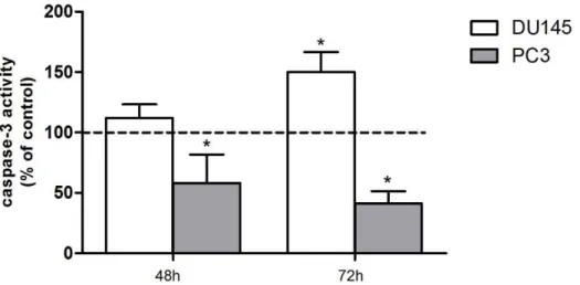
Figure IV.8. Caspase-3 activity in DU145 and PC3 cells after imatinib treatment for 48 and 72h. ... 36
Figure IV.9. Effect of imatinib on protein levels of caspase-8 (A) and caspase-9 (C) in DU145 and PC3 cells determined by WB analysis ... 37
Figure IV.10. Effect of imatinib on protein levels of Bcl-2 (A), Bax (C) and p53 (E) in DU145 and PC3 cells determined by WB analysis ... 38
Figure IV.11. Effect of imatinib on mRNA levels of p21 (A) and VEGF (B) in DU145 and PC3 cells determined by qPCR ... 39
Figure IV.12. Expression of full-length (145 kDa), and 50 and 30 kDa isoforms of c-KIT in DU145 and PC3 cells ... 40
Figure IV.13. Expression of c-KIT and its ligand SCF in the prostate of Tg-RGN rats comparatively with their wild-type counterparts ... 41
Figure IV.14. Expression of RGN in cell line models of HRPC, namely, DU145 and PC3 cells .. 42
List of Tables
Table I.1. Expression of SCF/c-KIT system in prostate cells ... 14
List of Abbreviations
ABL Abelson tyrosine kinase ADT Androgen-deprivation therapy
Apaf-1 Apoptotic protease activating factor-1
AR Androgen receptor
ATP Adenosine triphosphate Bcl-2 B-cell lymphoma 2
BPH Benign prostatic hyperplasia
Ca2+ Calcium
CZ Central zone
DHT 5-α-Dihydrotestosterone DTT DiThioThreitol
EGF Epidermal growth factor FDA Food and Drug Administration GIST Gastrointestinal stromal tumors GNNK Gly-Asn-Asn-Lys
GTPase Ran Ras-related nuclear protein HGPIN High-grade PIN
HRPC Hormone-refractory prostate cancer IGF-1 Insulin-like growth factor 1
IL Interleukin
KGF Keratinocyte growth factor
LHRH Luteinizing hormone-releasing hormone MAPK Mitogen-activated protein kinase mSCF Membrane isoform of SCF NF-kB Factor nuclear-kappa β PAP Prostatic acid phospathase
PBS Phosphate Buffer Saline
PCa Prostate cancer
PCR Polymerase Chain Reaction PDGF Platelet-derived growth factor
PDGFR Platelet-derived growth factor receptor
PFA ParaFormAldehyde
PI3K Phosphoinositide-3-kinase
PIA Proliferative inflammatory atrophy PIN Prostatic intraepithelial neoplasia PMSF PhenylMethylSulfonyl Fluoride pNA p-Nitro-Aniline
PSA Prostate-specific antigen
PTEN Tumor-suppressor phosphatase and tensin homolog PVDF PolyVinylidene DiFluoride
PZ Peripheral zone
qPCR Real-time Quantitative Polymerase Chain Reaction Rb Retinoblastoma protein
RGN Regucalcin
RIPA Radioimmunoprecipitation ROS Reactive oxygen species RTKs Tyrosine kinase receptors
SCF Stem cell factor
SDS-PAGE Sodium Dodecyl Sulfate-PolyAcrylamide Gel Electrophoresis SI Steel locus
S-KIT Soluble c-KIT protein
SMP30 Senescence marker protein-30 sSCF Soluble isoform of SCF
Tr-kit Truncated c-KIT protein
TZ Transition zone
VEGF Vascular endothelial growth factor W Spotted locus
I.
Introduction
Partially published in:
Cardoso HJ#, Figueira MI#, Correia S, Vaz CV, Socorro S (2014). The SCF/c-KIT system in the male: survival strategies in fertility and cancer. Molecular Reproduction and Development (in press). DOI: 10.1002/mrd.22430 IF: 2.812
Figueira MI#, Cardoso HJ#, Correia S, Maia CJ, Socorro S (2014). Hormonal regulation of c-KIT receptor and its ligand: implications for human infertility? Progress in Histochemistry and Cytochemistry (in press). DOI: 10.1016/j.proghi.2014.09.001 IF:5.909
1. Brief overview of anatomy and physiology of prostate
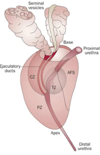
The prostate is considered the largest accessory gland organ of male reproductive tract, being, approximately, 4 cm long and 2 cm wide. It is located posterior to the symphysis pubis, anterior to the rectum, and inferior to the bladder. Prostate is constituted by a fibrous capsule containing smooth muscle cells and innumerous veins and nerves (Ali et al., 2004;Lee et al., 2011). Several pioneer studies of Mcneal and colleagues characterized prostate anatomy and divided it into 3 glandular zones: the central zone (CZ), transition zone (TZ), and peripheral zone (PZ) (Fig. I.1). These zones have different embryologic origins and can be distinguished by histology, anatomic landmarks, biological functions, and susceptibility to pathologic disorders (McNeal, 1972; 1981; 1984;Lee et al., 2011). TZ is situated in bilateral regions in the middle to the base of the gland that surrounds the first part of the urethra. CZ is surrounded for PZ and is constituted for a conical structure with ducts diverging from mid-prostate to the prostatic base. PZ, that is a major component of the mid-prostate, containing the ducts of urethra, in the mild-prostate, to the prostatic apex (McNeal, 1981). The PZ represents 70 % of prostate and is recognized as the zone most susceptible for development of prostate cancer (PCa) (Al-Ahmadie et al., 2008;Lee et al., 2011;Young et al., 2014). The prostate gland also includes a fourth non-glandular region that is constituted by fibromuscular stroma (Fig. I.1), which covers the urethra in the anteromedial prostate (Farnsworth, 1999;Young et al., 2014). The supporting stroma is a mixture of collagenous fibrous tissue and smooth muscle fibers (Farnsworth, 1999).
An alternative anatomical classification divides the prostate in an anterior lobe, a dorsal lobe, a lateral lobe and a ventral lobe (Young et al., 2014).
The normal function of prostate gland is fundamental to ensure sperm motility because this accessory gland produces a set of constituents of semen that provide the good environment for spermatozoa. These secretions empty into the prostatic urethra and make up about 30 % of the volume of semen (Frick and Aulitzky, 1991;Hayward and Cunha, 2000;VanPutte et al., 2014). The milky secretions of the prostate have a basic pH that possibly neutralizes the acidic environment of duct deferent and female vagina (VanPutte et al., 2014). Prostate epithelial cells produce other factors essential for proper sperm function, which includes citric acid and proteolytic enzymes (VanPutte et al., 2014).
The glandular prostatic epithelium is composed by the secretory epithelial cells, basal cells, stem cells and neuroendrocrine cells. The majority of epithelial cells, are columnar luminal cells, which are responsible for production of prostatic secretions including the prostate-specific antigen (PSA), the well-recognized indicator of PCa (Young et al., 2014). It is normally secreted into the ductal laminal and is removed by ejaculation. In normal situations PSA does not cross the epithelial basement membrane, however, in pathological conditions the histological organization of prostate is disrupted and PSA can be detected in the bloodstream (Cohen et al., 1992). The population of basal cells is present on the base of prostate gland in contact with basement membrane (Young et al., 2014). Prostate stem cells are confined to the basal compartment and represents a quiescent reserve that can divide
originating basal or luminal epithelial-like stem cells, uncommitted stem cells that differentiate to form semi-committed progenitors, or multiple populations of stem cells (Blum et al., 2010;Chen et al., 2013). The last cell type in the prostatic epithelium are the neuroendocrine cells that can secret neurosecretory products that promote prostate growth (Bonkhoff, 1998;Huang et al., 2006).
The human prostate is small in childhood, but in puberty rises in size to approximately 20-25 g in response to the increasing levels of serum testosterone. The prostate weight stabilizes until the individual has about 30 years and then prostatic weight turns to rise slowly, and this can mean the onset of Benign prostatic hyperplasia (BPH). The BPH nodules formed approximately upon 50 years can be transformed into PCa (Berry et al., 1984;Wong et al., 2003;Timms, 2008).
Figure I.1. Anatomy of Prostate gland. The prostate gland surrounds the bladder neck and also the first
part of urethra. The anterior and apical surfaces are bounded by anterior fibromuscular stroma (AFS), shaped by collagenous stroma and muscle fibers. Also the prostate is divided in four zones, transition (TZ) zone that surrounds proximal prostatic urethra, central zone (CZ) that surrounds the ejaculatory ducts, peripheral zone (PZ) that consists in approximately 70 % of prostate (Wadhera, 2013).
2. Prostate cancer
2.1. Etiology and development of prostate cancer
PCa is the most common cancer in men and represents the second leading cause of cancer deaths. In 2014, 233,00 new cases are expected to occur accounting for 27 % of total cancer cases diagnosed, which is estimated to represent 29,480 of deaths in the USA (Siegel et al., 2014). In Portugal, statistics indicate that the mortality by PCa has been growing, and that, only in 2011, 1764 men’s lost their life to this disease (Miranda, 2013).
PCa is mainly diagnosed upon 55 years and the prognostic of survival decreases in advanced ages (Whelan et al., 2013). Race and family history seem to contribute for development of PCa and, in fact, higher prevalence of disease is found in African descents. Also, in men whose first-degree family members are affected by disease the risk of PCa doubles (Ferlay et al., 2010).
Historically the human prostatic acid phosphatase (PAP) was the first serum biomarker for PCa, but it showed insufficient sensitivity, and later on, PSA was considered the ideal biomarker for screening of PCa, which still is the first line diagnosis marker (Ercole et al., 1987;Neal, 2010;Phillips, 2014). Nevertheless, it is also known that 15 % of men with normal or low levels of PSA had PCa (Mulders et al., 1990;Thompson et al., 2004). Therefore, a biopsy material is a fundamental piece for diagnosis, eliminating the presence of false positives and false negatives in PSA screening. The PCa is a heterogeneous and multifocal disease and the mechanisms driven its progression remain to be fully-elucidated. Normally, PCa is an asymptomatic disease that only manifest in latent stages of disease (Whelan et al., 2013). The prostatic epithelium can be damaged due to inflammation, infection and/or exposition to carcinogens, which can lead to the formation of proliferative inflammatory atrophy (PIA) (De Marzo et al., 1999). Alterations in this stage can conduct to the formation of histological lesions so-called prostatic intraepithelial neoplasia (PIN) (Putzi and De Marzo, 2000). PIN is characterized by the appearance of dysplasia of prostate luminal epithelial cells and a loss of distinct basal and secretory layers (Brawer, 2005;Wang et al., 2009;Davidsson et al., 2011). High-grade PIN (HGPIN) can be considered as the precursor of PCa (Brawer, 2005;Wang et al., 2009;Adamczyk et al., 2014).
Genomic lesions in PCa are common and can result in genomic rearrangement including amplification, alteration, deletion or translocation of segments of chromosomes (Saramaki and Visakorpi, 2007;Berger et al., 2011;Mao et al., 2011;Reid et al., 2012). The gain-of-function mutations in oncogenes and the deletion of tumor suppressor genes also are linked with carcinogenesis of prostate (Iurlaro et al., 2014). An uncontrolled cancer presents a combination of exacerbated proliferation, disrupted apoptosis and altered metabolic profiles (Lorenzo et al., 2007;Clarke et al., 2009;Vaz et al., 2012).
The phosphoinositide-3-kinase (PI3K) pathway is activated by several tyrosine kinases receptors and is the best characterized pathway in PCa. This pathway triggers cell proliferation through the activation of Akt and the tumor-suppressor phosphatase and tensin
homolog (PTEN) switches off the PI3K activity (Bitting and Armstrong, 2013). The overactivation of PI3K signaling in PCa is accompanied by PTEN deletion (Wang et al., 2006;Sun et al., 2009;Lonigro et al., 2011;Choucair et al., 2012). Also, alterations on the mitogen-activated protein kinase (MAPK) pathway seem contribute to the severity of PCa (Bakin et al., 2003;Mukherjee et al., 2011;Wang et al., 2012;Pavese et al., 2014).
Several other proteins involved in the control of cell cycle and cell survival are frequently mutated in PCa. Among other, this includes the widely recognized p53 and retinoblastoma protein (Rb). P53 is tumor-suppressor and a proapoptotic factor, and deletions and mutations on the p53 gene have been detected in approximately 40 % of PCa cases (Beltran et al., 2013). Also, the Rb protein, a tumor suppressor regulating cell cycle progression, is inactivated in advanced stages of this disease (Bookstein et al., 1990;Maddison et al., 2004;Thangavel et al., 2014). Other protein, the Homeobox protein Nkx-3.1, an important tumor suppressor in prostate, is frequently lost in PCa, by mechanisms of DNA-methylation and allelic deletion (Lei et al., 2006;Song et al., 2009;Akamatsu et al., 2010;Erbaykent-Tepedelen et al., 2011).
As described above, the inflammatory process may play a fundamental role triggering carcinogenesis. Leucocytes and macrophages migrate to inflammatory area and release reactive oxygen species (ROS). If high concentrations of ROS were produced can damage several cell structures including DNA and lipid-membranes (Ray et al., 2012). This event can contribute to carcinogenesis destructing cell-cell adhesion and cell-membrane functions and also contribute to mutations in DNA (Ishikawa et al., 2008;Kumar et al., 2008;Wong et al., 2009;Debelec-Butuner et al., 2014). Also, it has been shown that growth factors and cytokines released by macrophages in inflammatory response can contribute to suppress essential functions of androgen receptor (AR) in prostate (Debelec-Butuner et al., 2014). Factor nuclear-kappa β (NF-kB) seems to link inflammation to PCa. Despite its actions promoting apoptosis, NF-kB also can inhibit autophagy, promoting survival of cancer cells. In addition, NF-kB seems contribute to the progression of PCa progression increasing the expression of PSA (Chen and Sawyers, 2002;Ray et al., 2012). Moreover, it has been reported that several microorganism (Poutahidis et al., 2013) and chronic inflammation caused by sexually transmitted infection may promote the appearance of PCa (Sutcliffe et al., 2009;Chung et al., 2013).
The onset of PCa also depends on the activity of molecules produced and secreted by stromal cells, and the cross-talk between cancer cells and stroma has been identified as crucial aspect in carcinogenesis. This includes the secretion of a myriad of factor, such as, growth factors and cytokines. The stroma may sustain invasiveness by releasing products capable of digest the extracellular matrix (Bhowmick and Moses, 2005). Indeed, the key cell– cell binding regulator is the cadherin–catenin complex, whereas cell–matrix binding is largely mediated by integrins. Several evidences indicate that reduced expression of E-cadherin (Pontes et al., 2010;Liu et al., 2014) is related with advanced stages of PCa and similarly
important factors like vascular endothelial growth factor (VEGF), transforming growth factor-β (TGF-factor-β) and platelet-derived growth factor (PDGF) can be secreted by cancer cells and bind to its tyrosine kinase receptors on endothelial cells promoting angiogenesis (Russo et al., 2012;Roberts et al., 2013).
2.2. From androgen-responsive to castration-resistant prostate cancer
(HRPC)
As previously mentioned, pubertal development of the prostate is strictly linked with the circulating levels of androgens. In the adult prostate, androgenic actions, mediated by its cognized receptor, the AR, are primordial for its differentiation and function. The AR is expressed in all luminal cells, as well as, in stromal cells (Prins et al., 1996;Mirosevich et al., 1999), and AR signaling regulates the proliferation and inhibits apoptosis of prostate cells (Cunha, 1973;Wilson, 2011). The testosterone circulating in the blood stream is metabolized in prostate to 5 α-dihydrotestosterone (DHT), by the enzyme 5α-reductase. DHT displays higher activity and binds AR with more affinity than testosterone. The binding of DHT to the AR induces its dissociation from heat-shock proteins, and receptor phosphorylation and dimerization. AR dimers bind to the androgen-response elements in the promoter regions of target genes, which upon recruitment of regulatory proteins (activators or co-repressors) form the AR-transcriptional complex regulating the gene transcription (Socorro, 2014).
The actions of AR in the initial phases of PCa are similar to those in non-pathological prostate, namely, the control of PSA synthesis, lipid metabolism , growth and apoptosis (Cleutjens et al., 1996;Xu et al., 2006;Arnoldussen et al., 2011;Tennakoon et al., 2013). Because PCa is highly dependent on androgens, androgen-deprivation therapy (ADT) has been used as treatment for metastatic disease. ADT can be achieved surgically by orchiectomy or chemically with the use of anti-androgens, luteinizing hormone-releasing hormone (LHRH) agonists or LHRH antagonists (Schroder et al., 2012). Although the majority of tumors initially respond to these treatments, their effectiveness is limited and a complex process of resistance is developed (Karantanos et al., 2013). In fact, advanced-stages of metastatic PCa acquire several mechanisms that allow survival and invasiveness of cancer cells even in absence of androgens. At this stage patients develop the so-called hormone-refractory prostate cancer (HRPC). The mechanisms supporting the acquisition of this phenotype include amplification of AR expression, point mutations in the AR gene altering the AR activity, changes in cell signaling pathways that modulate the AR function, changes in the expression of coregulator proteins, and changes in steroid metabolism within tumor cells.
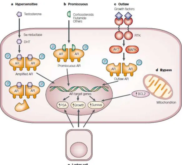
Hypersensitive Pathway. One possible mechanism by which PCa cells acquire resistance to ADT relies on the increasing sensitivity to low levels of androgens by increasing the AR synthesis and/or increasing AR sensitivity (Fig. I.2a). Both mRNA and protein levels of AR are augmented in PCa, and AR overexpression has been implicated in HRPC. Upon ADT therapy,
approximately 30% of HRPC cases display amplifications of the AR gene, resulting in increased AR expression, whereas no amplification were found before the ADT (Visakorpi et al., 1995;Koivisto et al., 1997). Complementing this information, it demonstrated that 80% of prostate tumors with AR amplification also displayed higher levels of AR protein (Edwards et al., 2003) (Fig. I.2a). Moreover, cancer cells survive continuing to proliferate even at low androgens levels, which may represent a second mechanism for development of HRPC (Waltering et al., 2009). Chen et al. (2004) verified that androgen-independent cells require 80% lower concentrations of androgens to grow than androgen-dependent cells. In another study, also was verified that the concentration of DHT stimulating growth of independent cells is four orders of magnitude lower than that required by androgen-dependent LNCaP cells (Gregory et al., 2001). Moreover, the increased stability of AR protein and the enhanced nuclear localization of AR found in androgen-independent cells complete the hypersensitive pathway in the acquisition of HRPC (Gregory et al., 2001). Finally, cancer cells might upregulate the expression of 5α-reductase enzymes increasing the production of DHT and maintaining AR signaling with lower levels of circulating androgens (Makridakis et al., 1997;Thomas et al., 2005;Das et al., 2010;Godoy et al., 2011). Supporting this mechanism are the data showing that ADT suppress testosterone levels in 95 %, while the concentration of DHT is reduced only in 60 % (Labrie et al., 1986), and the fact that ethnic groups with higher levels of activity this enzyme have a higher risk of PCa development (Makridakis et al., 1997). Moreover, castration resistant metastasis express higher levels of many enzymes responsible for the synthesis of adrenal androgens indicating genes involved in steroid biosynthesis are overexpressed (Holzbeierlein et al., 2004;Montgomery et al., 2008).
Promiscuous pathway. The specificity of AR in PCa has been questioned due to mutations present in ligand-binding domain. The frequency of genetic mutations in the AR loci are more frequent in advanced stages of disease (Taplin et al., 1995;Tilley et al., 1996;Ceraline et al., 2004;Sun et al., 2006). These mutations allow the AR to be activated by several non-androgenic molecules (Fig. I.2b), which maintains cell proliferation dependent on AR but stimulated by other ligands. Estrogens or anti-androgens have been pointed as the molecules that can bind to the mutant AR (Veldscholte et al., 1992;Zhao et al., 2000;Thin et al., 2003) (Fig. I.2b). The T7877A substitution is a good example of how a single mutation can alter the AR specificity, since in LNCaP cells, it resulted in cell growth in response to androgens and also to non-androgenic steroids (Suzuki et al., 1996). Again, this mechanism can be a defense of cancer cells, and Marcelli et al. (2000) described AR mutations in patients with lymph node metastasis that undergone ADT, whereas no mutations were found in the prostate glands of patients that received hormonal therapy. Also, other proteins that act as ativators and repressors of AR-induced transcription can be modified in HRPC. AR co-activators recruit other transcription factors to initiate transactivation of AR-regulated genes. Some of these co-activators, like ARA70, Tip60, TIF2 or SCR1 were increased in HRPC samples
repressors inhibit the transcription of AR-regulated genes, and the alteration of co-repressors like SMRT can be involved in progression of advanced stages of PCa (Godoy et al., 2012).
Outlaw Pathway. AR can be also activated by nonsteroid molecules, like growth factors
and cytokines. The growth factors, insulin-like growth factor 1 (IGF-1), keratinocyte growth factor (KGF) and epidermal growth factor (EGF), and cytokines, such as, interleukin (IL)-6, are capable of activating AR via signal transduction pathways (Culig et al., 1994;Hobisch et al., 1998). IGF-1, EGF and IL-6 are reported to be upregulated in HRPC (Di Lorenzo et al., 2002;Krueckl et al., 2004;George et al., 2005). Moreover, tyrosine kinase receptors (RTKs) also are connected with the activity of AR (Fig. I.2c). HER-2/neu, a member of the EGF receptor family of RTKs is overexpressed in androgen-independent cell lines generated from xenografts implanted in castrated mice (Craft et al., 1999), and this overexpression can activate AR-target genes independently androgens but not in the absence of AR (Craft et al., 1999;Yeh et al., 1999;Nishio et al., 2006;Ricciardelli et al., 2008). Moreover, this pathway can activate AR through the MAPK pathway. In fact, MAPK can phosphorylate the AR and leads to its activation (Yeh et al., 1999) Other pathway involved in cell survival is the Akt pathways and Akt activity is increased in independent cell lines compared with androgen-dependent cells (Graff et al., 2000;Morgan et al., 2009). Akt signaling can also alter cell cycle regulation by decreasing protein expression of p27, a known cell cycle inhibitor (Graff et al., 2000) (Fig. I.2c).
Bypass Pathway. It is also possible that alternative pathways to AR are capable to bypass and render cells with the ability to survive independently of AR activation. B-cell lymphoma 2) (Bcl-2) can be considered the candidate gene that can block apoptosis and which is not normally expressed in prostate (Colombel et al., 1993). In fact, several studies verified that Bcl-2 is overexpressed in cases of HRPC (Furuya et al., 1996;Fuzio et al., 2011). The blockage of Bcl-2 actions induces apoptosis in LNCaP cells (Yamanaka et al., 2006). Inactivation of the PTEN and subsequent activation of Akt is other bypass mechanism that cancer cells acquire in HRPC (Wang et al., 2006).
Lurker pathway. Other evidences point that HRPC develops because some cells that growth independently androgens were present before ADT. In fact, a minority sub-population of cells in prostate tumors that not express AR, has been identified as prostate cancer stem or progenitor cells (Collins et al., 2005). The cells can continue to proliferate and with ADT can exist a cancer cell selection. These cells are capable of self-renewal and are drug-resistant (Collins et al., 2005;Leong et al., 2008).
It is astonishing the number of different mechanisms underlying HRPC and conducting to more aggressive and lethal forms of PCa, which has hindered the development of specific and effective therapies for this stage of disease.
Figure I.2. Putative pathways in development of hormone refractory prostate cancer. a. In the
hypersensitive pathway, androgen receptor (AR) gene can be amplificated (more common) and/or enhanced sensitivity to androgens can occur. Also, augmented production of 5 α-dihydrotestosterone (DHT) due to increased activity of 5α-reductase has been indicated. b. In the promiscuous pathway, other molecules (like estrogens, corticosteroids, anti-androgens) can activate the AR pathway. c. In the outlaw pathway, others receptors, like receptor tyrosine kinases (RTKs) can be overactivated and, through Akt and/or the mitogen-activated protein kinase (MAPK) pathway, can phosphorylate AR (P) and activate it. d. In the bypass pathway other molecules involved in cell survival can control AR target genes, such as the Bcl-2 protein. e. In the lurker pathway, a minority sub-population of cells in prostate tumors that not express AR might be selected by the androgen deprivation therapy (ADT) (Feldman and Feldman, 2001).
3. The c-KIT receptor and its ligand stem cell factor (SCF) in
prostate pathophysiology
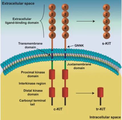
The c-KIT is a tyrosine kinase receptor belonging to type III RTKs family, which also includes the PDGFR and the macrophage colony stimulating factor receptor (Liu et al., 2007). It is also kwon like CD117, stem cell factor (SCF) receptor or KIT receptor. Firstly is described in 1986 as the transforming gene of the Hardy-Zuckerman 4 feline sarcoma virus, and identified as the proto-oncogene v-Kit (Yarden et al., 1987). The mouse c-KIT gene is located in the dominant white spotted locus (W) (Chabot et al., 1988;Geissler et al., 1988), while the human gene is located on Chromosome 4q11-q12 (d'Auriol et al., 1988). The main product of
considered (Fig. I.3): an extracellular ligand-binding domain, a transmembrane region, and an intracellular kinase domain (Qiu et al., 1988;Blechman et al., 1993;Blechman et al., 1995). The extracellular region, which comprises five immunoglobulin-like domains, recognizes the c-KIT ligand and also participates in receptor dimerization (Blechman et al., 1995;Yuzawa et al., 2007;Paulhe et al., 2009). In addition, the presence of the tetrapeptide Gly-Asn-Asn-Lys (GNNK) in the extracellular juxtamembrane domain of c-KIT (Fig. I.3) plays a relevant role regulating receptor activation and downstream signaling (Phung et al., 2013). A short chain of hydrophobic amino acids constitutes the transmembrane region, which allows fixation of c-KIT at the plasmatic membrane. The cytoplasmic region of c-c-KIT, containing proximal and distal kinase domains separated by an interkinase domain (Fig. I.3), is responsible for transduction of SCF/c-KIT signaling (Mol et al., 2003;Roskoski, 2005). Several c-KIT variants originated by distinct mechanisms have been identified in several cell types. Alternative promoter usage and transcription of a cryptic exon produces a truncated c-KIT protein (tr-KIT), with 30-50 kDa, which contains only the distal kinase domain and the carboxyl-terminal tail (Fig. I.3), and is located at cytoplasm (Rossi et al., 1992;Toyota et al., 1994). Moreover, the c-KIT protein can be proteolytically cleaved originating a soluble isoform (s-KIT, Fig. I.3) (Turner et al., 1995;Broudy et al., 1999).
Figure I.3. Schematic representation of c-KIT structure. The five immunoglobin-like domains of the
extracellular domain are involved in ligand-binding and receptor dimerization. The transmembrane domain anchors c-KIT in the cytoplasmic membrane. The intracellular region, responsible for signaling transduction, contains proximal and distal kinase domains separated by an interkinase region, and a carboxyl terminal tail. Some alternatively spliced forms of c-KIT are characterized by the presence of the tetrapeptide Gly-Asn-Asn-Lys (GNNK) in the extracellular juxtamembrane domain. The receptor can be cleaved and released from cell membrane originating a soluble c-KIT (s-KIT) only constituted by the extracellular domain. A truncated form of c-KIT (tr-KIT) originated by alternative promoter usage and lacks the extracellular and transmembrane domains retaining part of the kinase domain (Cardoso et al., 2014).
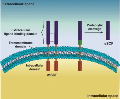
SCF is a growth factor/cytokine firstly identified in 1990 (Nocka et al., 1990;Williams et al., 1990;Zsebo et al., 1990). It is also known as c-KIT ligand or mast cell growth factor and it is codified on the Steel locus (Sl) of Chromosomes 12 and 10, respectively, in humans and mice (Zsebo et al., 1990;Geissler et al., 1991). The SCF protein, a 45 kDa glycoprotein is constituted by three distinct regions (Fig. I.4), the extracellular domain responsible for recognizing and binding of c-KIT (Langley et al., 1994), the hydrophobic transmembrane domain, and the intracellular domain (Langley et al., 1994;Zhang et al., 2000). The SCF is present at cell membrane as a noncovalent homodimer (mSCF) (Lu et al., 1991;Matous et al., 1996;Zhang et al., 2000), and the proteolytic cleavage of an alternatively spliced variant originates its soluble isoform (sSCF, Fig. I.4) (Flanagan et al., 1991;Pandiella et al., 1992;Majumdar et al., 1994) (Fig. 4). mSCF binds simultaneously two molecules of c-KIT in the membrane of receptor cell, which induces a conformational change that exposes a key dimerization site located in the fourth immunoglobulin-like domain of c-KIT (Fig. I.3) (Blechman et al., 1995;Lemmon et al., 1997;Paulhe et al., 2009). Receptor dimerization allows its autophosphorylation (Paulhe et al., 2009), which creates docking sites for several signal transduction molecules triggering the initiation of multiple signal transduction pathways (Mol et al., 2003;Roskoski, 2005).
Figure I.4. Schematic representation of stem cell factor (SCF) structure. The SCF display an
extracellular domain, responsible for recognizing and binding to c-KIT, a transmembrane domain and an intracellular domain. The SCF exists as a membrane-bound homodimer (mSCF) or as a soluble protein (sSCF). The sSCF is originated by the proteolytic cleavage of an alternatively spliced variant of SCF that contains the alternative exon 6 (green) (Cardoso et al., 2014).
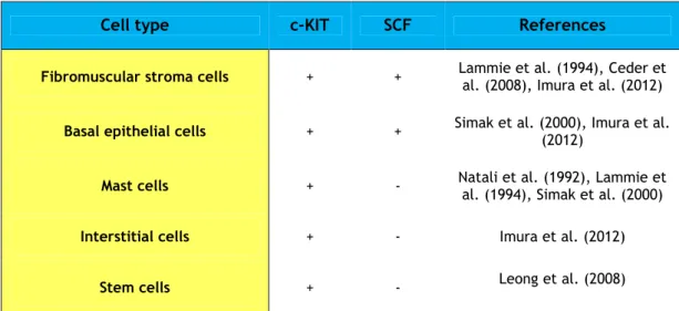
The expression of this system in prostate is poorly known, particularly in the case of rodents. Only one study has described the expression of c-KIT in rat prostate, which was regularly observed in the space between the smooth muscle layer and the glandular layer of the prostatic duct system (Kusljic and Exintaris, 2010). In human prostate c-KIT protein is
positive cells were identified as interstitial cells (Simak et al., 2000;Imura et al., 2012). SCF expression in human prostate is found in epithelial and stromal cells (Simak et al., 2000;Imura et al., 2012). However, c-KIT and SCF transcripts were detected at low levels in basal epithelial cells (Simak et al., 2000).
Considering the pathological conditions of prostate, it was found a significantly higher expression of SCF and c-KIT in cases of BPH comparatively to normal prostate (Imura et al., 2012). Moreover, the expression of c-KIT mRNA is higher during androgen deprivation compared to noncastrated clinical metastatic disease (Pfeiffer et al., 2011). The expression of c-KIT it is also higher in bone metastasis cancers (40%) relatively to primary PCa (14%) (Wiesner et al., 2008), suggesting an important function of c-KIT in metastatic forms of disease. Nevertheless, prostate tumors seem to specifically express the cytoplasmic KIT. tr-KIT is detected in tumoral tissues while the contralateral prostate not invaded by tumor cells, as well as, cases of BPH are negative, and its expression increases pronouncedly (~28% to ~66%) from less to more advanced stages of PCa (Paronetto et al., 2004). Studies in cases of human prostate adenocarcinoma strongly identified c-KIT in the cytoplasm and membrane of epithelial tumor cells (Simak et al., 2000), and in stromal cells (Di Lorenzo et al., 2004), while SCF showed to be frequently expressed in cytoplasm of tumor cells (Imura et al., 2012).
In human PCa cell lines the available literature has been controversial. If some studies revealed that c-KIT mRNA is not expressed in LNCaP, PC3 or DU145 cells, other reports showed its expression (Savarese et al., 1998;Wiesner et al., 2008). At protein level, some studies have reported c-KIT as being expressed in LNCaP and PC3 cells (Savarese et al., 1998), and in opposition others authors described that c-KIT is only present in DU145 cells (Wiesner et al., 2008;Brooks et al., 2012). Considering the tr-KIT, it was detected in LNCaP cells, but not in PC3 (Paronetto et al., 2004). It is liable to assume that, discrepancies verified on the expression of c-KIT and tr-KIT can be due to the use of different antibodies. Antibodies that recognized extracellular domain detected only the full-length c-KIT located at cell membrane, whereas antibodies that recognized the c-terminus of protein are capable of recognize both c-KIT and tr-KIT.
Relatively to SCF, transcripts were detected in all cancer cell lines already studied (Savarese et al., 1998;Simak et al., 2000), with DU145 cells displaying the highest expression comparatively with LNCaP and PC3 (Wiesner et al., 2008). The sSCF was detected at low levels in DU145 and PC3 cells (Savarese et al., 1998), which contradicts the findings of Wiesner et al. (2008) that reported only the expression of mSCF (Wiesner et al., 2008).
In vivo studies also demonstrated that in prostate adenocarcinoma, c-KIT was strongly identified in cytoplasm and membrane of epithelial tumor cells (Simak et al., 2000) and stromal cells (Di Lorenzo et al., 2004). On the other hand, SCF showed to be frequently expressed in cytoplasm of tumor cells (Imura et al., 2012).
Table I.1. Expression of SCF/c-KIT system in prostate cells (Cardoso et al., 2014).
+, positive expression; -, negative expression
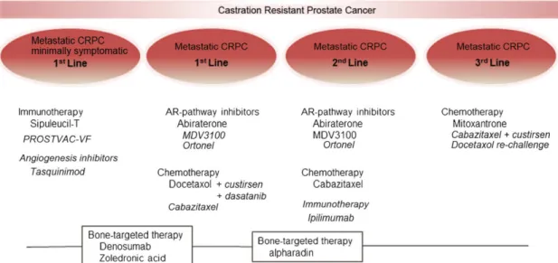
4. Prostate cancer treatment and the use of tyrosine kinase
inhibitor Imatinib
While localized PCa is potentially cured by surgery and radiation therapy. However, frequently this cancer advances to more aggressive stages, for which the most common treatment in locally and metastatic disease is ADT, as cited above. This therapy is effective at initial phases but in long-term, upon some months, resistance to therapy occurs and consequently the stage of HRPC emerges. Historically, treatment for HRPC was mainly palliative, and overall survival rates are in best of the cases approximately 35 months (Suzman and Antonarakis, 2014). In 2004, docetaxel became the first chemotherapy agent to extend survival in men with HRPC (Petrylak et al., 2004;Tannock et al., 2004). In last years, numerous studies evaluated the combination with docetaxel with new formulations but clinical benefit has not been demonstrated. However, in 2010, cabazitaxel, a taxane, was demonstrated to improve survival in patients (Galsky et al., 2010;Abidi, 2013). At present, cabazitaxel remains the only chemotherapy that shown survival benefits as a second-line treatment after docetaxel. But third-line options have been described (Fig. I.5). In fact, taxanes (like doxetaxel and carbizitaxel), that function by stabilizing the dynamic polymerization of microtubules in dividing cancer cells, are the principal drugs having success in treatment of HRPC. Nevertheless, other therapies have been tested in PCa (Fitzpatrick and de Wit, 2014). Is the case of immunotherapeutic approaches aiming to modulate the prostate cancer specific antigen and prolong the cytotoxic lymphocyte effects in tumor regression (Schweizer and Drake, 2014). Several approaches also have aimed to inhibit angiogenesis, more specifically the VEGF pathways involved in this process. However, trials with VEGF inhibitors do not improved survival (Kelly et al., 2012). Also, cell-signaling inhibitors have been tested like tyrosine kinase inhibitors. For example Desatinib, an inhibitor of Src kinases
Cell type c-KIT SCF References
Fibromuscular stroma cells + + Lammie et al. (1994), Ceder et al. (2008), Imura et al. (2012)
Basal epithelial cells + + Simak et al. (2000), Imura et al. (2012)
Mast cells + - Natali et al. (1992), Lammie et
al. (1994), Simak et al. (2000)
Interstitial cells + - Imura et al. (2012)
demonstrate that in combination with abiraterone and in II trials have interesting results (Araujo et al., 2012).
Figure I.5. Evolution of treatment of hormone refractory prostate cancer (HRPC). Therapies
currently approved are shown in regular font; those in ongoing phase III trials are shown in italic font. AR, androgen receptor; CRPC, castrate-resistant prostate cancer or HRPC (Toren and Gleave, 2013).
Imatinib mesylate is a selective inhibitor of the tyrosine kinases like Abelson tyrosine kinase (ABL), c-KIT and platelet-derived growth factor receptor (PDGFR). Imatinib was formulated in 1990 and approved by Food and Drug Administration (FDA) in 2001 (FDA, 2001). Since then, imatinib has been shown to be highly effective in treatment of chronic myeloid leukemia and gastrointestinal stromal tumors (GIST) (Druker et al., 1996;Tuveson et al., 2001). The active sites of these RTKs (c-KIT and PDGFR) are dependent of adenosine triphosphate (ATP) that, catalyzes the transfer of the phosphate to tyrosine kinase residues on its substrates. Imatinib competes with ATP for receptor binding sites and stops the phosphorylation and consequently the signaling pathways. Pharmacokinetics studies verified that the imatinib is well absorbed and is highly protein bound (95%) (Peng et al., 2005). Moreover, it is metabolized by P450 (CYP) isoenzymes present in gut and liver and the half-life time for elimination of imatinib is approximately 18 hours, while the elimination half-half-life of the active metabolite is 40 hours (range, 30-50 hours) (Peng et al., 2005).
The success of imatinib in treatment of chronic myeloid leukemia and GIST encouraged clinicians and the scientific community to explore its effects on other human cancers. Imatinib also has been tested for treatment of HRPC. Nevertheless, the administration of imatinib has shown modest therapeutic effectiveness into the clinical setting (Tiffany et al., 2004;Corcoran and Costello, 2005;Mathew et al., 2007;Lipton et al., 2010). These outcomes are discordant with experimental findings in cell and animal models, where imatinib had cytotoxic effects and improved chemo- and radiosensitivity of PCa cells (Kubler et al., 2005;Kimura et al., 2007;Choudhury et al., 2009).
The biochemical actions of imatinib include the inhibition of both PDGFR and c-KIT (Radford, 2002). Although PDGFR has been considered the potential target for imatinib in prostate (Uehara et al., 2003;Kim et al., 2004), it was shown that only 16 % of metastatic PCa
express the PDGFRβ (Hofer et al., 2004), which is the subunit associated with cell proliferation, migration and angiogenic effects (Claesson-Welsh, 1994). This finding strongly reinforces the importance of c-KIT in PCa and fosters the interest of scientific community to deeply understand the actions of c-KIT and imatinib in HRPC.
5. The regucalcin protein and its relationship with prostate
cancer
Regucalcin (RGN) is a calcium (Ca2+)-binding protein, that does not contains the typical
EF-hand Ca2+ -binding motif (Yamaguchi and Yamamoto, 1978;Shimokawa and Yamaguchi,
1993). It was also named senescence marker protein-30 (SMP30) due to its characteristic down-regulated expression with aging in rat kidney and liver (Fujita et al., 1995;Fujita et al., 1996;Kim et al., 2013). The RGN gene is localized on human chromosome Xq11.3-q11.2 and rat chromosome Xq11.1-11.2 (Fujita et al., 1995;Shimokawa et al., 1995). In both cases, the gene consists of seven exons and six introns, encoding a protein with approximately 33 kDa (Fujita et al., 1992;Maia et al., 2008). Alternative splicing of the RGN gene was described, and 3 transcripts were identified: the full-length mRNA with 897 bp, 681 and 549 bp transcripts corresponding to spliced variants without exon 4 (Δ4) and exons 4 and 5 (Δ4,5) respectively (Maia et al., 2009;Laurentino et al., 2012).
RGN was initially described as being highly expressed in the liver (Shimokawa and Yamaguchi, 1993) and kidney (Yamaguchi and Kurota, 1995), but its expression has been reported in several other tissues, namely, brain (Yamaguchi et al., 2000), heart (Yamaguchi and Nakajima, 2002), bone (Yamaguchi et al., 2004) and lung (Mori et al., 2004). RGN is also expressed in a broad range of male and female reproductive tissues, including ovary (Kagami et al., 2013), breast (Maia et al., 2008), testis (Laurentino et al., 2011), seminal vesicles (Laurentino et al., 2011), epididymis (Laurentino et al., 2011;Correia et al., 2013) and prostate (Maia et al., 2008;Maia et al., 2009). RGN protein is localized in cytoplasm, mitochondrial fraction, microsomal membranes, and also in cell nucleus (Mori and Yamaguchi, 1991;Kurota and Yamaguchi, 1997;Takahashi and Yamaguchi, 2000;Tsurusaki et al., 2000;Ishigami et al., 2003;Maia et al., 2008). The gene expression of RGN is regulated by various transcription factors that include the AP-1, NF1-A1, RGPR-p117 and β-catenin (Yamaguchi, 2011). Other hormonal and non-hormonal factors also regulate RGN tissue expression. Ca2+ modulates RGN expression (Nakajima et al., 1999) by a calmodulin or protein
kinase C-dependent mechanism (Hamano and Yamaguchi, 1999;Nakajima et al., 1999). Caloric restriction and oxidative stress also influence tissue levels of RGN (Marques et al., 2014a). At hormonal level RGN expression is modulated by thyroid and parathyroid hormones
(Yamaguchi et al., 2008), aldosterone, insulin, calcitonin, and sex steroids (Maia et al., 2008;Maia et al., 2009;Laurentino et al., 2011;Marques et al., 2014a;Vaz et al., 2014).
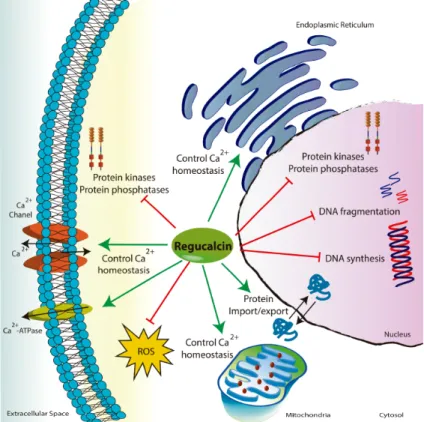
The biological functions of RGN (Fig. I.6) include a role in Ca2+ homeostasis by regulating
the activity of Ca2+-pumps at plasma membrane, endoplasmic reticulum and mitochondria
(Fujita et al., 1998;Yamaguchi and Nakajima, 2002;Yamaguchi and Daimon, 2005). The RGN’s role also has been linked to the control of intracellular signaling pathways and distinct experimental approaches demonstrated that RGN suppresses cell proliferation and apoptosis (Correia et al., 2014a;Marques et al., 2014a;Vaz et al., 2014). Also, the activity of several protein kinases and phosphatases were down-regulated with the overexpression of RGN (Katsumata and Yamaguchi, 1998;Inagaki and Yamaguchi, 2001;Fukaya and Yamaguchi, 2004). Another feature of RGN protein is its antioxidant activity by reducing the production of ROS and increasing the mechanisms of antioxidant defense (Handa et al., 2009;Kondo et al., 2014). Knockout animals for the RGN gene display higher levels of oxidative stress in the brain than their wild-type counterparts (Son et al., 2006). Accordingly, overexpression of RGN in different cell types and tissues is linked with increased antioxidant potential (Correia et al., 2013;Marques et al., 2014a).
At the nuclear compartment RGN is involved in regulation of protein transport, decreasing the expression of small GTPase Ran (ras-related nuclear protein), which is essential for protein export from the nucleus and protein import into the nucleus (Tsurusaki and Yamaguchi, 2001). When RGN is translocated to the nucleus mediates several nuclear functions decreasing DNA fragmentation, inhibiting endonuclease activity (Yamaguchi and Sakurai, 1991), and reducing DNA and RNA synthesis (Fig. I.6) (Yamaguchi and Kanayama, 1996;Yamaguchi and Ueoka, 1997).