Escherichia coli
AcrB Binding Pocket Phenylalanine
Mutants Using a 1,2
9
-Dinaphthylamine Efflux Assay
Ju¨rgen A. Bohnert*, Sabine Schuster, Magdalena Szymaniak-Vits, Winfried V. Kern
Department of Medicine, Center for Infectious Diseases and Travel Medicine, Albert-Ludwigs-University, University Hospital, Freiburg, Germany
Abstract
To evaluate the importance of phenylalanine residues for substrate transport in theEscherichia coliefflux pump protein
AcrB, we subjected Phe-to-Ala binding pocket mutants to a real-time efflux assay with the novel near-infrared lipophilic membrane probe 1,29-dinaphthylamine (1,29-DNA). All mutations, with the exception of F617A, led to considerable retardation of efflux. F610A was the point mutation with the most pronounced impact, followed by F628A, F615A, F136A, and F178A. This is the first study to demonstrate the importance of single phenylalanine residues within the AcrB binding pocket for real-time substrate transport.
Citation:Bohnert JA, Schuster S, Szymaniak-Vits M, Kern WV (2011) Determination of Real-Time Efflux Phenotypes inEscherichia coliAcrB Binding Pocket Phenylalanine Mutants Using a 1,29-Dinaphthylamine Efflux Assay. PLoS ONE 6(6): e21196. doi:10.1371/journal.pone.0021196
Editor:Katy C. Kao, Texas A&M, United States of America
ReceivedSeptember 28, 2010;AcceptedMay 23, 2011;PublishedJune 15, 2011
Copyright:ß2011 Bohnert et al. This is an open-access article distributed under the terms of the Creative Commons Attribution License, which permits unrestricted use, distribution, and reproduction in any medium, provided the original author and source are credited.
Funding:The work was funded by the University of Freiburg. The funders had no role in study design, data collection and analysis, decision to publish, or preparation of the manuscript.
Competing Interests:The authors have declared that no competing interests exist.
* E-mail: [email protected]
Introduction
The Escherichia coli AcrB multidrug resistance (MDR) efflux pump is a member of the resistance-nodulation-division (RND) family and extrudes a wide variety of structurally diverse dyes and antibiotics [1,2]. AcrB is part of a tripartite efflux machinery and cooperates with the membrane fusion protein AcrA and the TolC outer membrane protein.
AcrB X-Ray crystallography revealed an asymmetric trimer structure, where each protomer was suggested to correspond to a distinct functional state [3–5]. This led to the proposal of a 3-step transport cycle model in which substrates bind to a hydrophobic pocket, defined by phenylalanines 136, 178, 610, 615, 617, and 628, and are subsequently squeezed out towards the TolC funnel by a peristaltic-pump-like mechanism.
One of these studies could also show binding of minocycline and doxorubicin to different residues of the AcrB binding pocket [3]. Interestingly, most of the binding interactions were mediated by some of the above described phenylalanines. Minocycline interacted with F178 and F615 and doxorubicin with F615 and F617, respectively. Hence, it was proposed that the extremely broad substrate spectrum of AcrB was mainly due to the flexible interactions of various ligands with hydrophobic phenylalanines in the binding pocket.
The importance of binding pocket phenylalanines in substrate binding is also supported by several mutational studies. A study on mutations in thePseudomonas aeruginosaMexD pump demonstrated that the F608C mutation (homologous to F610C in AcrB) abolished the ability of the strain to transport pyronin Y [6].
Another study, concerned with theE. coliMdtF (YhiV) pump, showed that the binding pocket mutation V610F (homologous to V612F in AcrB) increased the linezolid MIC by 16-fold [7].
Moreover, it has recently been shown that replacing single binding pocket phenylalanines with alanine markedly reduces various substrate MICs, with the most effective mutation being F610A [8]. It seems that this phenotypic change results mostly from altered substrate recognition, although it cannot be ruled out that the phenotype is at least partly due to moderately lower (about 43% as determined by Western blot densitometric quantification) AcrB expression, compared with the F628F control strain.
While the F610A mutation affects most substrate MICs (with the exception of linezolid and Hoechst 33342), it does not markedly influence the accumulation of ethidium and phenylal-anine-arginine ß-naphthylamide (PAßN) in fluorescence-based assays. Instead, the most effective mutation in these accumulation assays was found to be F628A. The reason for this discrepancy remains unclear.
which would be highly desirable to derive kinetic information, including substrate competition, cannot be determined from MIC data.
If the influx rates of a substrate are not known or cannot be determined easily, the only other solution to determine the efficiency of substrate efflux would be to use a real-time efflux assay where efflux is triggered in bacterial cells preloaded with a substrate and efflux can be measured directly as a function of time. Such an assay has been recently published in a highly optimized form that allowed for the direct measurement of AcrAB-TolC Nile red efflux using spectrofluorometry [10].
To overcome the above problems with indirect efflux assays in phenotypic characterization of AcrB binding pocket mutants and to examine whether the suggested central role of residue F610 in substrate recognition could be reproduced in a real-time substrate transport assay, we opted to use such an assay in our previously described AcrB binding pocket Phe-to-Ala mutants. Instead of Nile Red we used the novel dye 1,29-dinaphthylamine (1,29-DNA) which has properties that make it very suitable for MDR efflux research. It is very lipophilic (logP6, according to http://www. sioc-ccbg.ac.cn/software/xlogp3) and like Nile red well retained in the membrane phospholipids. We chose 1,29-DNA instead of Nile red, since the latter is less lipophilic (log P 3.8) and we hypothesized that since the importance of the AcrB binding pocket phenylalanines in substrate recognition (due to lipophilic interactions) has been well established, 1,29-DNA might be the substrate of choice to characterize the phenotypes of single Phe-to-Ala mutants. Moreover, 1,29-DNA has the unique property of being capable of emission in the near-infrared range of the spectrum where the cellular autofluorescence is generally very low.
Materials and Methods
Strains and culture conditions
The AcrAB-TolC overexpressing E. coli strain 3-AG100 [11] and the 3-AG100-derived AcrB Phe-to-Ala binding pocket mutants [8] have been described previously. The strains were maintained at 280uC in CRYOBANK vials (Mast group, Merseyside, UK) for cryoprotection. They were grown at 37uC in modified Lysogeny Broth (LB), containing 1% tryptone, 0.5% yeast extract, and 1% NaCl.
MIC of 1,29-DNA
Serial dilutions of 1,29-DNA in LB were prepared in 96-well plates and MICs were determined by overnight cultivation of 3-AG100 in accordance with CLSI/NCCLS M100-S20 guidelines (available at http://www.clsi.org).
1,29-DNA efflux assay
To determine the optimal excitation and emission wavelengths for an 1,29-DNA efflux assay in whole cells, the AcrAB-TolC overexpressing E. coli strain 3-AG100 was deenergized by overnight incubation in LB and loaded with 4mM 1,29-DNA, obtained from TCI-Europe (Zwijndrecht, Belgium), in the presence of 5mM carbonyl cyanide m-chlorophenylhydrazone (CCCP) as described previously in the Nile red efflux assay protocol [10]. The only modification was that the dye loading was performed at room temperature (OD600 nmof the cells was 0.25) and the loading time reduced to 2.5 h. The emission spectra were then recorded within a 300 nm to 900 nm range, while varying the excitation wavelength of the deenergized cells at 10 nm intervals (slit width 10 nm) from 200 to 500 nm using a Perkin Elmer LS 55 spectrumfluorimeter. Thereafter, the spectral scans were repeated with energized cells (after addition of glucose to a
final concentration of 50 mM). The Phe-to-Ala 3-AG100-derived AcrB binding pocket mutants were then subjected to a 1,29-DNA efflux assay at dye loading concentrations (DLC) ranging from 0.25mM to 64mM (the dye was found to precipitate at higher concentrations).
To be able to compare the efflux properties of the mutants at a given DLC quantitatively (in the absence of a kinetic model), we determined the efflux half time (EHT), which is the time needed for a reduction of 50% of the dye fluorescence after energization (over a time course of 400 s).
Results and Discussion
1,29-DNA MICs
Using MIC microdilution assays we established that 1,29-DNA was non-toxic to the cells at concentrations of up to 64mM (higher concentrations led to precipitation of dye).
Determination of fluorescent properties of 1,29-DNA 1,29-DNA is an environment-sensitive membrane probe like the well-known structurally related N-phenyl-1-naphtylamine or NPN (almost non-fluorescent in aequous solution but strongly fluores-cent upon partitioning into the phospholipid bilayer), which has already been used in an AcrAB-TolC efflux assay [12]. However, in the latter study high concentrations of CCCP (100mM) were used to preload the cells with NPN. This normally leads to marked retardation of dye efflux after energization, since some CCCP is retained in the membranes even after repeated washing, as described in the Nile red efflux study [10].
We established that the highest fluorescence intensity ratio between the deenergized and energized state in the near-infrared band (,6-fold) could be obtained using an excitation wavelength of 370 nm and an emission wavelength of 810 nm. The signal-to-background ratio was,10. Apart from the near-infrared emission maximum at 810 nm, another maximum could be detected at 415 nm, similar to NPN, which has an emission maximum at 420 nm, but does not fluoresce in the near-infrared band. The only other near-infrared environment-sensitive lipophilic mem-brane probe we are aware of is the carbocyanine dye 1,19 -dioctadecyl-3,3,39,39-tetramethylindotricarbocyanine iodide (DiR) [13] which in comparison with 1,29-DNA has a smaller Stokes shift and lower emission maximum in the near-infrared band (absorption and emission maxima at 750 nm and 782 nm, respectively).
The novel membrane probe 1,29-DNA is an excellent dye for membrane and membrane transport research due to the second emission maximum within the near-infrared band of the light spectrum (810 nm) where low cellular autofluorescence and low interference with substrates or competitors is generally encoun-tered. Another advantage is that the dye is - like Nile red - well retained in the cell envelope while the bacteria are in the deenergized state, but is rapidly mobilized by the AcrAB-TolC efflux system upon energization.
We hypothesize that 1,29-DNA could also be used well for quenching or fluorescence resonance energy transfer (FRET) studies involving membranes or membrane proteins due to its unique properties.
Efflux phenotpyes of binding pocket mutants
the site-directed mutagenesis protocol introduced no systematic phenotypic changes, behaved like the 3-AG100 AcrB wildtype strain in the efflux assays (data not shown). Efflux was abolished in
the acrB deleted control strain 3-AG100 acrB:: rpsLneo
(EHT.400 s).
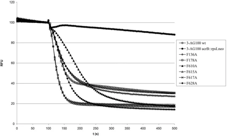
In our previous study on phenotypes of AcrB binding pocket mutants, the F610A mutation had the most pronounced effect on substrate MICs, but failed to demonstrate the same magnitude of phenotypic changes in our ethidium and PAßN accumulation assays [8]. In contrast, the very lipophilic 1,29-DNA clearly demonstrated the dominant role of the F610 residue in AcrB substrate efflux, in line with the previous MIC assays. We can only hypothesize that the observed discrepancy between the assays might be specific to ethidium and PAßN - probably due to their structural properties.
To compare the efflux efficiency of different mutants at a given DLC (using a concentration range from 0.25mM to 64mM), we derived the efflux half time (EHT), which is the time needed for the fluorescence to drop to 50% after energization with glucose over a time period of 400 s (Table 1). We used this descriptor since we could not derive Michaelis-Menten based kinetic constants, due to the fact that the relationship between the DLC and the preenergization relative fluorescence units (RFU) was not a linear one (Fig. 2), but rather followed an exponential function (with higher DLCs giving a dramatically lower increase in fluorescence intensity). There might be two possibilities that explain this observation: first, the phenomenon might be due to increased self-quenching or second, it might be due to membrane saturation effects. In either case the calculation of initial rates, normally used for the determination of kinetic constants, would lead to a dramatic error at higher DLCs.
Likewise, the EHT descriptor - although in theory somewhat more robust to the above described RFU/DLC non-linearity than initial rates - cannot be used to draw any conclusion about the
Figure 1. Representative 1,29-DNA efflux curves of 3-AG100-derived AcrB binding pocket mutants.After preloading with 4mM 1,29-DNA
the cells were energized at 100 s with 50 mM glucose. Fluorescence intensity is given as relative fluorescence units (RFU) with preenergization levels adjusted to 100 RFU.
doi:10.1371/journal.pone.0021196.g001
Table 1.Efflux half times (EHT) in AcrB binding pocket mutants derived from 1,29-DNA efflux assays over a duration of 400 s at different dye loading concentrations.
Strain EHT (s) at 1,29-DNA loading concentration
0.25mM 1mM 4mM 16mM 64mM
Meana SD Mean SD Mean SD Mean SD Mean SD
3-AG100 wtb 20 5.0 19 1.5 24 2.5 27 2.9 37 5.2
3-AG100 F136A
40 18.7 44 12.1 53 7.1 51 3.6 54 3.1
3-AG100 F178A
31 13.6 32 5.0 38 7.0 42 4.6 43 3.5
3-AG100 F610A
73 24.0 67 8.3 97 9.6 118 5.9 146 16.8
3-AG100 F615A
37 7.1 40 10.6 48 6.7 48 5.1 55 1.2
3-AG100 F617A
19 2.1 19 4.2 22 1.5 26 0.6 30 2.3
3-AG100 F628A
54 12.9 52 13.1 53 13.1 55 11.9 68 14.6
aAll measurements were done in triplicate. bwildtype strain for AcrB.
transport saturation characteristics of the AcrAB-TolC system, if the exact amount of self-quenching or membrane saturation effects is not known.
Nevertheless, this parameter is still useful to quantitatively compare the efflux efficiency of different mutants if the same DLC is used as a basis of comparison, since the preenergization RFU starting point (which is related to the intracellular dye concentra-tion) should in this case be the same for all mutants. Using our experimental data, it can be demonstrated that the ratio between the EHT for a given mutant and the EHT for the AcrB wildtype strain is relatively constant over a wide concentration range (Table 1). For instance, the EHT is 73 s at a DLC of 0.25mM in the F610A mutant and 20 s in the AcrB wildtype strain, respectively. The ratio is thus 73 s/20 s = 3.7. The EHT ratio at the higher DLC of 64mM amounts then to 146 s/37 s = 3.9.
If one assumes that the observed RFU/DLC non-linearity is entirely due to dye self-quenching (which is currently unclear), and one linearly reconstructs the self-quenching-adjusted fluorescence (Fsqa) curves using an Excel exponential curve fit (leading to the equation Fsqa(t) = 12e0.039F(t), where F stands for the experimen-tally determined RFU value) one can demonstrate that the EHT would not increase up to a DLC of 64mM. In the given example the calculated EHTs at 64mM would be 66 s for 3-AG100 F610A and 18 s for 3-AG100 (giving a ratio of 3.7 which is similar to the above described cases, again demonstrating the robustness of such an EHT ratio even under conditions of extreme self-quenching).
This would mean that the transport capacity of the pump would be far from saturation and that the experimentally observed EHT increase with higher DLCs would be due to dye self-quenching artifacts.
However, such a model is currently entirely hypothetical and experimental self-quenching data would be needed to verify it. This example simply serves to illustrate that the experimentally determined concentration-dependent increase in EHTs might not necessarily reflect saturation of substrate transport but might be due to other factors related to the physical properties of the dye and/or the cell envelope.
Although lipophilic dyes like 1,29-DNA, NPN or Nile red are currently the best tool to characterize the real-time behaviour of RND efflux pumps in whole cells, a limitation of these dyes has to be considered: their hydrophobicity limits the application to relatively low DLCs. However, using more hydrophilic dyes would lead to technical problems in a real-time efflux assay, since they would probably not be well retained in the membrane phospho-lipids. It would thus be interesting to generate derivates of 1,29 -DNA that are increasingly hydrophilic and to examine the amount of preenergization retainment in the membrane phospholipids.
Moreover, despite the fact that we could show that 1,29-DNA is an excellent dye for use with theE. coliAcrAB-TolC system, it has to be taken into account that the activation of efflux by means of glucose addition does not work well in non-fermenting bacteria likePseudomonas aeruginosa, although we have recently found that the 1,29-DNA efflux assay can be used successfully in an E. coli
strain expressing only the MexAB-OprM system (data not shown).
Conclusion
We have described a novel real-time efflux assay using the excellent AcrAB-TolC substrate 1,29-DNA. Since influx does not play a role in such an assay with dye-preloaded cells, we have been capable of directly comparing the efflux efficiency of phenylala-Figure 2. Increased quenching of 1,29-DNA fluorescence at higher dye loading concentrations (DLCs) in strain 3-AG100.Depicted are relative fluorescence units (RFU) of deenergized cells preloaded with 1,29-DNA after resuspension in fresh PPB. All experiments were done in triplicate. The error bars show the standard deviation.
nine point mutants over a wide concentration range. While we have not yet been able to derive a kinetic model due to a lack of knowledge about the relationship between the experimental RFU values and the intracellular dye concentration, conceiving such a model might in principle be possible if the amount of self-quenching and/or dye loading saturation could be determined experimentally.
Author Contributions
Conceived and designed the experiments: JAB WVK. Performed the experiments: JAB SS MSV. Analyzed the data: JAB. Contributed reagents/materials/analysis tools: JAB SS MSV. Wrote the paper: JAB.
References
1. Nikaido H (1998) Antibiotic resistance caused by gram-negative multidrug efflux pumps. Clin Infect Dis 27 Suppl 1: S32–S41.
2. Piddock LJ (2006) Clinically relevant chromosomally encoded multidrug resistance efflux pumps in bacteria. Clin Microbiol Rev 19: 382–402. 3. Murakami S, Nakashima R, Yamashita E, Matsumoto T, Yamaguchi A (2006)
Crystal structures of a multidrug transporter reveal a functionally rotating mechanism. Nature 443: 173–179.
4. Seeger MA, Schiefner A, Eicher T, Verrey F, Diederichs K, et al. (2006) Structural asymmetry of AcrB trimer suggests a peristaltic pump mechanism. Science 313: 1295–1298.
5. Sennhauser G, Amstutz P, Briand C, Storchenegger O, Grutter MG (2006) Drug export pathway of multidrug exporter AcrB revealed by DARPin inhibitors. PLoS Biol 5: e7.
6. Mao W, Warren MS, Black DS, Satou T, Murata T, et al. (2002) On the mechanism of substrate specificity by resistance nodulation division (RND)-type multidrug resistance pumps: the large periplasmic loops of MexD from
Pseudomonas aeruginosaare involved in substrate recognition. Mol Microbiol 46: 889–901.
7. Bohnert JA, Schuster S, Fa¨hnrich E, Trittler R, Kern WV (2007) Altered spectrum of multidrug resistance associated with a single point mutation in the
Escherichia coli RND-type MDR efflux pump YhiV (MdtF). J Antimicrob Chemother 59: 1216–1222.
8. Bohnert JA, Schuster S, Seeger MA, Fa¨hnrich E, Pos KM, et al. (2008) Site-directed mutagenesis reveals putative substrate binding residues in theEscherichia coliRND efflux pump AcrB. J Bacteriol 190: 8225–8229.
9. Nagano K, Nikaido H (2009) Kinetic behavior of the major multidrug efflux pump AcrB ofEscherichia coli. Proc Natl Acad Sci U S A 106: 5854–5858. 10. Bohnert JA, Karamian B, Nikaido H (2010) Optimized Nile Red efflux assay of
AcrAB-TolC multidrug efflux system shows competition between substrates. Antimicrob Agents Chemother 54: 3770–3775.
11. Jellen-Ritter AS, Kern WV (2001) Enhanced expression of the multidrug efflux pumps AcrAB and AcrEF associated with insertion element transposition in
Escherichia coli mutants Selected with a fluoroquinolone. Antimicrob Agents Chemother 45: 1467–1472.
12. Lomovskaya O, Warren MS, Lee A, Galazzo J, Fronko R, et al. (2001) Identification and characterization of inhibitors of multidrug resistance efflux pumps in Pseudomonas aeruginosa: novel agents for combination therapy. Antimicrob Agents Chemother 45: 105–116.