www.reumatologia.com.br
REVISTA BRASILEIRA DE
REUMATOLOGIA
* Corresponding author.
E-mail: [email protected] (E.Umay).
0482-5004/$ - see front matter. © 2013 Elsevier Editora Ltda. All rights reserved. www.reumatologia.com.br
Original article
Importance of cutaneous silent period in fi bromyalgia and
its relationship with disease characteristics, psychological
disorders and quality of life of patients
Ebru Umay
a,*, Umit Ulas
b, Ece Unlu
a, Hakan Akgun
b, Aytul Cakci
a, Zeki Odabasi
ba Ministry of Health Ankara Diskapi Yildirim Beyazit Training and Research Hospital, Department of Physical Medicine And Rehabilitation,
Ankara, Turkey
b Gulhane Military Medical Academy, Department of Neurology, Ankara, Turkey
a r t i c l e i n f o
Article history:
Received 26 September 2012 Accepted 9 December 2012
Keywords:
Fibromyalgia
Cutaneous silent period Disability
a b s t r a c t
Introduction: Cutaneous silent period (CSP) is an inhibitory spinal protective rel ex and its afferents consist of A-delta nerve i bers. We aimed to evaluate patients with i bromyalgia (FM) and healthy controls to determine any differences between the groups in terms of CSP duration and latency, and if present, to determine whether there is any relationship with disease characteristics, psychological disorders and quality of life.
Materials and methods: Thirty-two patients with FM and 32 healthy volunteers were
includ-ed in the study. The patient and control groups were comparinclud-ed in terms of CSP latency and duration in both upper and lower extremities. Disease characteristics, psychological disor-ders and quality of life of patients were assessed using the Fibromyalgia Impact Question-naire (FIQ) and Short Form-36 (SF-36). Patients with CSP measurements equal to or lower than those of the control group were compared with those with higher values than controls in terms of disease characteristics, psychological status and quality of life.
Results: Signii cantly prolonged CSP latencies in both upper and lower extremities were
determined in patients compared to controls. We found that prolongation of CSP latency in the lower extremity is associated with disease severity and functional disability.
Conclusions: CSP latencies in both upper and lower extremities in patients with FM are
lon-ger than in healthy volunteers. Moreover, prolongation of CSP latency in the lower extrem-ity is associated with disease severextrem-ity and physical functional disabilextrem-ity.
© 2013 Elsevier Editora Ltda. All rights reserved.
REVISTA BRASILEIRA DE
Importância do período de silêncio cutâneo na fi bromialgia e sua relação com as características da doença, distúrbios psicológicos e qualidade de vida dos pacientes
Palavras-chave:
Fibromialgia
Período de silêncio cutâneo Dei ciência
r e s u m o
Introdução: O período de silêncio cutâneo (PSC) é um rel exo protetor inibitório da coluna
vertebral e seus aferentes consistem em i bras nervosas A-delta. Nosso objetivo foi avaliar pacientes com i bromialgia (FM) e controles saudáveis para determinar as diferenças entre os grupos em relação à duração e latência do PSC, e quando presente, determinar se há al-guma relação com as características da doença, distúrbios psicológicos e qualidade de vida.
Materiais e métodos: Trinta e dois pacientes com FM e 32 voluntários saudáveis foram
in-cluídos no estudo. Os dois grupos foram comparados em relação à latência e duração do PSC em ambos os membros superiores e inferiores. Características da doença, distúrbios psicológicos e qualidade de vida dos pacientes foram avaliados utilizando o Fibromyalgia Impact Questionnaire (FIQ), e o Short Form-36 (SF-36). Os pacientes com medida de PSC igual ou inferior às do grupo controle foram comparados com aqueles com valores mais elevados do que os controles em termos de características da doença, estado psicológicos e quali-dade de vida.
Resultados: Latências signii cativamente prolongadas de PSC nos membros superiores e
in-feriores foram determinadas em pacientes comparados com os controles. Observou-se que a prolongamento da latência do PSC no membro inferior estava associado com a gravidade da doença e incapacidade funcional.
Conclusões: Latências do PCS nos membros superiores e inferiores em pacientes com FM
são mais longas do que em voluntários saudáveis. Além disso, o prolongamento da latên-cia do PSC no membro inferior está assolatên-ciado com a gravidade da doença e incapacidade funcional física.
© 2013 Elsevier Editora Ltda. Todos os direitos reservados.
Introduction
Fibromyalgia (FM) is a chronic pain syndrome characterized by widespread pain and tender points at specii c anatomic areas that cannot be understood regarding its etiology, de-spite all the new developments.1 Some symptoms and signs, including chronic fatigue, headache, sleep disturbance, psy-chological disorders, irritable bowel and bladder syndromes, dysmenorrhea, sensory disorders such as paraesthesia and dysesthesia without neuropathy, and Raynaud’s phenom-enon, are common in FM.1,2
Although the etiopathogenesis is not yet fully elucidated, studies have reported that various factors might be effective, such as neuroendocrine and autoimmune dysfunction and genetic predisposition.3 Further, studies have shown that hy-perexcitability of spinal and supraspinal neurons in FM play an important role in the development and maintenance of chronic pain.4,5
Studies that used the nociceptive l exion rel ex (NFR) to show the excitability of dorsal horn neurons of the spinal cord, which formed with peripheral C i bers (a nociceptive af-ferent), have reported that this excitability in patients with FM causes central sensitization and chronic pain.6,7
The assessment method of the A-delta i ber (the other no-ciceptive afferent) is the cutaneous silent period (CSP).8 The NFR and CSP are the excitatory and inhibitory parts of the same spinal protective rel ex, respectively.9 Although the CSP has been measured in various muscles using different meth-ods, there is only one study10 in the literature, and only the
upper extremity was evaluated in that study. To our knowl-edge, no study in the literature has evaluated the relationship between CSP and disease duration, pain level, numbers of to-tal symptoms and tender points, severity of FM, psychological disorders, and quality of life.
Therefore, we aimed to compare patients with FM and healthy controls to determine any difference in CSP duration and latency in the upper and lower extremities, and if pres-ent, to determine whether there is any relationship between CSP and disease characteristics, psychological disorders and quality of life.
Materials and methods
Study population
Thirty-two patients who were admitted to the Physical Medi-cine and Rehabilitation Clinic and were diagnosed with FM according to the American College of Rheumatology (ACR) 1990 classii cation criteria were included in the study.1 Thirty-two healthy volunteers consisting of hospital staff and rela-tives of patients were included in the study as controls.
pregnant or lactating or who had used any psychotropic and/ or antihistamine drugs in the last month were also excluded from the study.
Patients and volunteers with normal musculoskeletal and neurologic examinations including range of motion, muscle strength, superi cial sensation, and deep tendon rel ex, and who had normal laboratory parameters including complete blood count, complete urinalysis, erythrocyte sedimentation rate, vitamin B12, thyroid function tests, and biochemical tests including electrolytes and enzymes of liver, kidney and muscle were included in the study.
Patients and volunteers were informed about the study and their written consents were obtained at the start of the study. The study was approved by the local Ethical Board and was performed in accordance with the principles of the Dec-laration of Helsinki.
Electrophysiologic tests
Electrophysiologic evaluations were performed in the electro-physiology laboratory of Gülhane Military Medical Academy Department of Neurology using a 2+8 channel electromyo-gram (EMG) device (MEDELEC Synergy-Oxford, U.K.) and ac-cording to the protocol described by Oh.11 The room tempera-ture was 24 ± 1°C, and the skin temperatempera-ture of patients and volunteers was over 32°C. While measurements of the upper extremity were applied in the sitting position, measurements of the lower extremity were applied in the supine position.
Nerve conduction tests
Sensory nerve conduction tests were evaluated from the right median, left ulnar and right sural nerves. Motor nerve con-duction tests were evaluated from the right median, left ul-nar, right peroneal, and left tibial nerves. Distal motor latency (DML) (ms) and motor conduction velocity (MCV) in motor
nerves and sensory conduction velocity (SCV) (m/s) in sensory nerves were recorded.
CSP investigations
Sensory nerves were stimulated in the lower extremity using bar electrode and in the upper extremity using ring electrode. First, the sensory threshold was found. For this purpose, an electrical current of 0.5 ms duration starting from an inten-sity of 0.6 mA was performed laterally to the lateral malleolus in the lower extremity and to the second i nger of the up-per extremity. The lowest intensity, which was determined by gradually increasing the intensity until it was felt by the individual, was recorded as the sensory intensity threshold. CSP measurements were performed in the right upper and lower extremities using the abductor pollicis brevis (APB) and tibialis anterior (TA) muscles, respectively. The second i nger in the right upper extremity was stimulated, and recordings were obtained from the APB muscle. Before the recording, the patient was asked to perform thumb abduction with maximal effort, and the maximal motor unit action potential (MUAP) amplitude was measured on the screen. Subjects were asked to perform thumb abduction with MUAP amplitudes of at least 25% of the maximal MUAP amplitude. While the patient was constantly performing this abduction, the median nerve was stimulated at an intensity of 15 times the sensory thresh-old. Five recordings were obtained at 30-second intervals. The CSP latency and duration were measured by assessing the av-erage of 5 traces. The endpoint at which an observable clear-cut inhibition in muscle activity started was considered as the CSP latency (ms). The CSP duration (ms) was determined by measuring the time between the point of inhibition of muscle activity and the point at which it started to return to baseline muscle activity. The sural nerve was stimulated superi cially laterally to the lateral malleolus in the right lower extremity, and recordings were obtained from the TA muscle by using the same method described above.
Clinical tests
Disease characteristics including disease duration, symp-toms associated with FM and pain level were questioned. The numbers of tender points and symptoms were recorded. To-tal numbers of symptoms were calculated and recorded. The general pain level felt in the last 48 hours was assessed by visual analogue scale (VAS) with 0-10 cm.
To assess severity of disease, functional disability and spe-cii c quality of life, the Fibromyalgia Impact Questionnaire (FIQ)12 was used, according to which the total score was evalu-ated between 0-100, with a higher score showing a greater im-pact of the syndrome on the person.
To assess possible depression symptoms of the patients, the Beck Depression Inventory (BDI)13 was used, and to as-sess anxiety symptoms, the Beck Anxiety Inventory (BAI)14 was used. Twenty-one Likert-type questions were asked with these scales, and each question was evaluated between 0-3.
The general quality of life of patients was evaluated with Short Form-36 (SF-36).15 Accordingly, two sub-group scores were created as physical health and mental health. The total score was evaluated between 0-100.
Abbreviations
FM: i bromyalgia
NFR: nociceptive l exion rel ex CSP: cutaneous silent period EMG: electromyogram DML: Distal motor latency MCV: motor conduction velocity SCV: sensory conduction velocity APB: abductor pollicis brevis TA: tibialis anterior
MUAP: motor unit action potential VAS: Visual Analogue Scale
FIQ: Fibromyalgia Impact Questionnaire BDI: Beck Depression Inventory
BAI: Beck Anxiety Inventory SF-36: Short Form-36
SPSS: Statistical Package for the Social Sciences OR: odds ratio
Comparisons
The patient and control groups were compared in terms of CSP latency and duration in the upper and lower extremities. Sub-groups were formed according to the CSP latency and duration levels that were determined to be signii cantly dif-ferent between groups on the basis of the CSP mean of the control group. Patients with CSP measurements equal to or below values of the control group (group 1) were compared to patients with CSP measurements above the values of the control group (group 2) in terms of disease duration, number of total symptoms and tender points, pain level evaluated by VAS, FIQ score, levels of depression and anxiety, and quality of life.
Statistical analysis
Data analyses were made using the Statistical Package for the Social Sciences (SPSS Inc., USA) 11.5 for Windows. Descrip-tive statistics were shown as mean ± standard deviation and median for continuous variables and observation number (%) for nominal variables using chi-square tests. Statistically sig-nii cant differences between groups in terms of continuous variables were studied with Mann-Whitney U test (according to Kolmogorov-Smirnov test, continuous variables were not the distribution normal) and nominal variables with Pearson chi-square test. Signii cance of the difference in variables be-tween groups 1 and 2 was analyzed using Pearson chi-square test. Regression analysis was used for signii cant correlations by using group 1 values as the dependent variable. Values of P < 0.05 were considered as statistically signii cant.
Results
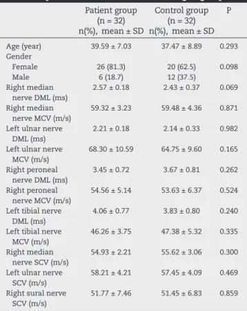
The median age of the 64 participants included in the study (46 [71.9%] females, 18 [28.1%] males) was 41.00 (38.53 ± 8.02) years. The distribution and comparison of demographic char-acteristics and motor and sensory nerve conduction values of patients (n = 32) and volunteers (n = 32) according to groups are presented in Table 1. There was no signii cant difference between groups in terms of age, gender and motor and sen-sory conduction values (P > 0.05).
The distribution and comparison of CSP latency and dura-tion measured from the APB and TA muscles of patients and controls according to groups are shown in Table 2.
While the mean CSP latencies in the upper and lower ex-tremities of patients were 87.25 and 107.75 ms and CSP dura-tions were 46.25 and 51.15 ms, respectively, these values in controls were 80.75 and 101.62 ms (latencies) and 48.75 and 54.50 ms (durations), respectively.
Signii cantly prolonged CSP latencies in both upper and lower extremities were determined in patients compared to the control group (P < 0.05).
The distribution of disease duration, numbers of total symptoms and tender points, VAS level, FIQ score, levels of depression and anxiety, and quality of life of patients are pre-sented in Table 3.
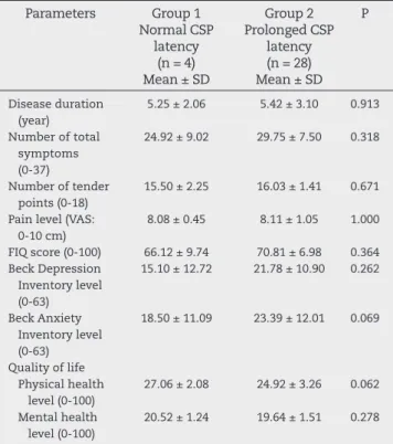
With respect to sub-groups formed according to mean CSP latency of the control group (patients with equal/lower
values versus higher values compared to controls), it was found that while the number of patients with normal CSP latency in the upper extremity (group 1 for upper extremity) was 4 (12.5%), the number of patients with normal latency in the lower extremity (group 1 for lower extremity) was 12 (37.5%).
The comparisons between group 1 and group 2 for upper and lower extremities in terms of disease duration, numbers of total symptoms and tender points, pain level evaluated by VAS, FIQ score, levels of depression and anxiety, and quality of life are shown in Tables 4 and 5.
Table 1 – Distribution and comparison of demographic characteristics and motor and sensory nerve conduction values of patients and volunteers according to groups.
Patient group (n = 32) n(%), mean ± SD
Control group (n = 32) n(%), mean ± SD
P
Age (year) 39.59 ± 7.03 37.47 ± 8.89 0.293
Gender Female Male
26 (81.3) 6 (18.7)
20 (62.5) 12 (37.5)
0.098
Right median nerve DML (ms)
2.57 ± 0.18 2.43 ± 0.37 0.069
Right median nerve MCV (m/s)
59.32 ± 3.23 59.48 ± 4.36 0.871
Left ulnar nerve DML (ms)
2.21 ± 0.18 2.14 ± 0.33 0.982
Left ulnar nerve MCV (m/s)
68.30 ± 10.59 64.75 ± 9.60 0.165
Right peroneal nerve DML (ms)
3.45 ± 0.72 3.67 ± 0.81 0.262
Right peroneal nerve MCV (m/s)
54.56 ± 5.14 53.63 ± 6.37 0.524
Left tibial nerve DML (ms)
4.06 ± 0.77 3.83 ± 0.80 0.240
Left tibial nerve MCV (m/s)
46.26 ± 3.75 47.38 ± 5.32 0.335
Right median nerve SCV (m/s)
54.93 ± 2.21 55.62 ± 3.06 0.300
Left ulnar nerve SCV (m/s)
58.21 ± 4.21 57.45 ± 4.09 0.469
Right sural nerve SCV (m/s)
51.77 ± 7.46 51.45 ± 6.83 0.859
SD, standard deviation; DML, distal motor latency; MCV, motor conduction velocity; SCV, sensory conduction velocity.
Table 2 – Distribution and comparison of CSP latency and duration measured from APB and TA muscles of patients and controls according to groups.
CSP (ms) Patient group (n = 32) Mean ± SD
Control group (n = 32) Mean ± SD
P
Upper extremity Latency Duration
87.60 ± 7.49 47.17 ± 7.33
79.77 ± 8.15 49.92 ± 9.74
0.001 0.151 Lower extremity
Latency Duration
108.85 ± 10.03 52.91 ± 13.20
103.42 ± 10.37 55.93 ± 9.11
0.037 0.692
As a result of the comparisons, while there was no re-lationship between prolongation of CSP latency in the up-per extremity and the evaluation parameters, we detected a relationship between prolongation of CSP latency in the lower extremity and numbers of total symptoms, FIQ score and physical health level. Accordingly, there was a positive
correlation between prolongation of CSP latency and num-ber of total symptoms and FIQ score and a negative cor-relation between prolongation of CSP latency and physical health level.
A regression analysis done for signii cant correlations by using group 1 values as the dependent variable demonstrated that prolongation of CSP latency in the lower extremity was associated with disease severity and functional disability measured with FIQ (odds ratio [OR]: 0.467, P = 0.002) and phys-ical health level measured with the physphys-ical health subscale of SF-36 (OR: −0.231, P = 0.024).
Discussion
Fibromyalgia (FM) is not directly associated with organ dys-function. Various gene polymorphisms, alterations of the hypothalamic-pituitary-adrenal (HPA) axis, abnormal concen-tration of neuropeptides and biogenic amines such as sero-tonin, norepinephrine, cortisol, and substance P, and altera-tions of activation of receptors such as N-methyl-D-aspartic acid (NMDA) and glutamate have been described in its etio-pathogenesis.16
A reduction in inhibitory mediators such as serotonin and an increase in excitatory mediators such as substance P induced by various factors of stress, trauma or infectious agents in genetically predisposed individuals may explain the symptoms, including psychological and sleep disorders and
Table 5 – Comparisons between group 1 and group 2 for lower extremities in terms of disease duration, number of total symptoms and tender points, pain level evaluated by VAS, FIQ score, levels of depression and anxiety, and quality of life.
Parameters Group 1 Normal CSP
latency (n = 12) Mean ± SD
Group 2 Prolonged CSP
latency (n = 20) Mean ± SD
P
Disease duration (year)
5.25 ± 2.95 5.66 ± 3.11 0.708
Number of total symptoms (0-37)
21.50 ± 8.45 27.95 ± 8.42 0.045
Number of tender points (0-18)
15.33 ± 2.30 15.70 ± 2.10 0.649
Pain level (VAS: 0-10 cm)
7.83 ± 1.11 8.10 ± 0.91 0.467
FIQ score (0-100) 61.50 ± 10.82 69.78 ± 7.23 0.015
Beck Depression Inventory level (0-63)
20.40 ± 11.56 21.83 ± 10.87 0.731
Beck Anxiety Inventory level (0-63)
21.83 ± 13.02 23.35 ± 11.40 0.732
Quality of life Physical health
level (0-100) Mental health
level (0-100)
27.85 ± 0.27
19.81 ± 1.54
26.16 ± 2.74
19.65 ± 1.48
0.043
0.769
CSP, cutaneous silent period; SD, standard deviation; VAS, visual analogue scale; FIQ, Fibromyalgia Impact Questionnaire.
Table 4 – Comparisons between group 1 and group 2 for upper extremities in terms of disease duration, number of total symptoms and tender points, pain level evaluated by VAS, FIQ score, levels of depression and anxiety, and quality of life.
Parameters Group 1 Normal CSP
latency (n = 4) Mean ± SD
Group 2 Prolonged CSP
latency (n = 28) Mean ± SD
P
Disease duration (year)
5.25 ± 2.06 5.42 ± 3.10 0.913
Number of total symptoms (0-37)
24.92 ± 9.02 29.75 ± 7.50 0.318
Number of tender points (0-18)
15.50 ± 2.25 16.03 ± 1.41 0.671
Pain level (VAS: 0-10 cm)
8.08 ± 0.45 8.11 ± 1.05 1.000
FIQ score (0-100) 66.12 ± 9.74 70.81 ± 6.98 0.364
Beck Depression Inventory level (0-63)
15.10 ± 12.72 21.78 ± 10.90 0.262
Beck Anxiety Inventory level (0-63)
18.50 ± 11.09 23.39 ± 12.01 0.069
Quality of life Physical health
level (0-100) Mental health
level (0-100)
27.06 ± 2.08
20.52 ± 1.24
24.92 ± 3.26
19.64 ± 1.51
0.062
0.278
CSP, cutaneous silent period; SD, standard deviation; VAS, visual analogue scale; FIQ, Fibromyalgia Impact Questionnaire.
Table 3 – Distribution of disease duration, numbers of total symptoms and tender points, VAS level, FIQ score, levels of depression and anxiety, and quality of life of patients.
Parameters Patient group (n = 32) Mean ± SD
Disease duration (year) 5.40 ± 2.97
Number of total symptoms (0-37)
25.53 ± 8.88
Number of tender points (0-18) 15.56 ± 2.15
Pain level (VAS: 0-10 cm) 8.03 ± 0.98
FIQ score (0-100) 66.71 ± 9.48
Beck Depression Inventory level (0-63)
20.93 ± 11.15
Beck Anxiety Inventory level (0-63)
22.78 ± 11.85
Quality of life
Physical health level (0-100) Mental health level (0-100)
26.79 ± 2.30 19.75 ± 1.49
muscle weakness.17 Inadequate levels of cortisol, growth hor-mone and insulin-like growth factor-1 due to dysfunction of the HPA may cause symptoms such as fatigue and exercise intolerance.18
However, these theories are not sufi cient to explain the chronic and widespread pain in FM. The pain threshold de-crease in FM and pain are not limited to tender point sites, and there is increased sensitivity to nonspecii c stimuli such as me-chanical pressure and cold/warm sensations in areas outside tender point sites or in areas without spontaneous pain. More-over, there is an aberration of the central pain mechanisms.7,19
Studies in the literature have reported that hyperexcitabil-ity of spinal and supraspinal neuron plays an important role in the development and maintenance of chronic pain.4,5
Indirect evidences such as regional increase in cerebral blood l ow of some brain areas, alterations of the nociceptive modulating system, central sensitization, increase in tempo-ral summation, late evoked potentials, sensitivity of C i bers, and alteration in levels of substance P, which are known to play an effective role in the transmission of pain in patients with FM, have been reported in the literature.20,21
Peripheral nociceptors can be stimulated with tissue trau-ma and/or up-regulation of nociceptor expression. Impulses from peripheral nociceptors are transmitted to the spinal cord by myelinated A delta and unmyelinated C i bers. First pain is mediated by A delta i ber, and chronic pain occurs by C i bers with following continued stimulus22. Although studies evaluat-ing C i bers by NFR are found in the literature,22,23 only one study has evaluated A delta i bers using the CSP measurement.10
Therefore, we aimed to compare patients with FM with healthy controls in order to evaluate any differences in CSP latency and duration in the upper and lower extremities, and if present, to determine whether any relationship exists be-tween CSP and disease characteristics, psychological disor-ders and quality of life.
Based on the results of our study, while signii cantly pro-longed CSP latencies in both upper and lower extremities were found in patients compared to the control group, there was no signii cant difference between groups in terms of CSP duration. In addition, we found that prolongation of CSP la-tency in the lower extremity was correlated with disease se-verity and physical functional disability of patients.
The CSP is a protective rel ex that causes a pause in vol-untary muscle contraction in the presence of painful stimuli of a cutaneous nerve. The afferent impulses that generate the CSP are carried by A delta i bers, but the central mecha-nism of CSP is not known.24 The CSP is useful to evaluate the components and segments of A delta i bers (not evaluated by modern electrodiagnostic methods) and to understand the central nervous system (CNS) diseases with motor and sen-sory disorders.25 Some studies have used CSP to evaluate the nociceptive pathway function at spinal and supraspinal levels in patients with neuropathic pain.26
Studies in the literature have shown that CSP was recorded in various sensory neuropathies including Friedreich’s ataxia, abetalipoproteinemia and Fabry disease, entrapment neurop-athies such as carpal and ulnar tunnel syndromes, spinal cord lesions including myelopathy, radiculopathy, syringomyelia, and root avulsion, and disorders of the CNS including Par-kinson’s disease and dystonia.25,27-31 In addition, studies have
reported that CSP measurements can be done with various muscles and with different methods. A study similar to our study in the literature, by Sahin et al.,10 showed that CSP la-tency recorded from the APB muscle with stimulation of the 5th i nger of patients with FM (n = 28) was longer than in the control group (n = 18), but there was no signii cant difference between groups in terms of CSP duration. Further, only the upper extremity was evaluated in their study.
In the present study, although we used different methods of stimulation from those in the literature, a signii cant pro-longation in CSP latencies (measured in APB and TA muscles) in both upper and lower extremities was found in patients compared to controls. Moreover, there was no difference in terms of CSP duration. This result is compatible with that re-ported by Sahin et al.10
Studies have reported that CSP latency occurs at three times: peripheral conduction time conducted by A delta i -bers, the time required for inhibition in the spinal cord, and the time from the spinal cord to muscle motor i bers.32 Our results are compatible with theories that cite changes in the pain pathway. Studies in the literature have reported that CSP duration is shortened and latency increased in peripheral neuron disorders such as neuropathy and loss of A delta i -bers. Furthermore, both CSP latency and duration are extend-ed in Parkinson’s disease and dystonia. None of our patients had evidence suggesting neuropathy in conduction velocity studies or evidence of loss of A delta i bers such as myelopa-thy, radiculopathy or root avulsion. Studies in Parkinson’s dis-ease have explained that prolonged CSP duration is related to longer-lasting activity in inhibitory circuits in the spinal cord.
According to our results, while there was a slight shorten-ing in CSP duration in patients when compared to the con-trol group, the difference was not statistically signii cant. Although our patients had no major disorder suggesting the loss of A delta i bers, Onal et al.,33 in their study performed in patients with no large i ber neuropathy with early stage diabetes mellitus, and Oz et al.34 in their study performed in patients with restless legs syndrome, reported that prolonged CSP latency is related to small i ber neuropathy. Moreover, Ulas et al.35 evaluated the presence of dysautonomia in FM and showed that latency of sympathetic skin response is lon-ger than in the control group, and they also reported that this result may be an indicator of small i ber neuropathy in pa-tients with FM.
In light of this information, we think that our patients may have had a small i ber neuropathy. However, except for the above-mentioned possibilities, the reason for normal CSP du-ration may be related to technical problems during the mea-surements.
of anxiety and depression were above normal values. As re-ported in the literature, these psychological disorders may be risk factors for the development of FM, and it is theorized that these disorders are present from the onset of the disease.38
Quality of life assessment instruments can be generic or specii c. We used SF-36 for the generic assessment and FIQ for specii c assessment. Studies in the literature have re-ported that quality of life levels assessed by FIQ and SF-36 in patients with FM were signii cantly higher than in healthy volunteers.39,40 Our results could not be compared, since there is no study in the literature that investigated CSP latency in patients with FM.
Pagano et al.41 evaluated the quality of life in patients with FM using FIQ and SF-36, and reported that FIQ is better for as-sessing the quality of life than SF-36. This study also showed a limitation in physical functioning in patients with FM, re-duced by 10-fold compared with the control group.
Our results, showing that prolongation of CSP latency is associated with the scores of FIQ and the physical health subscale of SF-36, demonstrate that the abnormality in pain pathways is rel ected in the physical function of the patients. Further, this result may be related to a potential small i ber neuropathy. Considering the results discussed above, the presence of a potential small i ber neuropathy in these pa-tients may explain the lack of difference in the mental health according to the SF-36 subscale and the levels of anxiety and depression. Although large-scale studies are needed, we think that the evaluation of CSP latency in patients with FM may shed light on the functional disability of patients.
The association of CSP latency in the lower extremity with disease severity and limitation of physical function may be explained by measurements in the upper extremity that were carried out in the sitting position, while measure-ments in the lower extremity were carried out in the supine position, which is more comfortable. Therefore, the mainte-nance of the voluntary muscle contraction may be easier in the supine position than in the sitting position. Studies have reported that muscle distance may be effective on latency and duration of CSP.28,42 The effect of this rel ex increases from the proximal to the distal muscles. The upper extrem-ity has a shorter rel ex pathway compared to the lower ex-tremity in terms of limb length; therefore, functional disabil-ity may be associated with the prolongation of CSP latency in the lower extremity.
Study limitations
This study is subject to several limitations. Clinical assess-ment scales were not impleassess-mented in the control group; thus, adequate comparisons could not be made. Further, no test such as skin biopsy or study of the autonomic nervous system was done to coni rm the diagnosis of small i ber neuropathy in our study, thereby precluding the statement of any dei ni-tive result.
Conclusion
CSP latencies in both upper and lower extremities in pa-tients with FM are longer than in healthy volunteers. We
think that this result supports the theory of abnormalities in the pain pathway at peripheral and spinal levels in the pathogenesis of FM. These abnormalities may be due to the changes in the posterior horn of the spinal cord as well as to a small i ber neuropathy due to a direct loss of A delta i bers. To determine the exact cause, studies evaluating the A delta i ber and utilizing several tests simultaneously are needed. As a secondary outcome, it was found that the prolongation of the CSP latency in the lower extremity is associated with disease severity and physical functional disability. Accord-ingly, we think that CSP latency may be used as an assess-ment method for evaluating the disease severity and physi-cal disability in FM. However, prior to its use as a standard measurement method, large-scale studies should be done and normal values created.
Conl icts of interest
The authors declare no conl icts of interest.
R E F E R E N C E S
1. Wolfe F, Smythe HA, Yunus MB, Bennett RM, Bombardier C, Goldenberg DL, et al. The American College of Rheumatology 1990 Criteria for the Classii cation of Fibromyalgia. Report of the Multicenter Criteria Committee. Arthritis Rheum. 1990;33(2):160-72.
2. Weir PT, Harlan GA, Nkoy FL, Jones SS, Hegmann KT, Gren LH, et al. The incidence of i bromyalgia and its associated comorbidities: a population-based retrospective cohort study based on international classii cation of diseases, 9th revision codes. J Clin Rheumatol. 2006;12(3):124-8.
3. Ablin J, Cohen H, Buskila D. Mechanisms of disease: genetics of i bromyalgia. Nat Clin Proct Rheumatol. 2006;2:671-8. 4. Jensen TS, Gottrup H, Kasch H, Nikolajsen L, Terkelsen AJ,
Witting N. Has basic research contributed to chronic pain treatment? Acta Anaesthesiol Scand. 2001;45:1128–35. 5. Woolf CJ, Salter MW. Neuronal plasticity: increasing the gain
pain. Science. 2000;288:1765–9.
6. Desmeules JA, Cedraschi C, Rapiti E, Baumgartner E, Finckh A, Cohen P, et al. Neurophysiologic evidence for a central sensitization in patients with i bromyalgia. Arthritis Rheum. 2003;48(5):1420-9.
7. Staud R, Cannon RC, Mauderli AP, Robinson ME, Price DD, Vierck CJ Jr. Temporal summation of pain from mechanical stimulation of muscle tissue in normal controls and subjects with i bromyalgia syndrome. Pain. 2003;102(1-2):87-95. 8. Leis AA. Cutaneous silent period. Muscle Nerve.
1998;21(10):1243-1245.
9. Leis AA, Stokic DS, Fuhr P, Kol er M, Kronenberg MF, Wissel J, et al. Nociceptive i ngertip stimulation inhibits
synergistic motoneuron pools in the human upper limb. Neurology. 2000;14;55(9):1305-9.
10. Sahin O, Yildiz S, Yildiz N. Cutaneous silent period in i bromyalgia. Neurol Res. 2011; 33(4):339-43.
11. Oh S. Principles of clinical electromyography. Normal values for common nerve conduction tests. In: Oh S (ed). 2nd ed. Baltimore: Williams and Wilkins Company, 1998; p.84-105. 12. Sarmer S, Ergin S, Yavuzer G. The validity and reliability of the
Turkish version of the Fibromyalgia Impact Questionnaire. Rheumatol Int. 2000;20:9-12.
14. Ulusoy M, Erkmen H, Sahin N. Turkish Version of the Beck Anxiety Inventory: Psychometric Properties. J Cog Psychother. 1998;12:163-72.
15. Kocyigit H, Aydemir O, Fisek G, Olmez N, Memis A. The validity and reliability of the Turkish version of the Short Form-36. Drug Ther J. 1999;12:102-6.
16. Ablin J, Neumann L, Buskila D. Pathogenesis of i bromyalgia - A review. Joint Bone Spine. 2008;75(3):273-9.
17. Yunus MB. Role of central sensitization in symptoms beyond muscle pain, and the evaluation of a patient with widespread pain. Best Pract Res Clin Rheumatol. 2007;21:481-97.
18. Dessein PH, Shipton EA, Joffe BI, Hadebe DP, Stanwix AE, Van der Merwe BA. Hyposecretion of adrenal androgens and the relation of serum adrenal steroids, serotonin and insulin-like growth factor-1 to clinical features in women with i bromyalgia. Pain. 1999;83(2):313-9.
19. Kosek E, Ekholm J, Hansson P. Sensory dysfunction in i bromyalgia patients with implications for pathogenic mechanisms. Pain. 1996;68:375-83.
20. Granot M, Buskila D, Granovsky Y, Sprecher E, Neumann L, Yarnitsky D. Simultaneous recording of late and ultra-late pain evoked potentials in i bromyalgia. Clin Neurophysiol. 2001;112:1881-7.
21. Lautenbacher S, Rollman GB. Possible dei ciencies of pain modulation in i bromyalgia. Clin J Pain. 1997;13:189-96. 22. Staud R, Bovee CE, Robinson ME, Price DD. Cutaneous C-i ber
pain abnormalities of i bromyalgia patients are specii cally related to temporal summation. Pain. 2008;139(2):315-23. 23. Lim EC, Sterling M, Stone A, Vicenzino B. Central
hyperexcitability as measured with nociceptive l exor rel ex threshold in chronic musculoskeletal pain: a systematic review. Pain. 2011;152(8):1811-20.
24. Koler M, Kumru H, Stetkarova I, Schindler C, Fuhr P. Muscle force up to 50% of maximum does not affect cutaneous silent periods in thenar muscles. Clin Neurophysiol. 2007;118:2025–30.
25. Floeter MK. Cutaneous silent periods. Muscle Nerve. 2003;28:391-401.
26. Truini A, Galeotti F, Biasiotta A, Gabriele M, Inghilleri M, Petrucci MT, et al. Dissociation between cutaneous silent period and laser evoked potentials in assessing neuropathic pain. Muscle Nerve. 2009;39(3):369-73.
27. Leis AA, Stokic DS, Fuhr P, Kol er M, Kronenberg MF, Wissel J, et al. Nociceptive i ngertip stimulation inhibits
synergistic motoneuron pools in the human upper limb. Neurology. 2000;14;55(9):1305-9.
28. Svilpauskaite J, Truffert A, Vaiciene N, Magistris MR. Electrophysiology of small peripheral nerve i bers in
man. A study using the cutaneous silent period. Medicina (Kaunas). 2006;42(4):300-13.
29. Leis AA, Kol er M, Ross MA. The silent period in pure sensory neuronopathy. Muscle Nerve. 1992;15:1345-8.
30. Pullman SL, Ford B, Elibol B, Uncini A, Su PC, Fahn S. Cutaneous electromyographic silent period i ndings in brachial dystonia. Neurology. 1996;46:503-8.
31. Serrao M, Parisi L, Valente G, Martini A, Fattapposta F, Pierelli F, et al. L-Dopa decreases cutaneous nociceptive inhibition of motor activity in Parkinson’s disease. Acta Neurol Scand. 2002;105:196-201.
32. Leis AA. Cutaneous silent period. Muscle Nerve. 1998;21(10):1243-1245.
33. Onal MR, Ulas UH, Oz O, Bek VS, Yucel M, Taslipinar A, et al. Cutaneous silent period changes in type 2 diabetes mellitus patients with small i ber neuropathy. Clin Neurophysiol. 2010;121:714-8.
34. Oz O, Erdogan C, Yucel M, Akgun H, Kutukcu Y, Gokcil Z, et al. Effect of pramipexole on cutaneous-silent-period parameters in patients with restless legs syndrome. Clin Neurophysiol. 2012;123:154-9.
35. Ulas UH, Unlu E, Hamamcioglu K, Odabasi Z, Cakci A, Vural O. Dysautonomia in i bromyalgia syndrome: sympathetic skin responses and RR interval analysis. Rheumatol Int. 2006;26(5):383-7.
36. Rainville P, Bao QV, Chretien P. Pain-related emotions modulate experimental pain perception and autonomic responses. Pain. 2005;118:306-18.
37. Yuen KC, Bennett RM, Hryciw CA, Cook MB, Rhoads SA, Cook DM. Is further evaluation for growth hormone (GH) dei ciency necessary in i bromyalgia patients with low serum insulin-like growth factor (IGF)-I levels? Growth Horm IGF Res. 2007;17:82-8.
38. Staud R. Biology and therapy of i bromyalgia: pain in i bromyalgia syndrome. Arthritis Res Ther. 2006;8:208-15. 39. Bennett RM, Bushmakin AG, Cappelleri JC, Zlateva G, Sadosky
AB. Minimal clinically important difference in the i bromyalgia impact questionnaire. J Rheumatol. 2009;36(6):1304-11. 40. Hoffman DL, Dukes EM. The health status burden of
people with i bromyalgia: a review of studies that assessed health status with the SF-36 or the SF-12. Int J Clin Pract. 2008;62(1):115-26.
41. Pagano T, Matsutani LA, Ferreira EA, Marques AP, Pereira CA. Assessment of anxiety and quality of life in i bromyalgia patients. Sao Paulo Med J. 2004;122(6):252-8.