Procalcitonin and C-reactive protein in differantiating to contamination from
bacteremia
Lutfiye Oksuz
1, Ayper Somer
2, Nuran Salman
2, Osman Erk
3, Nezahat Gurler
1 1Istanbul Faculty of Medicine, Department of Medical Microbiology, Istanbul, Turkey.
2
Istanbul Faculty of Medicine, Department of Pediatric Infectious Diseases, Istanbul, Turkey.
3
Istanbul Faculty of Medicine, Department of Emergency Internal Medicine, Istanbul, Turkey.
Submitted: October 22, 2013; Approved: April 17, 2014.
Abstract
Procalcitonin (PCT) and C-reactive protein (CRP) are important biological markers used in the diag-nosis of severe infections. The aim of this study was to evaluate the consistency of blood culture with PCT and CRP in differentiating contamination and non-bacteremia from true bacteremia. In this study blood samples were obtained from 809 febrile patients and analyzed using BACTEC 9120 sys-tem. All of positive blood cultures were performed Gram staining. The microorganisms were identi-fied with conventional methods and automated systems. Antibiotic susceptibility tests were made by disc diffusion. PCT levels were analyzed by mini VIDAS device and PCT kit. PCT and CRP levels were analyzed with blood cultures in same times. Kruskal Wallis test, Mann-Whitney U test, Spearman’s rho test and ROC curve were used for statistical analyses. The bacteremia group was found to be significantly different from non-bacteremia group and contamination group in terms of both PCT and CRP (p<0.0001). The p values of PCT and CRP in differentiating bacteremia from non-bacteremia were p<0.001 for PCT, p=0.002 for CRP and in differentiating bacteremia from con-tamination were p<0.001 for PCT, p<0.001 for CRP. PCT is a more useful marker than CRP in the differentiating of true bacteremia from contamination according to the results of this study
.
Key words: Procalcitonin, C-reactive protein, bacteremia, blood cultures, contamination.
Introduction
Microbiological diagnosis in the patients with bac-teremia is important for effective antimicrobial therapy (Llewelynet al., 2001). Although blood culture is known as the gold standard for the diagnosis of bacteremia, there are some problems, such as differentiating true infection from contamination, interpreting of the results of polymicrobial culture, interpreting the importance of microorganisms that normally has low virulence, etc (Cohenet al., 2004). Nec-essary treatment can be rapidly started in case contamina-tion is differentiated from bacteremia, unnecessary antibi-otic use can be prevented in case of contamination, and resistance can be prevented by decreasing selective pres-sure on microorganisms (Schuetzet al., 2007). Considering the necessity of experienced staff and long time for blood culture together with false negative and false positive
re-sults, a fast, sensitive and specific biological marker is needed for the identification of bacteremia. PCT and CRP are being widely used for this purpose in the recent years (Sakret al., 2008; Jeonget al., 2012).
PCT is the precursor of calcitonin hormone and is normally produced by C cell of thyroid gland, as well as by certain cell types in response to infection. Some of the strongest inducers of PCT include inflammatory cytokines (TNF-a, IL-6, IL-2) and bacterial endotoxins and
exotox-ins (Kristoffersenet al., 2009). PCT is considered to be a quite specific marker of severe bacterial infection in the pa-tients with suspicious sepsis/bacteremia (Sakr Y et al., 2008; Bouadmaet al., 2010). Comparing with widely used laboratory parameters, PCT has higher diagnostic accuracy (Schuetzet al., 2007). Increasing in plasma PCT concentra-tion occurs within post-infecconcentra-tion 2-4 hours and continues until appropriate treatment is initiated or the infection is
Send correspondence to L. Oksuz. Istanbul Faculty of Medicine, Department of Medical Microbiology, Capa-34093 Istanbul, Turkey. E-mail: [email protected].
taken under control. Half-life of plasma PCT is approxi-mately 24 hours (Kristoffersen et al., 2009). CRP is an acute phase reactant known to respond to inflammation, in-fection and tissue injury (de Beeret al., 1982). Increasings in PCT level in bacterial infections occurs faster than CRP. Whilst CRP is increased also in viral infections, PCT is in-creased in only bacterial infections (van Rossum et al., 2004). Chronic non-bacterial infections, autoimmune dis-eases and other systemic disdis-eases (vasculitis, SLE, etc.), and non-infectious and neoplastic diseases do not induce PCT, thus do not increase plasma PCT concentrations (Meisner M, 2000).
This prospective study aimed to investigation the consistency of blood culture with PCT and CRP, diagnostic performance of PCT and CRP, whether they are able to dif-ferentiate bacteremia from non-bacteremia, and difference in PCT and CRP in Gram positive and Gram negative bacteremia.
Methods
Blood samples were obtained from febrile patients (> 37 °C) in an 8-month period between November 2011 and June 2012. Blood samples taken from each patient were separated into two tubes (one was aerobic and the other one was anaerobic) and analyzed using BACTEC 9120 system (Beckton Dickinson, USA). In study, the term “bacteremia group” was used for positive blood cultures and the term “nonbacteremia group” for negative blood cultures. Posi-tive blood cultures were inoculated in 5% sheep-blood and chocolated agars and were incubated on 35-37 °C in seven days. Gram staining, morphology of the colony, biochemi-cal tests and automatic identification systems (API and VITEK 2 systems, bioMerieux, France), when needed, are used for bacterial identification. Antibiotic susceptibility tests were performed by disc diffusion in accordance with the recommendations of Clinical Laboratory Standards In-stitute (CLSI, 2011). Blood cultures without any growth at the end of 7thday were considered negative. Isolation of mi-croorganisms of skin flora (coagulase negative
staphylo-coccus-CNS, Corynebacterium spp, viridans
streptococcus, etc.), which have grown in a single blood culture bottle, was considered as contamination. Diagnosis of CNS-related bacteremia was done based on the isolation of strains from two blood cultures taken at two different times and their having similar antibiograms (Baron, 2005; Hallet al., 2006; CLSI, 2007).
PCT concentration was analyzed by mini VIDAS de-vice and PCT kit (bioMerieux, France) and CRP level was analyzed via Beckman Coulter AU Analyzer (USA) in blood samples taken simultaneously with blood cultures. Accepted cut-off values for PCT and CRP were 0.5 ng/mL and 5 mg/dL respectively. The lowest detection values for PCT and CRP were in turn 0.05 ng/mL and 0.05 mg/dL. Since the data have not been distributed normally, non-parametric Kruskal Wallis test was used for statistical
anal-ysis. Paired comparisons were done using Mann-Whitney U test and correlations were done using Spearman’s rho test. ROC curve was used to determine diagnostic value of PCT and CRP. In statistical analyses, p values of 0.05 and lower were considered significant.
Results
Patients were divided into three groups according to the results of blood culture: Group 1; bacteremia group with positive blood culture (n = 88), Group 2; non-bac-teremia group with negative blood culture (n = 672) and Group 3; contaminated blood culture group (n = 49). Bacte-remia group was further divided into three subgroups: Gram positive bacteria (n = 35), Gram negative bacteria (n = 49), and yeasts (n = 4). Age ranged between 19 and 92 years and the mean age was 52.22 ± 17.86 years in
adults, whereas the age ranged between 3 months and 18 years and the mean age was 6.34±5.57 years in
chil-dren.
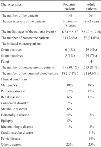
Demographic and clinical data of pediatric and adult patients are demonstrated in Table 1 and microorganisms isolated in bacteremia and contamination groups are dem-onstrated in Table 2 and 3. Since the number of pediatric patients and ratio of bacteremia were low, statistical analy-ses of these patients were evaluated together with that of the adults. A total of 809 patients from all groups underwent statistical analysis. Median, minimum and maximum PCT and CRP values according to the groups are demonstrated in Table 4. Both PCT and CRP were found significantly different in bacteremia group vs. non-bacteremia group (p < 0.0001). There was a difference between Gram posi-tive and Gram negaposi-tive bacteremia in terms of both PCT and CRP in the bacteremia group, but the difference was not statistically significant (p = 0.138 for PCT and p = 0.959 for CRP) (Table 5). Evaluating PCT and CRP according to Kruskal Wallis test, at least one of the three groups was found different from the others (p < 0.001). Based on Post-hoc tests performed after Kruskal Wallis test, Group 1 was found to be significantly different from Group 2 and Group 3 in terms of both PCT and CRP (p < 0.0001). Evalu-ating the difference between the groups according to Mann-Whitney U test with Bonferroni correction (0.05/3 = 0.0166), statistically significant difference was found be-tween Group 1 and Group 2 (p < 0.0001 for PCT and p < 0.002 for CRP) and between Group 1 and Group 3 (p < 0.0001 both for PCT and CRP). Both PCT and CRP were found significantly different in bacteremia groupvs.
(p < 0.001). PCT AUC was 0.755 (95% CI: 0.705-0.805) and CRP AUC was 0.601 (95% CI: 0.538-0.665) in differ-entiating bacteremia from non-bacteremia, and significan-ce was p < 0.001 for PCT and p = 0.002 for CRP. PCT AUC was 0.864 (95% CI: 0.799-0.929) and CRP AUC was 0.744 (95% CI: 0.652-0.835) in differentiating bacteremia from contamination, and significance was p < 0.001 for PCT and p < 0.001 for CRP. It was found that both PCT and CRP can be used in differentiating the groups but PCT is more effec-tive than CRP in differentiating bacteremia from both non-bacteremia and contamination (Figure 1 and Figure 2). When a cut-off value of 0.5 ng/mL was used for PCT and 5 mg/dL was used for CRP, sensitivity and specificity were 68.2% and 66.4% respectively for PCT and 93.2% and 9.5% respectively for CRP (Table 6).
Discussion
Both CRP and PCT are being used for a long time as biological markers for the diagnosis of severe infections. Whilst CRP is elevated in case of infection, inflammation and tissue damage, PCT is elevated only in bacterial infec-tions (Pepys et al., 2003; Sakr et al., 2008; Jeonget al., 2012). Since bacteria account for more than 90% of teremia cases, the use of PCT for the diagnosis of bac-teremia seems more realistic (Llewelyn et al., 2001). Although blood culture is known as the gold standard in de-tecting bacteremia, 24-48 hours are required for the results; thus, initiation of antibiotherapy is delayed (Riedelet al., 2011; Jeonget al., 2012). In addition, the present study was planned also considering that contamination, which is one of the most important problems encountered in evaluation of blood cultures, could be differentiated from bacteremia by the changes in PCT values.
Despite numerous studies that demonstrate the supe-riority of PCT over CRP in diagnosing bacteremia (Gia-marellouet al., 2004; Jimenoet al., 2004; von Lilienfeld-Toalet al., 2006; Schuttrumpfet al., 2006), there are a few studies that investigate the relation between PCT and con-taminated blood cultures (Schuetzet al., 2007; Jeonget al., 2012). The present study investigated the efficacy of PCT and CRP in differentiating true bacteremia from contami-nation and non-bacteremia. Based on our results, PCT is able to differentiate bacteremia from both non-bacteremia and contamination. Thus, decision for the initiation of anti-biotherapy would be made in a short time owing to the fact that PCT is able to differentiate contaminated blood cul-tures from true bacteremia or unnecessary antibiotic use would be prevented. Followings are the favorable conse-quences of this: both resistance to antibiotics would be de-creased, patients would be prevented against toxic effects of antibiotics, and economy of both the hospital and the country would have been protected (von Lilienfeld-Toalet al., 2006; Schuetzet al., 2011).
Many studies have reported higher PCT values in Gram negative bacteremia vs. Gram positive bacteremia Table 1- The demografic and clinical characteristics of the patients.
Characteristics Pediatric
patients
Adult patients
The number of the patients 148 661
The age intervals of the patients 3 months-18 years
19-92 years
The median ages of the patients (years) 6.34±5.57 52.22±17.86 The number of bacteremic patients 11 (7.4%) 77 (11.6%) The isolated microorganisms:
Gram positives 6 (4%) 29 (4%)
Gram negatives 5 (3%) 44 (7%)
Fungi 4
The number of nonbacteremic patients 119 (80.4%) 553 (84%) The number of contamined blood culture 18 (12.1% ) 31 (4.6% ) Clinical conditions:
Malignancy 30% 18%
Pulmoner disease 17% 17%
Renal disease 6% 11%
Congenital disorder 7%
Metabolic disorder 6%
Hematolojic disease 5% 2%
Epilepsy 5%
Rheumatologic disease 1% 2%
Cardiovascular disease 11%
Pelvic disease 14%
Other diseases 23% 25%
Table 2- The values of median, minimum and maximum of PCT and CRP Median (min-max)
Groups PCT (ng/mL) CRP (mg/dL)
Bacteremia (Group 1) (n = 88)
1.25 (0.05-157.7) 93 (0-594)
Nonbacteremia (Group 2) (n = 672)
0.20 (0.05-114.63) 64 (0-715)
Contamination (Group 3) (n = 49)
0.08 (0.05-6.77) 19 (0-331)
Table 3- The values of median, minimum and maximum of PCT and CRP for Gram positive bacteria, for Gram negative bacteria and for fungi in bacteremia group.
Bacteremia group (Group 1)
Median (min-max) PCT (ng/mL) CRP (mg/dL) Gram positive bacteria
(n = 35)
0.94 (0.05-103.15) 92 (0-552)
Gram negative bacteria (n = 49)
1.94 (0.05-157.73) 99 (3-594)
Fungi (n = 4) 0.43 (0.16-5.97) 70 (58-119)
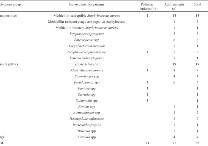
Table 4- The number of Gram positive bacteria, Gram negative bacteria and fungi in bacteremia group (n).
Bacteremia group Isolated microorganisms Pediatric
patients (n)
Adult patients (n)
Total
Gram positives Methicillin-susceptibleStaphylococcus aureus 1 14 15
Methicillin-resistant coagulase negative staphylococci 4 1 5
Methicillin-resistantStaphylococcus aureus 1 1
Streptococcus pyogenes 3 3
Enterococcusspp 6 6
Corynbacterium striatum 1 1
Streptococcus pneumoniae 1 2 3
Listeria monocytogenes 1 1
Gram negatives Escherichia coli 19 19
Klebsiella pneumoniae 1 8 9
Enterobacterspp 4 4
Pseudomonasspp 1 6 7
Pantoeaspp 1 1
Serratiaspp 1 1
Salmonellaspp 1 1
Proteusspp 1 1
A.cinetobacterspp 2 2
Haemophilus influenzae 2 2
Bacteroides fragilis 1 1
Brucellaspp 1 1
Fungi Candidaspp 4 4
Total 11 77 88
Table 5- The number of the isolated microorganisms in contamination group (n).
Contamination group Isolated microorganisms Pediatric patients(n) Adult patients(n) Total
Methicillin resistant-coagulase negative staphylococci 14 17 31
Methicillin susceptible-coagulase negative staphylococci 2 10 12
Difteroid basil 3 3
Alfa hemolytic streptococci 1 1 2
Bacillusspp 1 1
Total 18 31 49
Table 6- The sensitivity, specifity, positive and negative predictive values of PCT and CRP.
Cut off value Sensitivity Specifity Positive predictive value Negative predictive value
PCT 0.5 ng/mL 68.2 66.4 20.9 94.1
(Engelet al., 1999; Svaldiet al., 2001; Jeonget al., 2012). However, some studies (Al-Nawas et al., 1996; Giama-rellos-Bourbouliset al., 2001; von Lilienfeld-Toalet al., 2004) reported similar levels of PCT in Gram negative and Gram positive bacteremia. The present study failed to dem-onstrate statistically significant difference between Gram negative and Gram positive bacteremia in terms of PCT levels. This might have been resulted from various condi-tions. Bacteria such as Brucella spp, H.influenze, and
B.fragilis, which have been isolated from the patients with Gram negative bacteremia and low PCT, are the
microor-ganisms that grow late and difficult thereby induce PCT late. Moreover, there are patients with high PCT level and died before detection of any infectious agent other than contaminating bacteria. The blood samples might also have been obtained in early phase of infection.
In the recent years, there are numerous studies ex-pressing that PCT is beneficial not only in defining bacte-rial infection, but also in determining the severity of underlying disease, guiding treatment, and predicting the result. Meta analyses suggest that PCT is superior over CRP in differentiating bacterial infection from other causes of infection in critical patients (Sakret al., 2008). Similar with the results of the studies conducted by Engelet al., 1999 and Sakret al., 2008 the present study as well demon-strated that PCT is more effective than CRP in differentiat-ing bacteremia from non-bacteremia. In a meta-analysis Simonet al., 2004 reviewed 351 researches and reported that PCT has higher diagnostic accuracy as compared to CRP. Giamarellouet al.(2004) reported that PCT is a help-ful marker for the clinician in detecting severe sepsis, bacteremia and local infection but bacteremia due to CNS does not increase the level of PCT. This might have resulted from the authors’ considering every grown bacterium as an agent without differentiating CNS contamination from true bacteremia. Fleischhacket al.(2000) reported PCT was a more beneficial diagnostic parameter than CRP in cancer patients. Secmeeret al. (2007) reported that PCT, when measured periodically, was a more useful diagnostic pa-rameter than CRP in pediatric neutropenic-fever patients. Von Lilienfeld-Toal et al.(2004) reported that PCT is a more reliable marker than CRP in predicting bacteremia in the patients with febrile neutropenia. In addition to the stud-ies reporting high PCT levels in bacteremia (Giamarellouet al., 2004; Jimeno et al., 2004; von Lilienfeld-Toal et al., 2006; Schuttrumpfet al., 2006), there are studies defending just the opposite. de Bontet al.(2000) reported that PCT level showed no difference between bacteremia/sepsis group and the group with unknown fever among the pa-tients with neutropenic fever but that there was significant difference in terms of CRP level.
In the present study, PCT AUC value was 0.755 in differentiating bacteremia from non-bacteremia. Jeonget al.(2012), Bossinket al.(1999) and Kimet al.(2011) ob-tained similar results (respectively; 0.76; 0.70; 0.77) with that of the present study. The present study found PCT ROC-AUC value to be 0.86 in differentiating bacteremia from contamination. This is exactly the same with the result of the study conducted by Jeonget al.(2012).
Based on the recommendations of manufacturer com-pany, when a cut-off value of 0.5 ng/mL was used for PCT, sensitivity, specificity, and positive and negative predictive values were 68.2%, 66.4%, 20.9%, and 94.1% respectively. Other studies have found similar values for PCT (Kimet al., 2011; Jeong et al., 2012). Kim et al. (2011) used a cut-off value of 0.4 ng/mL and reached to a negative predic-Figure 2- The ROC curve of the PCT and CRP for discriminating
be-tween bacteremia group and contamination group.
tive value of 95.4% and found that bacteremia could be ex-cluded at a PCT level under 0.4 ng/mL. Likewise, the present study found that bacteremia could be excluded with an accuracy rate of 94.1% at a PCT level under 0.5 ng/mL. Using a cut-off value of 5 mg/dL, the sensitivity, specific-ity, and positive and negative predictive values for CRP were 93.2%, 9.5%, 11.8%, and 91.4% respectively. Low specificity of CRP despite high sensitivity might be ex-plained by the variety of reasons other than bacteremia by which the CRP level is increased.
One of the unfavorable situations in the present study is high levels of PCT found in some patients of non-bacteremia group leading to a decrease in positive predic-tive value and specificity of PCT. It has been reported that high PCT levels might be explained by likely use of antibi-otics or drugs that stimulate the release of proinflammatory cytokines, massive cell death, or probable failure in defin-ing causative microorganism (Pihuschet al., 2006; Schuetz
et al., 2011). It has been suggested that other clinical condi-tions may be in question in the patients with high PCT lev-els, or PCT may be induced by other reasons than bacteremia (pancreatitis, severe trauma, hepatic or renal disease, permanent shock and multi-organ failure, etc.) (Jeonget al., 2012). It has been also reported that PCT may be elevated in medullary thyroid carcinoma, small-cell lung carcinoma, and carcinoid tumors (Beckeret al., 2008). In this study, malignancy was detected by 30% in pediatric pa-tients and by 18% in adult papa-tients. Renal disease was pres-ent by 6% in pediatric patipres-ents and by 11% in adult patipres-ents. The present study displayed that PCT is more benefi-cial than CRP in diagnosing and excluding bacteremia. Moreover, PCT is more beneficial than CRP also in differ-entiating bacteremia from contamination. No statistically significant difference was found between Gram positive and Gram negative bacteria in the bacteremia group in terms of both PCT and CRP. Based on the results of this present study, early antibiotherapy can be initiated depend-ing on PCT result, which is measured concurrently with blood culture.
In conclusion, PCT is a more useful parameter than CRP in differentiating bacteremia from contamination and nonbacteremia in febril patients.
A part of this study was presented in 23rd European Congress of Clinical Microbiology and Infectious Diseases (ECCMID) in Berlin-Germany on April 27-30, 2013 (R2744).
Acknowledgement
This study was supported by the Research Fund of Is-tanbul University (Project Number: 2010/7541). The au-thors would like to thank to Mr Kamber KASALI, Msc, from Department of Biostatistics and Medical Informatics, Istanbul Faculty of Medicine for helpful studies on statisti-cal analysises.
Ethical Consideration
This project was approved by the Ethical Commitee of Clinical Researches of Istanbul Faculty of Medicine (2010/805-260).
References
Al-Nawas B, Krammer I, Shah PM (1996) Procalcitonin in diag-nosis of severe infections. Eur J Med Res 1:331-333. Baron EJ (coord ed) (2005) Blood Cultures, Cumulative
Tech-niques and Procedures in Clinical Microbiology (Cumi-tech), ASM Press, Washington, D.C.
Becker KL, Snider R, Nylen ES (2008) Procalcitonin assay in sys-temic inflammation, infection, and sepsis: clinical utility and limitations; Crit Care Med 36:941-952.
Bouadma L, Luyt CE, Tubach Fet al.(2010) Use of procalcitonin to educe patients’ exposure to antibiotics in intensive care units (PRORATA trial): a multicentre randomised contro-lled trial. Lancet 375:463-474.
Bossink A W, Groeneveld AB, Thijs LG (1999) Prediction of mi-crobial infection and mortality in medical patients with fe-ver: plasma procalcitonin, neutrophilic elastase-alpha1-antitrypsin and lactoferrin compared with clinical variables. Clin Infect Dis 29:398-407.
Clinical and Laboratory Standarts Institute (2007) Principles and Procedures for Blood Cultures. Approved Guideline. M47-A, Vol. 27 No. 17. Wayne, PA.
Clinical Laboratory Standards Institute (2011) Performance Stan-dards for Antimicrobial Suscebtibility Testing. 21st Informa-tional Supplement. M100-S21, Vol. 31, No. 1. Wayne, PA. Cohen J, Brun-Buisson C, Torres A, Jorgensen J (2004) Diagnosis
of infection in sepsis: An evidence bases review. Crit Care Med 32 Suppl (11):S466-494.
de Beer FC, Shine B, Pepys M (1982) Radiometric ligand binding assay for C-reactive protein. Complexed C-reactive protein is not detectable in acute phase serum. Clin Exp Immunol 50:231-237.
de Bont ES, Vellenga E, Swaanenburg J, Kamps W (2000) Pro-calcitonin: a diagnostic marker of bacterial infection in neu-tropenic cancer patients with fever? Infection 28:398-400. Engel A, Steinbach G, Kern P, Kern WV (1999) Diagnostic value
of procalcitonin serum levels in neutropenic patients with fever: comparison with interleukin-8. Scand J Infect Dis 31:185-189.
Fleischhack G, Kambeck I, Cipic D, Hasan C, Bode U (2000) Procalcitonin in paediatric cancer patients: its diagnostic rel-evance is superior to that of C-reactive protein, interleukin 6, interleukin 8, soluble interleukin 2 receptor and soluble tu-mour necrosis factor receptor II. Br J Haematol 111:1093-1102.
Giamarellos-Bourboulis EJ, Grecka P, Poulakou G, Anargyrou K, Katsilambros N, Giamarellou H (2001) Assessment of pro-calcitonin as a diagnostic marker of underlying infection in patients with febrile neutropenia. Clin Infect Dis 32:1718-1725.
neutropenia: experience from a multicentre study. Clin Mi-crobiol Infect 10:628-633.
Hall KK, Lyman JA (2006) Update review of blood culture con-tamination. Clin Microb Rev 19:788-802.
Jeong S, Park Y, Cho Y, Kim HS (2012) Diagnostic utilities of procalcitonin and C-reactive protein for the prediction of bacteremia determined by blood culture. Clin Chim Acta 413:1731-1736.
Jimeno A, Garcia-Velasco A, del Val O, Gonzalez-Billalabeitia E, Hernando S, Hernandez R, Sanchez-Munoz A, Lopez-Mar-tin A, Duran I, Robles L, Cortes-Funes H, Paz-Ares L (2004) Assessment of procalcitonin as a diagnostic and prognostic marker in patients with solid tumors and febrile neutropenia. Cancer 100:2462-2469.
Kim MH, Lim G, Kang SY, Lee WI, Suh JT, Lee HJ (2011) Utility of procalcitonin as an early diagnostic marker of bacteremia in patients with acute fever. Yonsei Med J 52:276-281. Kristoffersen KB, Sogaard OS, Wejse C, Black FTet al.(2009)
Antibiotic treatment interruption of suspected lower respira-tory tract infections based on a single procalcitonin mea-surement at hospital admission-a randomized trial. Clin Mi-crobiol Infect 15:481-487.
Llewelyn M, Cohen J (2001) Diagnosis of infection in sepsis. In-tensive Care Med 27 (Suppl 1):S10-S32.
Meisner M (2000) Procalcitonin: A new, innovative infection pa-rameter. Biochemical and clinical aspects. Georg Thieme Verlag, Stutgard Newyork.
Pepys MB, Hirschfield GM (2003) C-rective protein: a critical up-date. J Clin Invest 111:1805-1812.
Pihusch M, Pihusch R, Fraunberger Pet al.(2006) Evaluation of C-reactive protein, interlekin-6, and procalcitonin levels in allogeneic hematopoietic stem cell recipients. Eur J Haema-tol 76:93-101.
Riedel S, Melendez JH, An AT, Rosenbaum JE, Zenilman JM (2011) Procalcitonin as a Marker for the Detection of Bac-teremia and Sepsis in the Emergency Department. Am J Clin Pathol 135:182-189
Sakr Y, Sponholz C, Tuche F, Brunkhorst F, Reinhart K (2008) The role of procalcitonin in febrile neutropenic patients: Re-view of the literature. Infection 36:396-406.
Schuetz P, Mueller B, Trampuz A (2007) Serum procalcitonin for discrimination of blood contamination from bloodstream
in-fection due to coagulase-negative staphylococci. Inin-fection 35:352-355.
Schuetz P, Albrich W, Mueller B (2011) Procalsitonin for diagno-sis of infection and guide to antibiotic decisions: past, pres-ent and future. BMC Med 9:107
Schuttrumpf S, Binder L, Hagemann T, Berkovic D, Trumper L, Binder C (2006) Utility of procalcitonin concentration in the evaluation of patients with malignant diseases and elevated C-reactive protein plasma concentrations. Clin Infect Dis 43:468-473.
Secmeer G, Devrim I, Kara A, Ceyhan M, Cengiz B, Kutluk T, Buyukpamukcu M, Yetgin S, Tuncer M, Uludag AK, Tezer H, Yildirim I (2007) Role of Procalcitonin and CRP in dif-ferentiating a stable from a deteriorating clinical course in pediatric febrile neutropenia. J Pediatr Hematol Oncol 29:107-111.
Simon L, Gauvin F, Amre DK, Saint-Louis P, Lacroix J (2004) Serum Procalcitonin and C-Reactive Protein Levels as Mar-kers of Bacterial Infection: A Systematic Review and Meta-analysis. Clin Infect Dis 39:206-217.
Svaldi M, Hirber J, Lanthaler AI, Mayr O, Faes S, Peer E, Mitterer M (2001) Procalcitonin-reduced sensitivity and specificity in heavily leucopenic and immunosuppressed patients. Br J Haematol 115:53-57.
van Rossum AMC, Wulkan RW, Murphy AMO (2004) Pro-calcitonin as an early marker of infection in neonates and children. Lancet Inf Dis 4:620-630.
von Lilienfeld-Toal M, Dietrich MP, Glasmacher A, Lehmann L, Breig P, Hahn C, Schmidt-Wolf IG, Marklein G, Schroeder S, Stuber F (2004) Markers of bacteremia in febrile neu-tropenic patients with hematological malignancies: proca-lcitonin and IL-6 are more reliable than C-reactive protein. Eur J Clin Microbiol Infect Dis 23:539-544.
von Lilienfeld-Toal M, Schneider A, Orlopp K, Hahn-Ast C, Glasmacher A, Stuber F (2006) Change of procalcitonin pre-dicts clinical outcome of febrile episodes in patients with he-matological malignancies. Support Care Cancer 14:1241-1245.