Visual outcomes after implantation of a novel refractive toric multifocal intraocular lens
Resultado visual após implante de uma nova lente intraocular multifocal refrativa tórica
TaliTa Shimoda1, GilberTo Shimoda1, WilSon TakaShi hida2, CelSo TakaShi nakano2, anTônio FranCiSCo moTTa2, aline Silva GuimarãeS3, PaTriCk FrenSel m. TzelikiS3
Submitted for publication: August 15, 2013 Accepted for publication: December 1, 2013
Study conducted at Centro de Microcirurgia Ocular Atibaia (CEMOA), Atibaia, SP, Brazil. 1 Centro de Microcirurgia Ocular Atibaia (CEMOA), Atibaia, SP, Brazil.
2 Ophthalmology Department, Faculdade de Medicina, Universidade de São Paulo (USP), São Paulo, SP, Brazil.
3 Brasília Ophthalmologic Hospital (HOB), Brasília, DF, Brazil.
Funding: No specific financial support was available for this study.
Disclosure of potential conflicts of interest: None of the authors have any potential conflicts of interest to disclose.
Corresponding author: Patrick F. M. Tzelikis. SQN 203, Bloco G - Apto. 405 - Brasília (DF) - 70833-070 - Brazil - E-mail: [email protected]
ClinicalTrials.gov Identifier: NCT01505816 INTRODUCTION
The goal of modern cataract surgery is to gain spectacle inde-pendence for distance, intermediate, and near vision, which can be achieved with the implantation of multifocal intraocular lenses (IOLs). However, approximately 15%-29% cataract patients have more than 1.50 diopters (D) of corneal or refractive astigmatism(1-3). A residual
astigmatism error of 1.50 to 3.0 D after cataract surgery may decrease uncorrected visual acuity (UCVA) to 20/70 or 20/100 and may inter-fere with spectacle independence after multifocal IOL implantation(4).
To overcome this problem, various surgical options to control astigmatism during cataract surgery are available(4,5). Patients with a
considerable amount of corneal astigmatism who wish to undergo multifocal IOL implantation have several options, including a clear corneal cataract incision along the steep meridian(5), astigmatic
ke-ratotomy (AK)(6,7), opposite clear corneal incisions(8), limbal relaxing ABSTRACT
Purpose: To assess the postoperative outcomes of a novel toric multifocal in-traocular lens (IOL) in patients with cataract and corneal astigmatism.
Methods: This prospective nonrandomized study included patients with cata-ract, corneal astigmatism, and a motivation for spectacle independence. In all patients, a Rayner M-flex®
T toric IOL was implanted in the capsular bag. Three months after surgery, the distance, intermediate, and near visual acuities; spherical equivalent; residual refractive astigmatism; defocus curve; and contrast sensitivity were evaluated. A patient satisfaction and visual phenomena questionnaire was administered to all patients.
Results: Thirty-four eyes of 18 patients were included in this study. Three months after surgery, the mean corrected distance visual acuity (logMAR) was 0.00 ± 0.08 at 6 m, 0.20 ± 0.09 at 70 cm, and 0.08 ± 0.11 at 40 cm. Uncorrected distance vision acuity was 20/40 or better in 100% eyes. The preoperative mean refractive cylinder (RC) was -2.19 (SD: ± 0.53). After a 3-month follow-up, the average RC was -0.44 D (SD: ± 0.27; p<0.001). Contrast sensitivity levels were high. At the last follow-up, 87.5% patients were spectacle-independent for near, intermediate, and distance vision, and approximately 44% patients reported halos and glare.
Conclusion: Toric multifocal IOL implantation in patients with cataract and cor neal astigmatism using the Rayner M-flex® T toric IOL was a simple, safe, and accurate
option. This technology provides surgeons with a feasible option for meeting patient expectations of an enhanced lifestyle resulting from decreased spectacle dependence.
Keywords: Astigmatism/surgery; Lens implantation, intraocular; Lenses, intraocu-lar; Phacoemulsification; Polymethyl methacrylate; Refraction ocuintraocu-lar; Visual acuity
RESUMO
Objetivo: Avaliar o resultado pós-operatório de uma nova lente intraocular (LIO) multifocal tórica implantada em pacientes com catarata e astigmatismo corneano. Métodos: Estudo prospectivo não randomizado envolvendo pacientes com catarata, astigmatismo corneano, e motivação para eliminar a dependência dos óculos. Em todos os casos, a lente intraocular tórica Rayner M-flex®
T foi implantada dentro do saco capsular. Três meses pós-operatório foram avaliados: acuidade visual para longe, intermediário e perto; equivalente esférico; astigmatismo refracional residual; curva de defocus e sensibilidade ao contraste. Ao final do estudo um questionário referente à satisfação visual foi aplicado.
Resultados: Trinta e quatro olhos de 18 pacientes foram incluídos no estudo. Após 3 meses de pós-operatório, a acuidade visual corrigida para longe (logMAR) era de 0,00 ± 0,08 a 6 m, 0,20 ± 0,09 a 70 cm e 0,08 ± 0,11 a 40 cm. A acuidade visual sem correcão foi de 20/40, ou melhor, em 100% dos olhos. A média do cilindro refracional pré-operatório era de -2,19 (DP: ± 0,53). Após o seguimento de 3 meses a média do cilindro refracional era de -0,44 D (DP: ± 0,27) (p<0,001). Os níveis de sensibilidade ao contraste foram elevados. Ao final do seguimento, 87,5% dos pacientes estavam independentes dos óculos para perto, intermediário e para longe, e aproximadamente 44% dos pacientes relatavam halos e glare.
Conclusão: O implante de uma lente intraocular multifocal em pacientes com catarata e astigmatismo corneano utilizando a lente intraocular tórica Rayner M-flex® Tfoi
uma opção simples, segura e acurada. Essa nova tecnologia oferece ao cirurgião uma maneira passível de se atingir as expectativas dos pacientes em relação a qualidade de vida em razão de uma menor dependência dos óculos.
Descritores: Astigmatismo/cirurgia; Implante de lente intraocular; Lentes intraocu-lares; Facoemulsificação; Polimetil metacrilato; Refração ocular; Acuidade visual
incisions(9), and, of late, toric multifocal IOL implantation to decrease
astigmatism after cataract surgery(10).
Toric multifocal IOLs offer the opportunity to correct astigmatism and achieve spectacle independence at all distances in patients with corneal astigmatism with only one procedure. This study assessed the visual function after cataract surgery and implantation of a toric multifocal IOL (Rayner M-flex®
T toric) in a small single-site series of 34 eyes in 18 patients.
METHODS
for spectacle independence. Written informed consent was obtained from all patients before surgery, and the study was approved by the local ethics committee. All the procedures were in accordance with the ethical standards of the responsible committee on human expe-rimentation and the Helsinki Declaration of 1975. Exclusion criteria were previous ocular surgery and irregular corneal astigmatism.
Before surgery, patients underwent extensive ophthalmological examination, including the measurement of uncorrected distance visual acuity (UDVA), corrected distance visual acuity (CDVA), re-fraction, slit-lamp examination, fundoscopy, corneal topography (EyeSys unit, version 3.03; EyeSys Technologies, Houston, Texas), and biometry with the IOLMaster partial coherence interferometry device (Carl Zeiss Meditec AG, Jena, Germany). The targeted postoperative refractive error was the closest to 0.0 D.
IOL
All patients received a Rayner M-flex®
toric IOL (model 638F M-flex®
T, Rayner IOLs, Ltd., UK). This multifocal aspheric IOL is made of hydrophilic acrylate. It is a multizoned, refractive, aspheric IOL with either 4 or 5 annular zones (depending on IOL base power). The optic diameter is 6.25 mm and the overall diameter is 12.5 mm. Available spherical powers range from +14.0 D to +32.0 D in 0.50-D increments and include a near addition (add) of +3.00 D. Cylinder powers range from +1.00 to +6.00 D in 0.50-D increments. The IOL power was cal-culated using an online calculator.
S
URGICALTECHNIQUEAll patients were operated upon by the same surgeon using iden tical procedures. Before surgery, corneal reference marks were placed at 0° and 180° using a toric reference marker, with the patient sitting upright to correct for cyclotorsion. Next, the desired alignment axis for the toric IOL was intraoperatively marked with an angular graduation instrument. Phacoemulsification was performed using the Infinite (Alcon Surgical) Vision system. All IOLs were inserted in the capsular bag with the injector system. The toric IOL was implan-ted and rotaimplan-ted until the IOL markings agreed with the alignment marking. Postoperative follow-up was at 1 day and 1 and 3 months after surgery.
O
UTCOMEMEASURESPostoperative evaluation was performed at 3 months. UDVA and CDVA were assessed using the 100% contrast Early Treatment Diabe-tic Retinopathy Study chart. Uncorrected intermediate visual acuity (UIVA) for intermediate vision was assessed at 70 cm, and the un-corrected near visual acuity (UNVA) for near vision was assessed at 40 cm (Near reading chart, Presby Corp.). A binocular defocus curve was constructed using the Early Treatment of Diabetic Retinopathy Study (ETDRS) chart at 4 m. The cylinder axis of the IOL was measured at the slit lamp using the beam protractor after full mydriasis. The mean of the absolute IOL degrees that were off-axis was determined.
Contrast sensitivity was measured using the CSV-1000 HGT ins-trument (VectorVision, Inc. Greenville, OH, USA), which presents a translucent chart divided into 4 cycles with spatial frequencies of 3, 6, 12, and 18 cycles per degree (cpd). All measurements were obtained under mesopic (5 cd/m2) and photopic (85 cd/m2) conditions.
Exami-nations were unilaterally performed at a distance of 2.5 m, with the corrected visual acuity (BCVA) and an undilated pupil.
Patient satisfaction and quality of life were assessed by a simple questionnaire. All patients were interviewed 3 months after surgery. Patients were asked about visual disturbances, visual lifestyle activi-ties, spectacle use, and procedural satisfaction.
All data analyses were performed using SPSSX statistical programs (SPSS Inc, Chicago, IL, USA). The normality of all data samples was first checked using the Kolmogorov-Smirnov test. When parametric analysis was possible, Student’s t-test for paired data was used for all parameter comparisons between pre- and postoperative examina-tions. When parametric analysis was not possible, the Wilcoxon rank-sum
test was applied. Any differences showing a p-value of <0.05 (i.e., at the 5% level) were considered statistically significant.
RESULTS
The study enrolled 18 patients (34 eyes). Sixteen patients had bilateral Rayner M-flex®T toric multifocal IOL implantation; the other
2 received a Rayner M-flex® multifocal IOL because they had mild
astigmatism (<1.0 D). Three patients were men (16.7%) and 15 were women (83.3%), with a mean age of 69.44 years (range, 52-86 years; SD, ±8.9). All patients had mild cataract at presentation. The mean sphe-rical power of the implanted toric multifocal IOLs was +20.32 ± 3.1 D (range, 14.00-25.00 D). The mean preoperative axial length (AL) was 23.22 ± 0.8 mm (range, 21.39-26.19 mm). All patients were followed-up for 3 months.
V
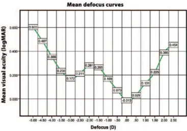
ISUALACUITYANDREFRACTIONTable 1 shows the distance, intermediate, and near visual acuities. The postoperative UDVA was 20/40 (0.3 logMAR) or better in 34 eyes (100%), 20/30 (0.18 logMAR) or better in 31 eyes (91.2%), and 20/25 (0.10 logMAR) or better in 30 eyes (88.2%), while CDVA was 20/40 or better in 34 eyes (100%) and 20/25 or better in 32 eyes (94.1%). Figure 1 shows the binocular defocus curve with 2 peaks of optimum CDVA at 0.00 D and -2.75 D.
Table 1. Mean preoperative and postoperative visual acuity and refraction (logMAR)
Mean ± SD
Preop Postop p-value
UDVA 0.51 ± 0.33 0.03 ± 0.09 <0.001 CDVA 0.13 ± 0.13 0.00 ± 0.08 <0.001
UIVA - 0.22 ± 0.09
-DCIVA - 0.20 ± 0.09
-UNVA - 0.10 ± 0.13
-DCNVA - 0.08 ± 0.11
-SE (D) -0.24 ± 1.59 0.11 ± 0.40 0.21 Refractive cylinder (D)
Mean -2.19 ± 0.53 -0.44 ± 0.27 <0.001 Range -1.25 to -3.25 0.00 to -1.00
UDVA= uncorrected distance visual acuity, CDVA= corrected distance visual acuity, UIVA= uncorrected intermediate visual acuity, DCIVA= distance-corrected intermediate visual acuity, UNVA= uncorrected near visual acuity, DCNVA= distance-corrected near visual acuity, SE= spherical equivalent.
The average SE of these eyes was -0.24 D (range, -2.75 – +3.00 D; SD, ±1.59) at referral and 0.11 (range, -1.00 – +1.25 D; SD, ±0.40;
p=0.21) at 3 months after surgery. The average refractive cylinder of these eyes was -2.19 D (range, -1.25 – -3.25 D; SD, ±0.53) at referral and -0.44 D (range, 0.00 – -1.00 D; SD, ±0.27; p<0.001) at 3 months. Almost 95% eyes had a postoperative residual refractive astigmatism of -0.75 D or less.
Corneal topography values at presentation were recorded for all eyes. The average flatter corneal meridian topography was 43.21 D (range, 40.40-45.96 D; SD, ±1.20), and the average steeper corneal meridian topography was 45.57 D (range, 42.74-47.74 D; SD, ±1.27). The average topographic astigmatism was 2.29 D (range, 1.36-3.19 D; SD, ±0.52), which increased to 43.25 D (range, 40.32-45.93 D; SD, ±1.25) at 3 months after surgery. The average steeper corneal me-ridian topography was 45.51 D (range, 42.64-47.74 D; SD, ±1.26), and the average topographic astigmatism was 2.16 D (range, 1.38-3.16 D; SD, ±0.54; p>0.05).
M
ISALIGNMENTThree months after surgery, the mean error in IOL alignment was 2.34 ± 2.33° (range, 0-9°). The mean error in IOL alignment was more than 5° in 5 eyes (17.6%). No IOL was misaligned by 10° or more.
C
ONTRASTSENSITIVITYFigure 2 shows the mean log monocular contrast sensitivity va lues under photopic and mesopic conditions at 3 months after surgery. The mean contrast sensitivity levels in photopic and mesopic conditions were 1.60 ± 0.11 and 1.39 ± 0.16, respectively, at 3 cpd, 1.69 ± 0.12 and 1.44 ± 0.21, respectively, at 6 cpd, 1.12 ± 0.36 and 0.93 ± 0.41, respectively, at 12 cpd, and 0.57 ± 0.42 and 0.37 ± 0.41, respectively, at 18 cpd.
P
ATIENTSATISFACTIONANDSPECTACLEDEPENDENCYAll 16 patients with bilateral Rayner M-flex® T toric IOLs completed
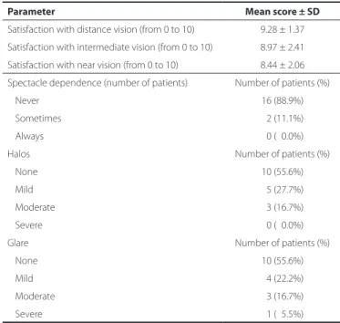
the questionnaire. Table 2 shows patient-reported spectacle depen-dence; satisfaction with distance, intermediate, and near vision; and the incidence of halos and glare. Three months after cataract surgery, no eye required a second procedure. At the last follow up, 14 patients (87.5%) were spectacle-independent for near, intermediate, and distance vision and were very satisfied with their quality of vision without glasses. Good distance and near vision were reported by all patients.
DISCUSSION
Clinical trials evaluating the clinical, optical, functional, and quality-of-life outcomes after implantation of these new-generation IOLs found that the participants had improved near vision and
good distance vision(10-13). Although patients with these IOLs report
less limitation in visual function and less spectacle dependency compa red with patients with monofocal IOLs(14), some have reported
dis sa tis faction with the outcomes that are mostly related to visual aberrations such as halos, glare, and dysphotopsia(15).
After cataract surgery, even a relatively low amount of uncorrected astigmatism can significantly decrease visual acuity in eyes with mul tifocal IOLs, which will further decrease the ability to perform low-contrast tasks(16,17).However, today’s cataract patients are more
demanding, and they usually seek total visual rehabilitation and total independence from spectacles. These requirements have been the motivating factor in the development of toric multifocal IOLs. These IOLs were designed to compensate for corneal astigmatism and the loss of accommodative ability after cataract extraction.
In the current study, we evaluated visual and refractive outcomes, contrast sensitivity, and patient satisfaction after Rayner M-flex toric multifocal IOL implantation. At present, there are only 4 types of toric multifocal IOL models available: diffractive ReSTOR IQ toric IOL (Alcon Laboratories, Inc.), diffractive AT Lisa toric IOL (Carl Zeiss Meditec AG), Lentis Mplus toric IOL (Oculentis GmbH), and refractive M-flex T IOL (Rayner Intraocular Lens Ltd.)(18). So far, to our knowledge, no study of
the Rayner M-flex T IOL has been published.
Nontoric Rayner multifocal IOL implantation has been evaluated in a few studies(19).This IOL has the same platform as the Rayner M-flex
T IOL. Cezón Prieto and Bautista(19) studied the Rayner M-flex 630F
+3.00 D IOL and found a mean monocular CDVA of 0.03 logMAR (ap-proximately 20/20) 12 months after surgery; 100% eyes had a CDVA of 0.30 logMAR (20/40) or better, while 100% eyes had a CDVA of 0.00 logMAR (20/20) or better. The toric Rayner monofocal IOL has also been evaluated in a few studies(20).In a study of the Rayner T-flex
623T toric IOL, some authors(20) found a mean CDVA of 0.19 logMAR
(approximately 20/30) 4 months after surgery; 96.6% eyes had a CDVA of 0.30 logMAR (20/40) or better, 81.8% eyes had a CDVA of 0.18 logMAR (20/30) or better, and 21.1% eyes had a CDVA of 0.00 logMAR (20/20) or better. Our study found a good CDVA (mean 0.00 logMAR), with all patients having a CDVA of 20/25 or better at 3 months. There are few published studies describing refractive
Figure 2. Mean log contrast sensitivity values at 3 months.
Table 2. Subjective symptoms and spectacle-dependence 3 months after surgery
Parameter Mean score ± SD
Satisfaction with distance vision (from 0 to 10) 9.28 ± 1.37 Satisfaction with intermediate vision (from 0 to 10) 8.97 ± 2.41 Satisfaction with near vision (from 0 to 10) 8.44 ± 2.06 Spectacle dependence (number of patients) Number of patients (%)
Never 16 (88.9%)
Sometimes 02 (11.1%)
Always 00 (00.0%)
Halos Number of patients (%)
None 10 (55.6%)
Mild 05 (27.7%)
Moderate 03 (16.7%)
Severe 00 (00.0%)
Glare Number of patients (%)
None 10 (55.6%)
Mild 04 (22.2%)
Moderate 03 (16.7%)
Severe 01 (05.5%)
outcomes after toric multifocal IOL implantation(10,13,21). Other
au-thors(10,13) reported a binocular logMAR CDVA of -0.06 and 0.05 after 3
months of follow-up using the AT Lisa toric multifocal IOL.
Uncorrected outcomes were also good, with 90% patients achie ving a UDVA of 20/25 or better in our study. Cezón Prieto and Bautista(19) found a mean UDVA of 0.09 logMAR (approximately 20/25)
12 months after surgery using the Rayner M-flex 630F IOL, with 100% eyes achieving a UDVA of 0.30 logMAR (20/40) or better and 75% eyes achieving a UDVA of 0.00 logMAR (20/20) or better. Similar results were reported by Viesser et al.(10) using the AT Lisa toric multifocal IOL
with a mean monocular logMAR UDVA of 0.04 3 months after surgery; 98% eyes had a UDVA of 0.30 logMAR (20/40) or better and 71% eyes had a UDVA of 0.10 logMAR (20/25) or better.
Recently, there has been a trend toward decreasing the power of the near add (from +4.00 D to +3.00 D) in some models of multifocal IOLs to improve intermediate vision or expand reading distance(19,21-23).
At 3 months, the mean monocular UIVA and distance-corrected inter-mediate visual acuity (DCIVA) in our study were 0.22 logMAR and 0.20 logMAR, respectively. These findings are slightly better than those of Viesser et al.(10), who reported a 3-month monocular intermediate
vision (at 60 cm) with toric multifocal IOL of 0.40 logMAR with and without correction. For intermediate distances, different models of multifocal IOL with a +3.00-D add power have shown similar results. Alfonso et al.(22), in a study of the AcrySof ReSTOR SN6AD1, found
mean binocular UIVA and DCIVA logMAR values of 0.165 and 0.147 at 70 cm after 6 months of follow-up. Muñoz et al.(23) studied the Lentis
Mplus LS-312 multifocal IOL and found a logMAR UIVA and DCIVA of 0.13 and 0.14 at 1 m 6 months after surgery. Cezón Prieto and Bautista(19) reported a mean UIVA and DCIVA value of 0.15 logMAR
in eyes with Rayner M-flex IOL. This was consistent with our finding that the majority of patients did not use spectacles for intermediate vision because of the excellent visual acuity between 50 and 70 cm.
In our study, the Rayner M-flex T IOL with +3.00 D add provided high performance for near vision; the mean monocular UNVA was 0.10 logMAR and the mean DCNVA was 0.08 logMAR at 3 months. The near vision achieved with the Rayner M-flex T IOL was comparable to that with other multifocal IOLs(10,12,13,22,23).In the study by Viesser et
al.(10), the mean UNVA was 0.20 logMAR. In another study of toric
mul-tifocal IOL by Alió et al.(13) the mean postoperative UNVA and CDNVA
was 0.24 and 0.24 logMAR, respectively, at 3 months. Cezón Prieto and Bautista(19) reported a UNVA value of 0.28 logMAR in eyes
implan-ted with Rayner M-flex IOL. Similar results (binocular logMAR DCNVA, approximately 0.10) were reported by Fujimoto et al.(24), who used the
multifocal refractive Array SA40N IOL (Abbott Medical Optics, Inc.) and by Alió et al.(25), who used the multifocal refracti ve-diffractive Acri.
LISA 366D (Carl Zeiss Meditec AG) and the AcrySof ReSTOR SN6AD3 (Alcon Laboratories, Inc.).
The defocus curve for the Rayner M-flex T IOL was consistent with the above findings, with a V-shaped curve with 2 peaks of ma-ximum vision, 1 at distance (around a 0.00-D defocus level) and 1 at near (approximately -2.75 D defocus level). Between these 2 peaks, acceptable vision was maintained, resulting in acceptable interme-diate vision. IOL performance for distance was similar to that with a monofocal IOL, with a logMAR acuity of approximately 0.00. The IOL provided a logMAR intermediate acuity of approximately 0.20-0.30 between 50 cm and 70 cm and had the best logMAR near vision between 33 cm and 40 cm. When considering the defocus curve for other multifocal IOLs, the range of focus for functional vision exten-ded approximately from +1.0 D to -3.5 for +3.0-D IOL(10,22).
According to slit-lamp examination, the mean misalignment of Rayner M-flex T IOL was 2.34 ± 2.33°, with no IOL off-axis by more than 10° at 3 months. We found the Rayner M-flex T IOL to be stable once positioned in the capsular bag. The amount of rotation of different toric IOL models is reported to be between 0° and 20°(26). The FDA trial
of the AcrySof SA60T toric IOL compared 244 patients with a control group of 250 patients who received the nontoric AcrySof SA60AT
IOL. The former exhibited excellent rotation stability, with a mean rotation of less than 4° from the initial alignment at 12 months after surgery. Rotation misalignment was 10° or less in 97% patients and 5° or less in 81% patients. Zuberbuhler et al.(27) subsequently
repor-ted a large series of AcrySof SN60T toric IOLs in 44 eyes. The mean toric IOL axis rotation was 2.2 ± 2.2°, and 95%IOL were within 5° of the targeted axis. The current study of the rotational stability of the Rayner M-flex T toric IOL showed nearly identical results; the mean rotation was less than 4°, with 100% IOL showing a rotation of 10° or less. Our results were comparable with those in studies of the AcrySof toric IOL. Intraoperative positioning of toric IOLs with grea-ter accuracy was necessary to achieve the most optimum cylinder correction in all patients(21,26,27).
The Rayner M-flex T is a refractive toric multifocal IOL with a mul-tizone design that can cause loss of contrast sensitivity because of the distribution of total available light between several focal points. Patients with multifocal IOLs may therefore be more sensitive to changes in contrast and have been reported to have lower contrast sensitivity compared with the normal population(28). Several
stu-dies(22,23,25,28) have reported lower photopic contrast sensitivity with a
multifocal IOL than with a monofocal IOL; however, the contrast sen-sitivity was still in the normal range. The results in our study confir med these results. We obtained contrast sensitivity values under mesopic and photopic conditions that were similar to those obtained by Cezón Prieto and Bautista(19), Montés-Micó et al.(28), and de Vries et al.(29).
In a normal population, contrast sensitivity levels measured with the CSV-1000 system were 1.56 ± 0.15 at 3 cpd, 1.80 ± 0.16 at 6 cpd, 1.50 ± 0.15 at 12 cpd, and 0.93 ± 0.25 at 18 cpd(30). This indicated that the
contrast sensitivity after Rayner M-flex T implantation was compa-rable with that in the normal population at 2 spatial frequencies (3 and 6 cpd) and decreased at high spatial frequencies (12 and 18 cpd) under photopic and mesopic conditions.
CONCLUSIONS
The results of our study showed that implantation of the Rayner M-flex T IOL effectively decreased visually significant keratometric astigmatism and spectacle-dependence after cataract surgery, with acceptable low rates of IOL misalignment. Further studies on the effi-cacy of this toric multifocal IOL in cataract surgery are recommended with larger sample sizes and longer follow-ups.
REFERENCES
1. Hoffmann PC, Hütz W. Analysis of biometry and prevalence data for corneal astigmatism in 23239 eyes. J Cataract Refract Surg. 2010;36(9):1479-85. Comment in: J Cataract Refract Surg. 2010;36(9):1447-8; J Cataract Refract Surg. 2011;37(3):621; author reply 621. 2. Hoffer KJ. Biometry of 7,500 cataractous eyes. Am J Ophthalmol. 1980;90(3):360-8. 3. Grabow HB. Intraocular correction of refractive errors. In: Kershner RM, editor.
Refracti-ve keratotomy for cataract surgery and the correction of astigmatism. New Jersey: Slack Thorofare; 1994. p.79-115.
4. Frohn A, Dick HB, Thiel HJ. Implantation of a toric poly(methylmethacrylate) intrao-cular lens to correct high astigmatism. J Cataract Refract Surg. 1999;25(12):1675-8. 5. Koch D, Lindstrom RL. Controlling astigmatism in cataract surgery. Sem Ophthalmol.
1992;7:224-33.
6. Shepherd JR. Correction of preexisting astigmatism at the time of small incision cataract surgery. J Cataract Refract Surg. 1989;15(1):55-7.
7. Osher RH. Paired transverse relaxing keratotomy: a combined technique for reducing astigmatism. J Cataract Refract Surg. 1989;15(1):32-7.
8. Lever J, Dahan E. Opposite clear corneal incisions to correct pre-existing astigmatism in cataract surgery. J Cataract Refract Surg. 2000;26(6):803-5. Comment in: J Cataract Refract Surg. 2000;26(6):789-90; J Cataract Refract Surg. 2001;27(1):7-8; J Cataract Refract Surg. 2000;26(12):1697-8.
9. Müller-Jensen K, Fisher P, Slepe U. Limbal relaxing incision to correct astigmatism in clear corneal cataract surgery. J Refract Surg. 1999;15(5):586-9.
10. Viesser N, Nuijts RM, de Vries NE, Bauer NJ. Visual outcomes and patient satisfaction after cataract surgery with toric multifocal intraocular lens implantation. J Cataract Refract Surg. 2011;37(11):2034-42.
VII Congresso Baiano de Oftalmologia
10 e 11 de outubro de 2014
Hotel Sheraton
Salvador - BA
Informações:
Site: www.sofba.com..br
12. Pepose JS, Qazi MA, Davies J, Doane JF, Loden JC, Sivalingham V, et al. Visual perfor-mance of patients with bilateral vs combination Crystalens, ReZoom, and ReSTOR in traocular lens implants. Am J Ophthalmol. 2007;144(3):347-57. Comment in: Am J
Ophthalmol. 2008;145(4):593-4.
13. Alió JL, Piñero DP, Tomás J, Plaza AB. Vector analysis of astigmatism changes after ca-taract surgery with implantation of a new toric multifocal intraocular lens. J Caca-taract Refract Surg. 2011;37(7):1217-29.
14. Souza CE, Mucciolli C, Soriano ES, Chalita MR, Oliveira F, Freitas LL, et al. Visual perfor-mance of AcrySof ReSTOR apodized diffractive IOL: a prospective comparative trial. Am J Ophthalmol. 2006;141(5):827-32.
15. Woodward MA, Randleman JB, Stulting RD. Dissatisfaction after multifocal intraocular lens implantation. J Cataract Refract Surg. 2009;35(6):992-7.
16. Hayashi K, Manabe S, Yoshida M, Hayashi H. Effect of astigmatism on visual acuity in eyes with diffractive multifocal intraocular lens. J Cataract Refract Surg. 2010;36(8): 1323-9.
17. Wolffsohn JS, Bhogal G, Shah S. Effect of uncorrected astigmatism on vision. J Cata-ract RefCata-ract Surg. 2011;37(3):454-60.
18. Lane SS, Morris M, Nordan L, Packer M, Tarantino N, Wallace RB 3rd. Multifocal intra-ocular lenses. Ophthalmol Clin North Am. 2006;19(1):89-105.
19. Cezón Prieto J, Bautista MJ. Visual outcomes after implantation of a refractive multi-focal intraocular lens +3.00 D addition. J Cataract Refract Surg. 2010;36(9):1508-16. 20. Entabi M, Harman F, Lee N, Bloom PA. Injectable 1-piece hydrophilic acrylic toric
intraocular lens for cataract surgery: efficacy and stability. J Cataract Refract Surg. 2011;37(2):235-40.
21. Liekfeld A, Torun N, Friederici L. [A new toric diffractive multifocal lens for refractive surgery]. Ophthalmologe. 2010;107(3):256;258-61. German.
22. Alfonso JF, Fernández-Vega L, Amhaz H, Montés-Mico R, Valcárcel B, Ferrer-Blasco T. Visual function after implantation of an aspheric bifocal intraocular lens. J Cataract Refract Surg. 2009;35(5):885-92.
23. Muñoz G, Albarrán-Diego C, Ferrer-Blasco T, Sakla HF, García-Lázaro S. Visual function after bilateral implantation of a new zonal refractive aspheric multifocal intraocular lens. J Cataract Refract Surg. 2011;37(11):2043-52.
24. Fujimoto K, Honda K, Wada YR, Tanaka M, Irie T. Four-year experience with a silicone refractive multifocal intraocular lens. J Cataract Refract Surg. 2010;36(8):1330-5. 25. Alió JL, Plaza-Puche AB, Piñero DP, Amparo F, Rodríguez-Pratas JL, Ayala MJ. Quality
of life evaluation after implantation of 2 multifocal intraocular lens models and a monofocal model. J Cataract Refract Surg. 2011;37(4):638-48.
26. Viestenz A, Seitz B, Langenbucher A. Evaluating the eye’s rotational stability during standard photography: effect on determining the axial orientation of toric intraocular lenses. J Cataract Refract Surg. 2005;31(3):557-61.
27. Zuberbuhler B, Signer T, Gale R, Haeflinger E. Rotational stability of the AcrySof SA60TT toric intraocular lenses: a cohort study. BMC Ophthalmology. 2008;8:8. 28. Montés-Micó R, España E, Bueno I, Charman WN, Menezo JL. Visual performance with
multifocal intraocular lenses; mesopic contrast sensitivity under distance and near conditions. Ophthalmology. 2004;111:85-96.
29. de Vries NE, Webers CAB, Montés-Micó R, Ferrer-Blasco T, Nuijts RM. Visual outcomes after cataract surgery with implantation of a +3.00 D or +4.00 D aspheric diffractive multifocal intraocular lens. Comparative study. J Cataract Refract Surg. 2010;36(8): 1316-22.