344
Rev Bras Oftalmol. 2013; 72 (5): 344-7 The authors declare no conflicts of interest
Received for publication: 19/3/2012 - Accepted for publication: 27/5/2012
C
ASER
EPORTSupplementary pseudopakic IOL Sulcoflex
®
Toric over Toric Acrysof
®
IOL
LIO pseudofácica suplementar - Sulcoflex Toric
- sobre LIO Acrysof Toric
Hilton Arcoverde Gonçalves de Medeiros
1, João Eugenio Gonçalves de Medeiros
1, Jorge Luiz Silveira Baldiotti
11Dr. João Eugenio Eye Clinic - Brasilia/DF, Brazil
Work conducted at Dr. João Eugenio Eye Clinic - Brasilia/DF, Brazil
R
ESUMOPara correção de alta hipermetropia e astigmatismo irregular secundário a múltiplas cirurgias refrativas em uma mulher de 45 anos, foi utilizada a sutura das incisões radiais, com implante de lente intraocular (LIO) Acrysof® Toric. Para correção do residual de erro
refracional, foi implantada LIO pseudofácica tórica suplementar sobre LIO Primária. A acuidade visual (AV) inicial era de LogMAR 0,9 e a final de LogMAR 0,3. O implante da LIO suplementar tórica sobre LIO tórica primária mostrou-se uma boa opção, proporcionando melhora da acuidade visual.
Descritores: Hiperopia; Ceratotomia radial; Lentes intraoculares; Acuidade visual; Relatos de casos
A
BSTRACTTo correct a high hyperopia and irregular astigmatism secondary to multiples refractive surgeries in a 45 years old female, radial incisions suture and intraocular (IOL) Acrysof Toric lens was performed. To correct the residual refractional error a pseudophakic toric supplementary IOL was implanted over the primary one.The initial visual acuity (VA) was LogMAR 0,9 and the final VA was LogMAR 0,3. The implant of IOL supplementary over a primary toric IOL appears to be a good option, increasing the visual acuity.
345
Rev Bras Oftalmol. 2013; 72 (5): 344-7
I
NTRODUCTIONE
ye glasses are not satisfactory for correction of hyperopia secondary to radial keratotomy (RK) because they do not correct irregular astigmatism or higher order aberrations(1).Treating such high hyperopias is a challenge for refractive surgeons. They usually consist of anisometropias leading to low quality of vision and patient dissatisfaction. Several therapeutic options are suggested, such as circular suture, suture of incisions, photorefractive keratectomy (PRK), laser-assisted in situ keratomileusis (LASIK), and phacorefractive surgery(2).
Phacoemulsification has been gaining support among ophthalmologists, especially after the introduction of advances that provide better biometric calculation and intraocular lenses (IOLs) with special designs. Phacoemulsification leads to a marked and predictable reduction of high hyperopia to a degree that allows further treatment with excimer laser.
Good results are almost always obtained but additional correction may be required due to unidentified preoperative astigmatism, surgically-induced astigmatism, use of multifocal lenses, or biometric error. The patient may also be unfit for corneal surgery due to low corneal thickness. In these patients, implantation of an additional lens is an alternative.
The Sulcoflex™ (Rayner, East Sussex, UK) IOL is designed specifically for implantation in the ciliary sulcus over another IOL implanted in the capsular bag. It can be used to correct optical aberrations and other optical/visual changes which were previously difficult to treat.
It is made of hydrophilic acrylic polymer with excellent uveal biocompatibility. It has a large optical diameter, reducing the risk of iris capture, and its large curved haptics with rounded edges reduce trauma to adjacent tissues and provide excellent rotational stability(3). The haptics have a posterior angulation of
10 degrees, increasing separation with the iris anteriorly and the primary IOL posteriorly and reducing the risk of interlenticular opacification (a frequent complication of piggyback plano-convex IOLs, especially if both IOLs are in the capsular bag)(4).
Even though it is primarily indicated for correction of spherical or cylindrical pseudophakic ametropias, or even pseudophakic presbyopia, it has been suggested preoperatively for surgical correction of high refractive errors, congenital cataract and corneal astigmatism, among others(2).
C
ASER
EPORTANDM
ÉTHODSOur patient was a 45-year-old female who had had undergone radial keratotomy (RK) 25 years earlier. She had received 4 radial incisions combined with 02 astigmatic incisions in each eye.
The patient reported that 10 years after RK she underwent excimer laser (LASIK) twice to correct a residual refractive error in the left eye, which remained with a lower visual acuity.
On examination, LogMAR visual acuity (VA) was 0.18 in the right eye (RE) and 1.0 in the left eye (LE). Refraction after cycloplegia was +1.00 -1.25 30° in the RE and +10.50 -5.25 175° in the LE. Corrected LogMAR VA was 0.1 in the RE and 0.9 in the LE. Slit lamp examination showed RK and LASIK scars bilaterally.
Topography showed severe corneal flattening with a mean central curvature of 35.87 mm (Figure 1), and ultrasonic pachymetry showed a central thickness of 439ìm. We performed a double concentric interrupted suture (modified Grene-Lasso
suture) of the radial incisions in the LE and adapted a toric soft contact lens.
Sutures were removed after 1 year.
An improvement in corneal curvature was then observed, with reduction of the refractive error in the LE to +8.00 -4.50 175°, a mean central curvature of 36.59 and a corrected LogMAR VA of 0.8; keratometry was 37.62 × 35.95.
Refractive phacoemulsification with implantation of a toric IOL was then indicated in the LE. The procedure was performed three months later after the corneal sutures were removed, with implantation of an AcrySof™ Toric T5 IOL with a refractive power of 24.5 D in the 155° axis, based on Alcon’s nomogram (www.acrysoftoriccalculator.com).
The patient’s corrected LogMAR VA improved to 0.5, with residual correction of +3.00 -2.50 155°. After 1 year using the toric soft contact lens (Focus Toric, Ciba, UK) the patient complained of persistent discomfort with the contact lens, despite having tried other brands. Implantation of an additional Sulcoflex™ Toric IOL of +2.50 D and +3.00 D cyl in the 165° axis (second Rayner nomogram, available at www.toriciol.rayner.com) was suggested and conducted in June 2010 (Figures 2 and 3). Mean central curvature was 36.75, with a keratometry of 37.65 × 36.12.
The outcome was satisfactory with improvement of refraction to +0.50 -0.75 145° and a LogMar VA of 0.3 after a 4-month follow-up.
D
ISCUSSIONIn many eyes submitted to RK the procedure is associated with the astigmatic technique using transverse incisions, further accentuating corneal instability and irregular astigmatism(2) due
to the coupling effect in which the cornea is flattened in the incision’s meridian with compensatory bulging in the perpendi-cular meridian(5).
Corneal suture has been used as an alternative to correct undesirable flattening, either by simple interrupted sutures, simple circular suture, double circular suture, or a combination thereof, aiming to flatten the peripheral cornea and thus curve its central region. However, the outcome is insufficient in severe cases as it is unstable and unpredictable(6).
Other alternatives include PRK or LASIK, although the latter is controversial because it may cause the incisions to open, reducing the corrective effect of surgery(1); this can happen even
when a femtosecond laser is used(7). An advantage of PRK or
Figure 1: Topographic aspect after refractive surgeries, before phakectomy and implantation of the Sulcoflex™ IOL.
346 Medeiros HAG, Medeiros JEG, BaldiottiJLS
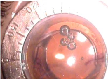
Figure 2: Trans-operative aspect during implantation of the Sulcoflex™ IOL. Note the haptics at different positions from the primary IOL.
Figure 3: Immediate postoperative period. Note the overlap of the Sulcoflex™ IOL haptics with the primary IOL haptics.
LASIK that they induce reduced fluctuations in daily vision(8)
because they can correct higher order aberrations. However, several authors agree that these alternatives are only valid for mild and moderate hyperopia (up to approximately 5.00D)(9-12).
Phacoemulsification with implantation of AcrySof™ Toric IOLs has been used in high astigmatism(13,14). Studies show
reductions of up to 95% in astigmatism after implantation of AcrySof™ Toric IOLs(13), especially if supplemented with
treatment of high-order aberrations through excimer laser. However, there are limitations to the correction of astigmatism with AcrySof™ Toric IOLs. Although controversial, phacoemulsification has been indicated for high hyperopia and presbyopia in patients with a clear lens(15).
Residual refractive disorders may occur postoperatively to IOL implantation, requiring a secondary surgical intervention(7). Implantation of an additional IOL can be easier
and is one of the safest alternatives, since exchanging the IOL is associated with an increased risk for zonular or capsular rupture(16). In addition, the calculation for the additional IOL
depends mainly on the current refraction of the operated patient. If the IOL is exchanged, one can not be sure that the new IOL will be implanted exactly in the same plane as the original one(14).
Implantation of an additional IOL over a primary piggyback IOL, a technique advocated in the early part of this decade, leads to certain long-term complications, especially opacification between the two lenses. This requires surgical treatment since YAG-laser is almost never sufficient to clean the surfaces, especially if the two IOLs were implanted within the capsular bag.
The Sulcoflex™ IOL is mainly indicated for correction of spherical or cylindrical pseudophakic ametropias or pseudophakic presbyopia, but it has been suggested preoperatively to surgical correction of high refractive errors, congenital cataract, and corneal astigmatism, among others(2).
Calculation of the refractive power of the IOL is done through the website www.toriciol.rayner.com, and the IOL has a single adding power of +3.50 D for presbyopia.
Since in the case reported here the possibility to correct the refractive error was limited, and despite the fact that the
patient had irregular astigmatism, implantation of a Sulcoflex™ IOL seemed to be the safest and least traumatic option compared with other alternatives.
C
ONCLUSIONImplantation of a toric IOL over a primary toric IOL led to a good functional outcome. Correct positioning of the secondary IOL over the primary IOL was critical to therapeutic success. The secondary implantation proved to be a good alternative, however more cases need to be assessed to validate the procedure.
R
EFERENCES1. Matos L, Carvalho LAV. Resultados preliminares de um algoritmo para ablação de lente de contato personalizada. Arq Bras Oftalmol. 2009;72(2):174-9.
2. Ghanem VC, Ghanem RC, Ghanem EA, Souza DC, Souza GC. Ceratectomia fotorrefrativa baseada em topografia para correção da hipermetropia secundária à ceratotomia radial. Arq Bras Oftalmol. 2007;70(5):803-8.
3. Clauoé C, Amom M, Daniel R, Körber N, Smith R. Sulcoflex® pseudophakic supplementary IOLs. Cataract Refract Surg Today Europe. 2009; Supplement.
4. Werner L, Mamalis N, Stevens S, Hunter B, Chew JJ, Vargas LG. Interlenticular opacification:dual-optic versus piggyback intraocu-lar lenses. J Cataract Refract Surg. 2006;32(4):655-61. 5. Netto AL, Fioravanti GA, Lui ACF, Lui TAF, Andrade MR.
Ceratectomia fotorrefrativa (PRK) com mitomicina C a 0,02% para correção de grau acentuado de astigmatismo hipermetrópico composto secundário a cirurgia de ceratotomia radial. Rev Bras Oftalmol. 2009;68(3):156-60.
6. Damiano RE, Forstot SL, Frank CJ, Kasen WB. Purse-string sutures for hyperopia following radial keratotomy. J Refract Surg. 1998;14(4):408-13.
7. Muñoz G, Albarrán-Diego C, Sakla HF, Javaloy J. Femtosecond laser in situ keratomileusis for consecutive hyperopia after radial keratotomy. J Cataract Refract Surg. 2007;33(7):1183-9. 8. Mimura T, Fujimura S, Yamagami S, Usui T, Honda N, Shirakawa
R,et al. Severe hyperopic shift and irregular astigmatism after radial keratotomy. Eye Contact Lens. 2009;35(6):345-7.
347
Corresponding author:
Hilton Arcoverde Gonçalves de Medeiros SHIS QI 05 Conj. 09 casa 02
Brasília, DF, 71615-090. E, Brazil E-mail: [email protected]
Supplementary pseudopakic Sulcoflex Toric IOL over an Acrysof Toric IOL: case report
Rev Bras Oftalmol. 2013; 72 (5): 344-7 9. Azar DT, Tuli S, Benson RA, Hardten DR. Photorefractive
kera-tectomy for residual myopia after radial keratotomy. PRK After RK Study Group. J Cataract Refract Surg. 1998;24(3):303-11. 10. Francesconi CM, Nosé RA, Nosé W. Hyperopic laser-assisted in
situ keratomileusis for radial keratotomy induced hyperopia. Oph-thalmology. 2002;109(3):602-5.
11. Ghanem RC, Ghanem EA, Kara-José N. Segurança da ceratectomia fotorrefrativa com mitomicina-C para o tratamento de hipermetropia após ceratotomia radial. Arq Bras Oftalmol. 2010;73(2):165-70.
12. Clausse MA, Boutros G, Khanjian G, Wagner C, Garabet AL. A retrospective study of laser in situ keratomileusis after radial keratotomy. J Refract Surg. 2001;17(2 Suppl):S200-1.
13. Tsinopoulos IT, Tsaousis KT, Tsakpinis D, Ziakas NG, Dimitrakos SA. Acrylic toric intraocular lens implantation: a single center experience concerning clinical outcomes and postoperative rota-tion. ClinOphthalmol. 2010;4:137-42.
14. Amon M. Correcting refractive surprises following cataract sur-gery. Cataract Refract Surg Today Europe. 2009;56-9.
15. Kolahdouz-Isfahani AH, Rostamian K, Wallace D, Salz JJ. Clear lens extraction with intraocular lens implantation for hyperopia. J Refract Surg. 1999;15(3):316-23.Erratum inJ Refract Surg. 1999;15(6):620.