135
IMAGE IN MEDICINE
Rev Assoc Med Bras 2012; 58(2):135-137
Exogenous lipoid pneumonia in children: a disease to be reminded of
IZABEL ATHAYDE SALGADO1, CHARLENE CAVALCANTE SANTOS2, JOÃO VICTOR SALGADO3, PATRÍCIA CONSORTE FERRAZ4, DENISE MARIA HAIDAR5, HILDA ALMEIDA PEREIRA6
1 Specialist in Pediatry; Pediatric Infectology Resident, Universidade Federal do Maranhão (UFMA), Parasitological and Infectious Diseases Service, Hospital Universitário
Materno-infantil, UFMA, São Luís, MA, Brazil
2 Specialist in Neonatology, UFMA, Neonatology Service, Hospital Universitário Materno-infantil, UFMA, São Luís, MA, Brazil 3 MSc in Health Sciences, Universidade de Brasília (UnB); Clinical Analysis Service, Hospital Universitário, UFMA, São Luís, MA, Brazil 4 Medicine Student, UFMA, Hospital Universitário Materno-infantil, São Luís, MA, Brazil
5 MSc in Health Sciences, UFMA; Pediatric Pneumology Service, Hospital Universitário Materno-infantil, UFMA, São Luís, MA, Brazil 6 Specialist in Neonatology, UFMA; General Pediatry Service, Hospital Universitário Materno-infantil, UFMA, São Luís, MA, Brazil Study conducted at Hospital Materno-infantil, Universidade Federal do Maranhão, São Luís, MA, Brazil
Correspondence to: Izabel Athayde Salgado – Rua das Limeiras, qd.D, casa 17, São Francisco, CEP: 65075-260, São Luís – MA, Brazil, Phone: +55 (98) 3235-2888 – Fax: +55 (98) 2109-8026 – [email protected]
©2012 Elsevier Editora Ltda. All rights reserved.
BACKGROUND
Lipoid pneumonia (LP), also known as pulmonary ste-atosis or pulmonary lipidosis, is an uncommon chronic inlammation of the lung parenchyma with interstitial in-volvement caused by the accu mulation of oily material in the alveoli. Chronic constipation is a common symptom in pediatrics, and physicians oten use mineral oil to treat intestinal constipation in children. he accidental aspira-tion of lipid formulaaspira-tions is the most frequent cause of ex-ogenous lipoid pneumonia in pediatrics1.
he radiological feature of the lipoid pneumonia can mimic many other lung diseases, including infections and carcinoma. Diagnosis of the lipoid pneumonia is oten de-layed or missed due to the nonspeciic clinical manifesta-tion and radiological appearances1. Given this scenario, a
report on a patient with a history of chronic constipation and mineral oil ingestion is presented.
CASEREPORT
A four-month-old boy was admitted to Hospital Univer-sitário Materno-infantil (Universidade Federal do Mara-nhão) presenting diarrhea (frequency of 10 evacuations a day with mucus, without blood), increasing of abdominal volume, fever, and vomiting for 10 days. he patient had a history of meconium ileus, evacuating for the irst time within ten days. Ater the initial 10 days, the patient had persistent diiculty in evacuation. Due to these circum-stances, his mother had been administering mineral oil.
In addition, he was presenting cough without se-cretion and tachypnea for two days. He was exclusively breastfed, and his scheduled vaccines were completed. At the physical exam he was slightly pale, with tachy-pnea (requiring 5 L/min of mask oxygen 60%), dehydra-tion, cyanosis, and jaundice; although feverless. Pulmo-nary auscultation showed a rude vesicular breath sound with soter rhonchi. In the laboratory exams, leukocy-tosis (66.8% neutrophils, 26.0% lymphocytes), positive C-reactive protein, 8.85 g/dL of hemoglobin, and serum
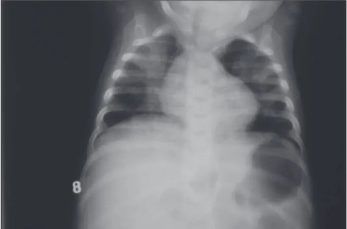
protein of 4.1 g/dL (albumin: 2.9 g/dL and globulin: 1.2 g/dL) were found. Chest radiography showed an inil-trate in the apical regions in both lungs (Figure 1). he irst diagnosis was community-acquired pneumonia. hus, empiric antibiotic therapy was started with ampicillin gentamicin (5 mg/kg/day). Four days later, his intestinal presentation had improved, but he still presented crackles and rhonchi in the pulmonary auscultation. hirteen days later, he started presenting febrile peaks and a new chest radiograph was requested, still showing opacities in the parenchymal of both lungs (Figure 1).
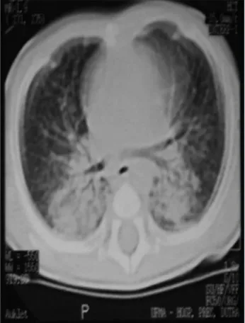
One week later, he was having daily febrile peaks de-spite the antibiotics. Requests were made for a chest com-puted tomography (CT); a gastric lavage for tuberculosis research (three samples), which came negative; and an-other hemogram and CRP, that were normal. CT showed extensive consolidations and geographic ground glass opacities in both lungs, without any hilar lymphadenopa-thy, volume loss, mass, or airway compression (Figure 2).
Bronchoscopy with bronchoalve olar lavage (BAL) was performed and 10 mL of an opalescent luid were divided in three samples. he samples were centrifuged and then analyzed using hematoxylin and eosin staining.
136
IMAGEINMEDICINE
Rev Assoc Med Bras 2012; 58(2):135-137
he microscopic exam showed amorphous material, with inlammatory, red blood, cylindrical, and epithelial cells isolated and grouped. he exam also displayed frequent macrophages, some of then xanthomized and with lipid vacuoles (Figure 3). hus, with the bronchoscopy, the diag-nosis of LP was made. Our patient was in a stable condition and therefore discharged from hospital one month later, but with instructions to take prednisone 1 mg/kg/day for 30 days. Follow-up was conducted at the hospital pulmon-ology service. he follow-up chest radiographies showed a partial decrease of the parenchymal lesion, and the child was in good condition, without any clinical manifestations.
DISCUSSION
Lipoid pneumonia can be classiied as endogenous, idio-pathic or exogenous. Endogenous LP, also called choles-terol pneumonitis, is less frequent and generally observed in patients with chronic bronchial obstruction of the air-ways by tumors, bronchiolitis obliterans, and lipid storage diseases such as Gaucher’s disease and Niemann-Pick dis-ease. Idiopathic LP is a rare disorder and has been associ-ated with smoking in healthy individuals. Although exog-enous LP is well documented in both pediatric and adult medical literature, it also occurs in the pediatric popula-tion as a rare condipopula-tion caused by aspirapopula-tion of mineral, vegetable, or animal oils. he use of mineral oil in the treatment of constipation or as an adjuvant treatment for partial bowel obstruc tion by Ascaris lumbricoides has
been well accepted because of its eicacy and infrequent side efects1-3.
In general, children present a widespread acute aspi-ration condition which progresses to respiratory failure and, sometimes, death. he presence of increasing dys-pnea, cough, chest pain, fever, and vomit, together with alveolar iniltrates in the chest X-rays and the previous ac-cidental ingestion of an oily substance, should be cause for suspicion of exogenous lipoid pneumonia. Moreover, the presence of oil in the lungs predisposes to recurrent infec-tions, and in rare cases can be complicated by atypical my-cobacterial and cryptococcal infection. hese infections result in a bronchoalveolar macrophage response, fol-lowed by the release of inlammatory mediators, leading to fever, leukocytosis, neutrophilia, and elevated eryth-rocyte sedimentation rate. Due to its nonspeciic clinical presentation and radiographic signs, exogenous LP can mimic other pulmonary diseases, such as bacterial pneu-monia, tuberculosis, cystic ibrosis, bronchiectasis, and tumors2,4,5.
Currently, analysis of the bronchoalve olar lavage (BAL) luid is consid ered the diagnostic method of choice for suspected cases of LP, and only the cytochemical ex-amination using staining can conirm the diagnosis6. Both
Figure 2 – Computed tomography (CT) of the chest showing extensive consolidations and geographic ground glass opaci-ties in both lungs.
137
EXOGENOUSLIPOIDPNEUMONIAINCHILDREN: ADISEASETOBEREMINDEDOF
Rev Assoc Med Bras 2012; 58(2):135-137
high resolution computed tomography (HRCT) and mag-netic resonance imaging have showed suggestive signs of lipid iniltration of pulmonary parenchyma. However, HRCT is the best imaging modality for the diagnosis of exogenous LP5,7,8.
In a recent review article, Marchiori et al.5 reported that
there are still no studies in the literature that establish the best therapeutic option for LP. Nevertheless, the key pro-cedure is interrupting the use of mineral oil, which leads to signiicant clinical improve ment. Due to the fact that the depuration of the inhaled oil is a slow process, the best therapeutic strategy would be to withdraw the oil as soon as possible through bronchoscopy with multiple BALs6.
Gondouin et al.9, in a retrospective multicenter study,
found that corticosteroid therapy has not been very ef-fective for all cases of lipoid pneumonia. he use of sys-temic corticosteroids in patients without clinical symp-toms remains controversial. However,whole lung lavage, thoracoscopy with surgical debridement, and systemic corticosteroids have been used in patients with difuse pulmonary damage. Corticosteroid therapy has also been suggested in cases in which functional and radiologi-cal alterations continue despite cliniradiologi-cal improvement5,10.
Even though some retrospective case series demonstrated no beneit in the use of corticosteroids, the patient had a good response ater this therapy, and remained asymp-tomatic at follow-up visits.
he present case emphasizes the need for increased awareness among caregivers and pediatricians about the potential hazards of mineral oil use for chronic constipa-tion. Currently, despite no consensus on treatment, bron-choalveolar lavage seems to be an eicient therapeutic measure for the clearance of mineral oil from the lung parenchyma. his procedure may contribute to preventing ibrosis and reducing the morbidity of lipoid pneumonia, which remains a rare diagnosis.
REFERENCES
1. Sias SM, Ferreira AS, Daltro PA, Caetano RL, Moreira JS, Quirico-Santos T. Evolution of exogenous lipoid pneumonia in children: clinical aspects, ra-diological aspects and the role of bronchoalveolar lavage. J Bras Pneumol. 2009;35(9):839-45.
2. Khilnani GC, Hadda V. Lipoid pneumonia: an uncommon entity. Indian J Med Sci. 2009;63(10):474-80.
3. Hadda V, Khilnani GC. Lipoid pneumonia: an overview. Expert Rev Respir Med. 2010;4(6):799-807.
4. Schwaiblmair M, Berghaus T, Haeckel T, Wagner T, Scheidt W. Lip-oid pneumonia - an underestimated syndrome. Dtsch Med Wochenschr. 2010;135(1-2):27-31.
5. Marchiori E, Zanetti G, Mano CM, Hochhegger B. Exogenous lipoid pneumo-nia. Clinical and radiological manifestations. Respir Med. 2011;105(5):659-66. 6. Sias SM, Daltro PA, Marchiori E, Ferreira AS, Caetano RL, Silva CS, et al.
Clinic and radiological improvement of lipoid pneumonia with multiple bron-choalveolar lavages. Pediatr Pulmonol. 2009;44(4):309-15.
7. Marchiori E, Zanetti G, Mano CM, Irion KL, Daltro PA, Hochhegger B. Lipoid pneumonia in 53 patients ater aspiration of mineral oil: comparison of high-resolution computed tomography indings in adults and children. J Comput Assist Tomogr. 2010;34(1):9-12.
8. Betancourt SL, Martinez-Jimenez S, Rossi SE, Truong MT, Carrillo J, Erasmus JJ. Lipoid pneumonia: spectrum of clinical and radiologic manifestations. AJR Am J Roentgenol. 2010;194(1):103-9.
9. Gondouin A, Manzoni P, Ranfaing E, Brun J, Cadranel J, Sadoun D, et al. Exog-enous lipid pneumonia: a retrospective multicentre study of 44 cases in France. Eur Respir J. 1996;9(7):1463-9.