First trimester pregnancy abnormalities: iconographic
essay*
Anormalidades do primeiro trimestre da gravidez: ensaio iconográfico
Lívia Teresa Moreira Rios1, Ricardo Villar Barbosa de Oliveira2, Marília da Glória Martins3, Kemuel Pinto Bandeira4, Olga Maria Ribeiro Leitão4, Graciete Helena Nascimento Santos5, Márcia Silva Sousa5
First trimester pregnancy abnormalities may be detected by transvaginal ultrasonography at routine examinations or in cases where abnormal vaginal bleeding is present. Threatened miscarriage is frequently observed in the first trimester, occurring in more than one-third of pregnancies. The advent of high-resolution transvaginal ultrasonography has revolutionized the understanding of the pathophysiology and the management of early pregnancy. This method represents an essential tool for determining the pregnancy viability in cases of threatened miscarriage. An expectant approach in the management of miscarriage could significantly reduce the number of unnecessary evacuations of retained products of conception, depending on the adopted criteria. Keywords: Miscarriage; Pregnancy abnormalities; Transvaginal ultrasonography.
As anormalidades do primeiro trimestre da gravidez são detectadas pela ultrassonografia transvaginal em exame de rotina ou em caso de sangramento vaginal anormal. A ameaça de abortamento é uma afecção comum no primeiro trimestre da gestação, ocorrendo em mais de um terço dos casos. O advento de sondas vaginais de alta resolução vem revolucionando nossa compreensão da fisiopatologia e o manejo da gestação inicial. Trata-se de ferramenta essencial para determinar a viabilidade da gestação nos casos de ameaça de abortamento. Uma conduta expectante no abortamento poderia reduzir significativamente o número de es-vaziamentos desnecessários de produtos retidos, dependendo dos critérios utilizados.
Unitermos: Abortamento; Anormalidades; Ultrassonografia transvaginal. Abstract
Resumo
* Study developed at Unit of Obstetrics and Gynecology, Hos-pital Universitário da Universidade Federal do Maranhão (HU-UFMA), São Luís, MA, Brazil.
1. Master, Coordinator for the Imaging Clinic at Unit of Obstet-rics and Gynecology, Hospital Universitário da Universidade Fe-deral do Maranhão (HU-UFMA), São Luís, MA, Brazil.
2. Master, MD, Imaging Clinic at Unit of Obstetrics and Gyne-cology, Hospital Universitário da Universidade Federal do Maranhão (HU-UFMA), São Luís, MA, Brazil.
3. PhD, Associate Professor, Head of Unit of Obstetrics and Gynecology, Hospital Universitário da Universidade Federal do Maranhão (HU-UFMA), São Luís, MA, Brazil.
4. Specialists, Titular Members of Colégio Brasileiro de Radio-logia e Diagnóstico por Imagem (CBR), MDs, Imaging Clinic of Unit of Obstetrics and Gynecology at Hospital Universitário da Univer-sidade Federal do Maranhão (HU-UFMA), São Luís, MA, Brazil. 5. Masters, MDs, Unit of Obstetrics and Gynecology at Hospi-tal Universitário da Universidade Federal do Maranhão (HU-UFMA), São Luís, MA, Brazil.
Mailing address: Dra. Lívia Teresa Moreira Rios. Avenida do Vale, L-10, Q-35, Ed. Costa Rica, ap. 801, Jardim Renascença. São Luís, MA, Brazil, 65075-820. E-mail: [email protected] Received July 28, 2009. Accepted after revision February 22, 2010.
early or partial gestational trophoblastic disease, when other diagnoses may be sug-gested.
The present iconographic essay is aimed at demonstrating the main sonographic cri-teria already established in the literature that characterize most conditions at the first trimester of pregnancy, besides discussing their possible diagnostic difficulties.
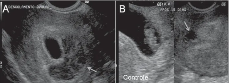
PARTIAL OVULAR DETACHMENT
The maternal circulation inside the pla-centa starts peripherally (in the plapla-cental margins) and is associated to physiological oxidative phenomena that may lead to membranes rupture and formation. The abnormal development of such membranes may result in subchorionic hemorrhage, enhancing the predisposition to an adverse gestational outcome at the third trimester (PPROM and PTL)(4).
Such abnormality is common and also denominated as subchorionic hemorrhage
Rios LTM, Oliveira RVB, Martins MG, Bandeira KP, Leitão OMR, Santos GHN, Sousa MS. First trimester pregnancy abnor-malities: iconographic essay. Radiol Bras. 2010;43(2):125–132.
one third of pregnancies(1). Even in the
presence of fetal heart activity, bleeding in the period between the 7th and 12th weeks is associated to 5–10% of gestational loss at the first trimester, particularly in cases occurring before the 9th week, and in cases where the mother’s age is > 35 years(2,3). A
relation between adverse gestational out-comes such as preterm premature rupture of membranes (PPROM) and preterm labor (PTL) has been reported in cases where the bleeding is observed at the second half of the third trimester of pregnancy(4).
Transvaginal ultrasonography is the technique of choice for evaluating the ges-tational viability(5). Sonographic criteria for
characterization of most conditions at the first gestational trimester are well estab-lished in the literature. A consensus is still to be reached in relation to the cut-off point to be adopted in the measurement of en-dometrial thickness for identifying the presence of retained gestational product. Diagnostic difficulties are usual in cases of INTRODUCTION
hematoma resorption.
The main differential diagnosis for ovu-lar detachment is incomplete fusion of pa-rietal and capsular deciduas, whose texture is anechoic, homogeneous, as a function of
The image identification at the first tri-mester brings confusion with ovular de-tachment, especially in the presence of vaginal bleeding.
remain to be effectively validated . Endometrial thickness < 15 mm, with no evidence of significant endometrial het-erogeneity associated with absence of ab-dominal pain and cessation of the vaginal
Figure 2. Sonographic finding of incomplete decidual fusion. Anecoic, homogeneous image. The gestational sac is not deformed at the 7th (A) and 11th gestational weeks (B).
A
B
in a spontaneous resolution will be higher, requiring surgical evacuation(7).
EARLY EMBRYO DEATH
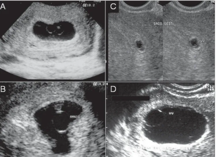
Some sonographic findings characterize an embryo death in the first half of the first trimester in early phases, before the crown-rump length can be measured. The follow-ing aspects are highlighted: small, hype-rechoic yolk sac (Figure 5), or hydropic yolk sac increased in volume with diameter > 7 mm, or even small amniotic cavity dispro-portionate to the gestational sac size. Before the 9th week, small gestational sac may be associated with aneuploidy (Figure 6)(4).
ANEMBRYONIC GESTATION
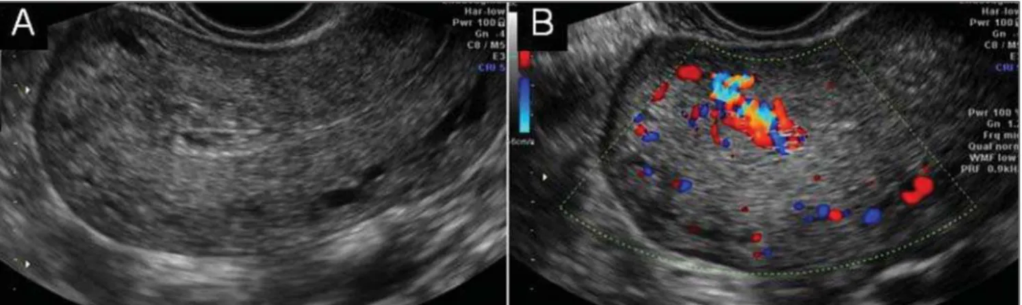
At transvaginal ultrasonography, the yolk sac should be visualized in a gesta-tional sac with mean diameter ≥ 10 mm. Figure 3. Absence of retained products of conception. A: Endometrial thickness of 5 mm with small intracavitary contents corresponding to coagula. B: Focally increased vascularization represented by a small group of vessels extending deeply into the myometrium, site of previous connection.
bleeding should be interpreted as complete miscarriage, without signs of retained prod-ucts of conception(1,4).
Retained products of conception are characterized by a thickened, disorganized and heterogeneous endometrium, with
ill-defined mucosal layers and cavitary line, either with or without the presence of ges-tational sac. Clinically, the women presents abdominal pain and relative vaginal bleed-ing(1,4). In the presence of an intact
gesta-tional sac and closed cervix, the difficulty
Figure 5. Sonographic signs of early embryo death. A: Topic pregnancy with no sign of an embryo, with small, hyperechoic yolk sac. B: Monochorionic, diamniotic twin gestation with early death of one of the embryos (vanishing twin syndrome).
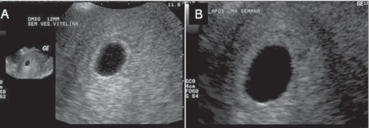
The absence of a yolk sac within the ges-tational sac with ≥ 10 mm in mean diam-eter, or the absence of a yolk sac within the gestational sac with ≥ 16 mm in diameter characterize anembryonic gestation (Fig-ure 7)(1,5).
GESTATIONAL TROPHOBLASTIC DISEASE
The typical sonographic finding in most of cases of complete hydatidiform mole is a echogenic, intracavitary solid mass with intermingled, small cystic loci(8)
resem-bling a “snow storm”, corresponding to the vesicles that macroscopically characterize this condition.
The higher the gestational age, the larger the vesicles visualized as homoge-neous anechoic images, increasing the method specificity. The ultrasonography
sensitivity will depend on the gestational age at the moment of the diagnosis. Ultra-sonography can detect vesicles with > 2 mm in diameter (Figure 8). In early preg-nancies with trophoblastic disease, the sonographic method accuracy is limited, hindering the differentiation of gestational trophoblastic disease from other conditions involving the endometrial cavity.
Partial hydatidiform mole offers higher diagnostic difficulty by ultrasonography. In a reasonable number of cases, this disease presents as an empty gestational sac corre-sponding to anembryonic gestation, or as early embryo death (Figure 9). However, two criteria have been described in the lit-erature: gestational sac transverse/antero-posterior diameter ratio > 1,5 and cystic changes, irregularity of increase in echo-genicity of decidual/placenta or myometrial reaction (Figure 10)(9).
Myometrial nodules similar to intrac-avitary findings can be identified, demon-strating a low impedance flow velocity wave. Low resistivity indices are correlated with high levels of β-hCG. In cases of tro-phoblastic disease, velocimetric changes may precede by weeks the peak serum lev-els of β -hCG(10,11). Doppler US studies of
the uterine arteries and of the myometrial lesion site must be performed.
Uterine artery Doppler velocimetry is useful in the diagnosis, prognosis and fol-low-up of trophoblastic disease. In case of persistent disease, the vascular resistivity remains reduced in association with high hormone levels. On the other hand, color or amplitude Doppler US plays a signifi-cant role in facilitating the identification and determination of myometrial implants extent, besides allowing the diagnosis of arteriovenous malformations associated Figure 6. Sonographic signs of early embryo death. A,B: Small amniotic cavity. C: Delayed growth of the gestational sac with disproportionate yolk sac. D:
Figure 9. Partial hydatidiform mole. Thick, irregular trophoblast, with sonographic signs suggesting anembryonic gestation. Histopathological study demon-strated the presence of molar tissue in the evacuation material.
Figure 7. Sonographic signs of anembryonic gestation. A: Gestational sac with 12 mm in mean diameter, without yolk sac. B: One week later, the gestational sac remains without a yolk sac.
with trophoblastic disease that usually de-velop with low serum levels of β-hCG (Fig-ure 11).
Despite the attempts to define the main sonographic findings of gestational tropho-blastic disease, the gold standard in the diagnosis is the histopathological study of the product of conception following the surgical evacuation(9).
ECTOPIC PREGNANCY
Sonographic findings of ectopic preg-nancy will vary as a function of the gesta-tional age and site.
Classically, the following sonographic findings are described: tubal ring sign, ad-Figure 8. Sonographic signs of complete hydatidiform mole. Abdominal and transvaginal US study
Figure 10. Sonographic signs of partial hydatidiform mole. Focal thickening of the placental bed with predominance of cystic areas and irregularity. Embryo or embryonic remains (arrows) can be visualized.
nexal disorganized mass molded to the adnexa and/or cul de sac, solid, organized mass with regular margins mimicking a pediculated myomatous nodule, clinically progressing with low β-hCG levels (Fig-ures 12 and 13), and presence of a live
ex-trauterine conceptus. Uncommon gesta-tional sites may be observed such as ab-dominal ectopic pregnancy, cervical ec-topic pregnancy and ecec-topic pregnancy in a previous Cesarean section pregnancy (Figure 14).
CONCLUSION
Figure 12. Sonographic findings of ectopic pregnancy. A: Tubal ring sign (gestational sac in the adnexa). B: Adnexal mass.
Figure 13. Sonographic finding of ectopic pregnancy. Solid, isoechoic adnexal, organized mass mimicking a pediculated myomatous nodule.
Figure 14. Uncommon sites of ectopic pregnancy. A: Gestational sac implanted in the cervical region. B: Gestational sac implanted on a previous Cesarean section scar.
REFERENCES
1. Condous G, Okaro E, Bourne T. The conservative management of early pregnancy complications: a review of the literature. Ultrasound Obstet Gynecol. 2003;22:420–30.
2003;21:62–5.
6. Mantoni M. Ultrasound signs in threatened abor-tion and their prognostic significance. Obstet Gynecol. 1985;65:471–5.
7. Nielsen S, Hahlin M, Möller A, et al.