7 artigo 626
ORIGINAL ARTICLE
1 – Master’s degree and Doctor’s degree in Medicine; Coordinator, Knee Surgery Department, Professor Nova Monteiro Orthopedics and Traumatology Clinic, Hospital Municipal Miguel Couto (SOT-HMMC) – Rio de Janeiro, RJ, Brazil.
2 – Collaborating doctor at the SOT-HMMC – Rio de Janeiro, RJ, Brazil.
3 – Master’s degree in Medicine; Coordinator, Medical Residency Program of the SOT-HMMC – Rio de Janeiro, RJ, Brazil. 4 – Master’s degree in Medicine; Head of the SOT-HMMC – Rio de Janeiro, RJ, Brazil.
5 – Master’s degree and Doctor’s degree in Medicine; Head, Orthopedics Clinic, Santa Casa da Misericórdia do Rio de Janeiro – Rio de Janeiro, RJ, Brazil.
Study conducted at the Professor Nova Monteiro Orthopedics and Traumatology Clinic of the Hospital Municipal Miguel Couto (SOT-HMMC) – Rio de Janeiro.
Correspondence: Av. Henrique Dodsworth, 83/105, Copacabana – Rio de Janeiro, RJ. Email: [email protected]
Received for publication: 11/15/2011, accepted for publication: 2/7/2012.
ePidemiological sTudy on Tendon ruPTures of The knee
exTensor mechanism aT a leVel 1 hosPiTal
Rodrigo Pires e Albuquerque1, Juliano Prado2, Rafael Hara2, Evaldo Ferreira2, Leonardo Schiavo2, Vincenzo Giordano3, Ney Pecegueiro do Amaral4, João Mauricio Barretto5
ABSTRACT
Objectives: The purpose of the present study was to review the epidemiological aspects of tendon ruptures of the knee extensor apparatus at a level 1 hospital. Methods: We retrospectively analyzed 76 lesions of the knee extensor apparatus that were treated surgically at the Miguel Couto Municipal Hospital be-tween March 2004 and March 2011. We took into consideration age, sex, trauma mechanism, anatomical classification of the lesion, affected side, comorbidities and associated lesions. Re-sults: Among the patients studied, 68 were male and the mean
The authors declare that there was no conflict of interest in conducting this work
This article is available online in Portuguese and English at the websites: www.rbo.org.br and www.scielo.br/rbort
Rev Bras Ortop. 2012;47(6):719-23 age was 36 years. Regarding the trauma mechanism, 62 lesions occurred due to direct trauma; the right side was affected in 21 cases; eight presented comorbidities and four presented associa-ted lesions. Conclusion: The majority of the patients were male, at an economically active age (young people), and were victims of direct trauma. Ruptures of the patellar ligament were the most frequent lesions. Associated lesions were rare and comorbidities were infrequent in our sample.
Keywords– Epidemiology; Knee; Rupture
INTRODUCTION
Extensor mechanism injuries of the knee are rare. There are numerous comorbidities that predispose to the occurrence of this type of injury. Excessive sports activity and the chronic use of certain medications also facilitate tendon ruptures.
Epidemiological studies are fundamental tools for understanding the occurrence of the injury. In this stu-dy, we observe age, sex, and the most frequent type of injury, as well as the risk factors that should be observed in the prevention of extensor mechanism injuries of the knee. The aim of this work is to carry out a retrospective epidemiological study of tendon ruptures of the extensor mechanism of the knee at a level I trauma hospital.
METHODS
Seventy-six knee extensor mechanism injuries, treated surgically at a level I trauma hospital in the
Figure 1 –Distribution by sex.
Source: Hospital Municipal Miguel Couto, 2011.
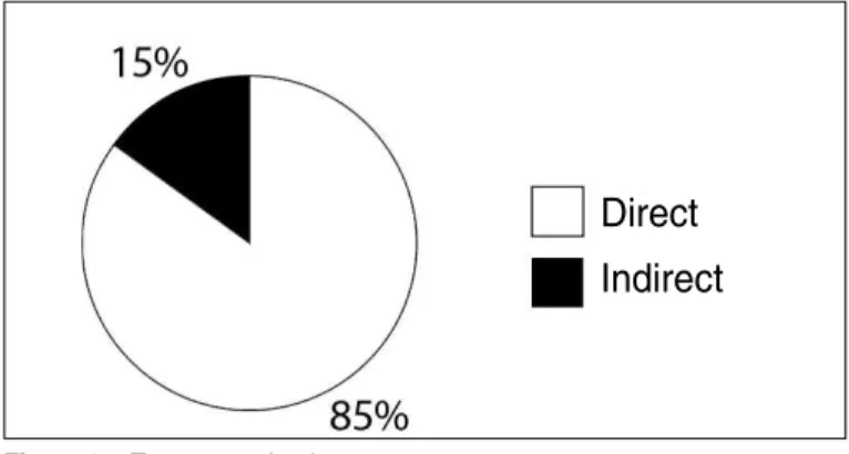
Figure 2 –Trauma mechanism.
Source: Hospital Municipal Miguel Couto, 2011.
Figure 3 –Affected region.
Source: Hospital Municipal Miguel Couto, 2011.
Figure 4 –Affected side.
Source: Hospital Municipal Miguel Couto, 2011.
Figure 5 –Site of the injury.
Source: Hospital Municipal Miguel Couto, 2011.
and anatomical classification of the lesion were taken into consideration. Extensor mechanism injuries of the knee are classified as: rupture of the quadriceps tendon, fracture of the patella, rupture of the patellar ligament, and fracture of the tibial tuberosity(1). All the patient records and radiographs in anteroposterior and profile projections of the knee were assessed by a doctor with a PhD, member of the Brazilian Society of Knee Surgery. The results were assessed by mean and frequency of occurrence.
Of the 76 patients assessed, three were excluded from the analysis because their medical records and/or imaging exams had incomplete data in relation to the topics considered in the present study. This exclusion was due to failure to obtain information relating to the trauma mechanism of the injury. During the period of the study, there were no individuals treated by non-surgical means. In all, 73 patients were effectively included in our analysis. This study is in accordance with the Helsinki Declaration of the World Medical Association.
RESULTS
Of the 73 patients, 68 were male and five were female (Figure 1), and the mean age was 36 years (minimum of 10 and maximum of 80 years). In rela-tion to the mechanism of the injury, 62 were by direct trauma and 11 were by indirect mechanism (Figure 2); 39 patients presented rupture of the patellar ligament, versus 34 with rupture of the quadriceps tendon (Figu-re 3); 52 cases we(Figu-re on the left side, and 21 we(Figu-re on the right side (Figure 4); 29% of the injuries occurred in the muscle belly, 26% in the lower patellar pole, 22% in the osteotendinous junction, 12% in the tibial tuberosity, 7% in the tendon substance, and 4% were sleeve fractures (Figure 5); eight patients presented
Male
Female
Direct
Indirect
Patellar
Quadriceps
Left
Right
Muscle belly 29% Lower patellar pole 26%
Osteotendinous junction 22% Sleeve fracture 4% Tibial tuberosity 12%
Rev Bras Ortop. 2012;47(6):719-23 measurements were not included in the analysis. Our view was that these measurements would be inade-quately collected, generating a research bias.
The ruptures of the patellar ligament were classi-fied according to the site of the lesion: lower patellar pole, ligament substance, and tibial tuberosity(5). Our findings demonstrated that the lower patellar pole was the most frequent site, which corroborates with the findings in the literature(6). In the quadriceps lesions, the most frequent site was the muscle belly. Ilan et al(7) mention the osteotendinous junction and the mus-cle belly as the most common sites, corroborating with our findings.
Injuries in younger patients are generally related to physical activities, generating repetitive microtrau-mas. In elderly patients, meanwhile, the main causal factor is degeneration of the tendon, causing it to be-come weak(5). The findings in group also agreed with this assertion. In our opinion, tendinopathies caused by excessive use, in young patients, should be better assessed and treated.
Ruptures of the patellar ligament occur in patients aged under 40 years, while ruptures of the quadriceps tendon are more common in patients over 40 years(5). Our research confirms these data.
Bilateral lesions of the knee extensor apparatus are largely related to the presence of comorbidities(7). We did not observe this fact in this study.
There have been studies on structural changes in the tendon arising from microtraumas or degenera-tion of the tendon, leading to traumatic ruptures(8,9). On the other hand, other researchers defend the view that direct traumatism of the knee is the cause of pa-tellar lesions in healthy patients(10). In this research, we observed a higher occurrence of patellar ligament rupture in young adults with no previous complaints or systemic diseases. For this reason, we defend direct trauma as the mechanism of injury, as demonstrated by Cree et al(11). However, we agree that structural changes increase the risk of these injuries.
In the immature skeleton, the muscles, ligaments and tendons are generally stronger than the growth plates. For this reason, it is rare to find rupture of the tendon substance in children or adolescents. In the proximal region of the patellar ligament, the most common lesion is sleeve fracture, while in the distal region, we saw tibial tuberosity avulsion(12). At our hospital, because it is a level I emergency hospital, we comorbidities, two with bilateral lesions, and four
with associated lesions. Chart 1 shows the general sample of the case series.
DISCUSSION
The knee extensor mechanism injuries include patellar fractures and tendon lesions of the extensor apparatus. Patellar fractures are more frequent than tendon ruptures, with rates of 17/1 and 43/1, respecti-vely(2). For this reason, patellar fractures were exclu-ded, in order to better understand the epidemiology of tendon ruptures of the extensor mechanism of the knee. There is no literature to date on the epidemio-logy of knee extensor mechanism injuries.
Clayton and Court-Brown(3) carried out an epide-miological study on ligament and tendon lesions of the musculoskeletal system. This research observed a percentage of 0.6 of rupture of the patellar ligament and 1.3 of rupture of the quadriceps tendon. These results confirm our findings that these are very rare injuries, even in a level I trauma clinic.
Rupture of the patellar ligament is the third most common cause of injury of the knee extensor mecha-nism, surpassed in number only by patellar fractures and rupture of the quadriceps tendon. It is estimated that a force of 17.5 times the body weight is needed to cause a rupture of the patellar ligament in healthy indi-viduals(4). In our study, we found a higher occurrence of rupture of the patellar ligament compared with that of the quadriceps.
In our case series, males were more commonly affected, corroborating with the literature in regard to the prevalence of males over females(3). Our view is that males, because they are physically stronger, are more susceptible to rupture of the knee extensor me-chanism. Meanwhile, females have greater ligament la-xity and hormonal changes due to the menstrual cycle.
Chart 1 – General sample of the case series.
Patient number Age Sex Affected side Injury Injury mechanism Location Comorbidities Associated injuries
1 35 M R Patellar Direct trauma Lower patellar pole No No
2 40 M L Quadriceps Direct trauma Osteotendinous junction No No
3 65 M R Patellar Direct trauma Lower patellar pole No No 4 34 M R Patellar Direct trauma Lower patellar pole No No
5 34 M L Patellar Direct trauma Lower patellar pole No No
6 48 M L Quadriceps Direct trauma Muscle belly No No 7 45 M R Quadriceps Direct trauma Osteotendinous junction Renal failure No
8 45 M L Quadriceps Direct trauma Osteotendinous junction Renal failure No
9 19 M R Patellar Direct trauma Tibial tuberosity No No
10 42 F L Quadriceps Direct trauma Osteotendinous junction No No
11 56 M R Quadriceps Direct trauma Osteotendinous junction No No
12 28 M L Quadriceps Direct trauma Osteotendinous junction No No
13 33 M L Patellar Direct trauma Lower patellar pole No No
14 48 F R Patellar Direct trauma Lower patellar pole No No 15 42 M R Quadriceps Direct trauma Osteotendinous junction No No
16 52 M L Patellar Direct trauma Tendon substance No No
17 57 M L Quadriceps Direct trauma Osteotendinous junction No No
18 54 M L Patellar Direct trauma Lower patellar pole No No
19 80 M L Quadriceps Direct trauma Muscle belly No No
20 49 M L Patellar Indirect trauma Tibial tuberosity No No
21 10 M R Patellar Direct trauma Sleeve fracture No No
22 15 M L Patellar Direct trauma Tibial tuberosity No No
23 25 M R Quadriceps Direct trauma Muscle belly No No
24 35 M L Quadriceps Indirect trauma Osteotendinous junction No No
25 34 M L Quadriceps Direct trauma Muscle belly No No
26 74 M L Quadriceps Indirect trauma Osteotendinous junction Diabetic No
27 37 M L Quadriceps Direct trauma Muscle belly No No
28 46 M L Quadriceps Direct trauma Osteotendinous junction No No
29 22 M L Patellar Direct trauma Tendon substance Exposed Ulnar fracture
30 42 M R Quadriceps Direct trauma Muscle belly No No
31 36 M L Quadriceps Indirect trauma Muscle belly No No
32 38 M R Patellar Indirect trauma Lower patellar pole No No
33 30 M R Quadriceps Indirect trauma Osteotendinous junction No No
34 35 M L Patellar Direct trauma Lower patellar pole No No
35 52 M L Quadriceps Indirect trauma Osteotendinous junction Diabetic No
36 45 M L Quadriceps Direct trauma Muscle belly No No
37 26 M R Patellar Direct trauma Muscle belly No Tibial fracture
38 24 M R Quadriceps Direct trauma Muscle belly No Exposed
39 34 F R Patellar Direct trauma Lower patellar pole No No 40 20 M L Patellar Direct trauma Lower patellar pole No No
41 66 M L Quadriceps Indirect trauma Osteotendinous junction Diabetic No
42 48 M R Quadriceps Indirect trauma Muscle belly No No
43 40 M L Patellar Direct trauma Lower patellar pole No No 44 49 M L Patellar Direct trauma Lower patellar pole Renal failure No
45 49 M R Quadriceps Direct trauma Muscle belly Renal failure No
46 62 M L Quadriceps Indirect trauma Muscle belly Diabetic No 47 47 M L Patellar Direct trauma Lower patellar pole No No
48 38 M L Quadriceps Direct trauma Osteotendinous junction No No
49 29 M L Patellar Direct trauma Muscle belly Exposed Femoral + R tibial fracture
50 23 M L Quadriceps Direct trauma Muscle belly No Femoral + L tibial fracture
51 24 M L Patellar Direct trauma Tendon substance No No
52 47 M L Patellar Direct trauma Lower patellar pole No No
53 69 M L Patellar Direct trauma Tibial tuberosity Diabetic No
54 27 M L Patellar Direct trauma Tendon substance No No
55 27 M R Patellar Direct trauma Tendon substance No No
56 11 M L Patellar Direct trauma Sleeve fracture No No
57 11 M L Patellar Direct trauma Lower patellar pole No No
58 15 M L Patellar Direct trauma Tibial tuberosity No No
59 25 M R Patellar Direct trauma Lower patellar pole No No
60 42 M L Quadriceps Direct trauma Muscle belly No No
61 36 M L Patellar Direct trauma Lower patellar pole No No
62 36 M L Quadriceps Direct trauma Muscle belly No No
63 62 M L Quadriceps Direct trauma Muscle belly Diabetic No
64 23 M L Patellar Direct trauma Lower patellar pole No No
65 12 M L Patellar Direct trauma Sleeve fracture No No
66 53 M L Quadriceps Indirect trauma Muscle belly Lupus No
67 13 M L Patellar Direct trauma Tibial tuberosity No No
68 14 M L Patellar Direct trauma Tibial tuberosity No No
69 13 F L Patellar Direct trauma Tibial tuberosity No No
70 13 F R Patellar Direct trauma Tibial tuberosity No No 71 13 M L Quadriceps Direct trauma Muscle belly No No
72 52 M L Patellar Direct trauma Osteotendinous junction Diabetic No
73 43 M L Quadriceps Direct trauma Muscle belly No No
M: male; F: female; R: right; L: left.
Rev Bras Ortop. 2012;47(6):719-23 saw extremely rare injuries in the immature skeleton
(bilateral avulsion fracture of the tibial tuberosity in a 13-year-old girl, rupture of the patellar ligament and contralateral sleeve fracture in an 11-year-old boy, and rupture of the quadriceps tendon in a 13-year--old youth.
In relation to the imaging exams, the knee radio-graphy (trauma series) provides good accuracy in the diagnostic confirmation, as well as being low in cost. We did not use ultrasound, as it is an examiner--dependent exam. On the other hand, Heyde et al(13) recommend ultrasound in injuries of the knee extensor mechanism. In our view, magnetic resonance imaging is a high-cost imaging exam that is not available in all Brazilian hospitals. As this exam becomes more widespread, it will contribute greatly to the analysis of the condition of the tendon and the structures around the knee. We emphasize that the diagnosis of ruptures of the extensor apparatus of the knee is basically
cli-nical. Imaging exams are complementary exams that assist in the surgical planning.
We believe our case series is representative, and this research is aimed at improving our understanding of the occurrence of this type of lesion, and based on these data, enabling effective prevention. Ramseier et al(14) carried out a study on postoperative functional assessment of extensor apparatus injuries. They report on a small case study, and affirm that future research on this subject is necessary, a view with which we agree and corroborate.
CONCLUSION
The majority of the patients were male, of an eco-nomically active age (young), and victims of direct trauma, with ruptures of the patellar ligament as the most frequent injuries. Associated injuries are rare, and comorbidities were infrequent in our case series.
REFERENCES
1. Newberg A, Wales L. Radiographic diagnosis of quadriceps tendon rupture. Radiology. 1977;125(2):367-71.
2. Kellersmann R, Blattert TR, Weckbach A. Bilateral patellar tendon rupture without predisposing systemic disease or steroid use: a case report and review of the literature. Arch Orthop Trauma Surg. 2005;125(2):127-33.
3. Clayton RA, Court-Brown CM. The epidemiology of musculoskeletal tendinous and ligamentous injuries. Injury. 2008;39(12): 1338-44.
4. Zernicke RF, Garhammer J, Jobe FW. Human patellar tendon rupture. J Bone Joint Surg Am. 1977;59(2):179-83.
5. Enad JG. Patellar tendon ruptures. South Med J. 1999;92(6):563-6. 6. Munakata T, Nishida J, Shimamura T, Ichinohe S, Abe M, Ehara S. Simultaneous
avulsion of patellar apexes bilaterally in a hemodialysis patient. Skeletal Radiol. 1995;24(3):211-3.
7. Ilan DI, Tejwani N, Keschner M, Leibman M. Quadriceps tendon rupture. J Am Acad Orthop Surg. 2003;11(3):192-200.
8. Rosenberg JM, Whitaker JH. Bilateral infrapatellar tendon rupture in a patient
with jumper’s knee. Am J Sports Med. 1991;19(1):94-5.
9. Kannus P, Jozsa L. Histopathological changes preceding spontaneous rupture of a tendon. J Bone Joint Surg Am. 1991;73(10):1507-25.
10. Quintero Quesada J, Mora Villadeamigo J, Abad Rico JI. Spontaneous bilateral patellar tendon rupture in an otherwise healthy patient a case report. Acta Orthop Belg. 2003;69(1):89-92.
11. Cree C, Pillai A, Jones B, Blyth M. Bilateral patellar tendon ruptures: a missed diagnosis. Knee Surg Traumatol Arthrosc. 2007;15(11):1350-4.
12. Moretti B, Notarnicola A, Moretti L, Garofalo R, Patella V. Spontaneous bilateral patellar tendon rupture: a case report and review of the literature. Chir Organi Mov. 2008;91(1):51-5.
13. Heyde CE, Mahfeld K, Stahel PF, Kayser R. Ultrasonography as a reliable diagnostic tool in old quadriceps tendon ruptures: a prospective multicentre study. Knee Surg Traumatol Arthrosc. 2005;13(7):564-8.