ISSN 1517-7076 artigo 11530, pp.16-23, 2014
Autor : PEREIRA, B.L Data de envio: 31/10/13 Data de aceite: 30/06/2014
Titanium bioactivity surfaces obtained
by chemical/electrochemical treatments
Bruno Leandro Pereira 1, Paola Tummler 2, Cláudia E. B. Marino 3, Paulo César Soares4, Neide K. Kuromoto5
1
Laboratório de propriedades nano mecânicas-LabNano – PIPE/UFPR, CP: 81530-900 Curitiba, PR e-mail: [email protected]
2 PGMEC/UFPR, CP: 81530-900 Curitiba, PR; e-mail: [email protected] 3
Departamento de Engenharia Mecânica/ UFPR, CP: 81530-900 Curitiba, PR; [email protected], 4Engenharia Mecânica/ PUC, Curitiba, PR; [email protected]
5
Departamento de Física/UFPR, Curitiba, PR.; [email protected]
ABSTRACT
There are various surface treatments used to modify titanium surfaces to render it bioactive. In this study commercially pure titanium surfaces (cp Ti), grade 2 were modified by acid etching (AE) and anodic oxida-tion (OA) in order to evaluate the bioactivity in vitro of these surfaces using the simulated body fluid (SBF). The AE was realized using a mixture of acids and AO using 1 mol.L-1 sulfuric acid. The anodic films were obtained under potentiostatic mode, during 60s using as anode a bar of titanium. All the surfaces that means cp Ti, AE and AO were analyzed concerning to morphology, rugosity, structural changes before in vitro bio-activity tests. It was observed by scanning electron microscopy (SEM) that all surfaces presented different morphologies: those with AE showed a surface with peaks and rounded valleys, with Ra = (564±80) nm, the oxidized surfaces with sulfuric acid showed a morphology with small pores uniformly distributed over the surface and Ra = (177±0,02) nm. X-rays diffraction results showed the presence of titanium hydride on the samples with AE and the anatase and rutile phases on the anodic films after heat treatment at 600°C/1h. Bio-activity tests in vitro using SBF at 37°C showed that small aggregates containing Ca and P were observed on surfaces with AE after 30 days soaked in SBF and the surfaces oxidized were fully coated with an apatite layer, identified by SEM.
Keywords: Titanium, Anodic oxidation, Bioactivity, acid etching.
1. INTRODUCTION
The titanium (Ti) and Ti alloys are broadly used as dental screws and orthopedic implants due low tendency to corrosion, allied to their good mechanical strength biocompatibility and high osseointegration rate [1, 2]. Ti and its alloys are generally defined as bioinert materials, due to the lack of direct chemical bonding be-tween the bone tissues and the implant [3].
To enhance the anchoring power of the osteoblasts between implant and human body in way to accelerate the apatite nucleation, surface modifications are performed on titanium surface [4-5]. Among the surface treat-ments has been highlighted acid etching and anodic oxidation. Both treattreat-ments results in rough morphology. Anodic oxidation technique presents some advantages such as easily well-adhered oxide film deposited on titanium surface through an electrochemical process. Porous and roughness films can be formed by applying high voltage to produce dielectric breakdown [6-12]. Extensive research in titanium shows that the morphol-ogy, surface chemistry, roughness, rutile and anatase phases significantly influences the apatite-inducing ability [13-18]. The titanium bioactivity can be evaluated using simulate body fluid (SBF) [8]. According to Kokubo and Takadama [8], a surface is bioactive if an apatite layer is formed at the Ti surface soaked in SBF at 37oC during several days. Thus, when the implant is soaking in the body fluid (pH 7.4) the interaction of the bioactive surface and SBF begins, giving origin to Ti-OH groups and the surface becomes slightly nega-tive charged. The neganega-tive surface attracts Ca2+ ions from SBF solution and forms calcium titanate on the Ti surface [6-7], which acts as the nucleation point for apatite deposition.
17
The morphology of the films, before and after soaked in SBF, was observed using a scanning electron mi-croscopy (SEM) (Jeol JSM-6360LV). The average surface roughness (Ra) was obtained using a perfilometer (Veeco Dektak 150). The crystalline phases were analyzed using X-ray diffractometer (Shimadzu XRD-7000) using Bragg Brentano geometry, 40kV, 20mA and radiation Cu Kα. XRD data was investigated using the Crystallographica software.3. RESULTS AND DISCUSSION
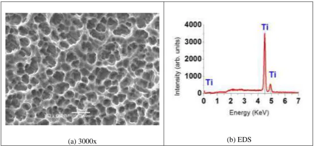
The figure 1.a shows the Ti surface after acid etching process. The SEM image shows a surface with many round craters. The elements present on this surface is showed in EDS spectrum (fig 1.b), where oxygen is not observed, only Ti. The ausence of O is due to acid etching.
(a) 3000x (b) EDS
Figure 1: Titanium surface morphology after acid etching (a), and EDS spectrum identifying the elements present on the
surface (b).
18
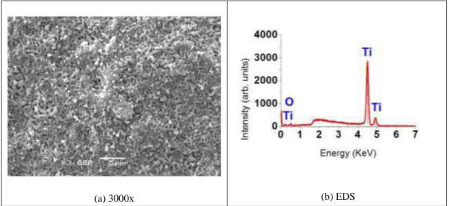
(a) 3000x (b) EDS
Figure 2: Ti Surface morphology after anodic oxidation in 1 mol.L-1 sulfuric acid electrolyte (160V- 60s) plus heat
treatment (a) with the respective EDS (b).
The results concerning to rugosity measurements are showed in table 1. It can be observed that the rugosity values increased with the AE. According to Curtis et.al, the rugosity is an important parameter in the orienta-tion of the cells in bone osseointegraorienta-tion process once the contact area is increased [18], and the rugosity affects adhesion strength of apatite layer grown over treated surface [19].
Table 1: Rugosity (Ra) measurements on the Ti surface with different treatments.
SAMPLE RUGOSITY UNIT
Grounded Ti 2880.04 nanometers (nm)
Acid etching 5640.08 nanometers (nm)
Anodic oxidation + heat treatment 1770.02 nanometers (nm))
19
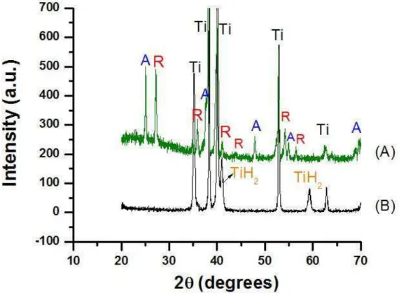
Figure 3: XRD patterns of (A) Anodic oxidation in H2SO4 1Mol.L-1, 160 V, during 60 s, followed by heat treatment (B)
Acid etched titanium surface. A= anatase R= rutile TiH2= titanium hydride.
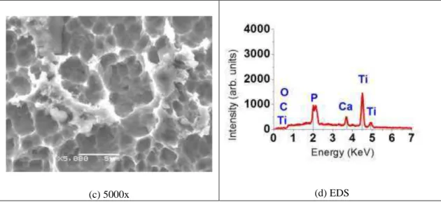
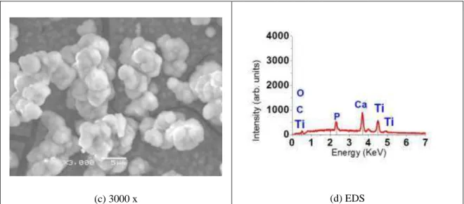
After soaked in SBF during 30 days at 37°C, the samples was evaluated using SEM, EDS and x-ray diffrac-tion. In the acid etching surface, it was observed a small precipitates nucleated on the porous surface (figure 4a-c). The EDS analysis showed that these precipitates contain Ca and P (fig. 4d).
20
(c) 5000x (d) EDS
Figure 4: Surface morphology of acid etching in titanium, after 30 days soaking with the respective EDS.
SEM images of the anodized surface soaked in SBF showed a new layer covering the entire surface (fig. 5). The EDS results indicated the presence of the Ca and P in this layer. This layer corresponds to apatite, as identified by X-ray diffraction technique, showed in figure 6. According to Kokubo, a surface may be con-sidered bioactive, when an apatite layer grows on the surface when soaked in SBF [8]. Nucleation of apatite occurs from the consumption of Ca and P ions that are in the SBF solution. The responsible for the nucleation start process are the Ti-OH groups on the TiO2 layer. These groups exhibit amphoteric character when they are in different pH range 5-6 (isoelectric point) [4]. Thus, when soaking in the SBF (pH 7.4), the surface is slightly negative charged. The negative surface attracts Ca2+, and then the Ca ions absorbed PO4 ions from the solution to form apatite on the surface. These results indicate that the oxidized surface is more bioactive than the acid etching surface, as was to be expected because surface features like as thicker oxide layer, with crystalline phases.
21
(c) 3000 x(d) EDS
Figure 5: Surface morphology of anodic oxidation in sulfuric acid (1 Mol.L-1 - 160V- 60s) soaking in SBF during 30
days (a-c) with the respective EDS (d).
Figure 6: XRD patterns of anodized Ti surface in H2SO4 1Mol/L, 160 V/60s plus heat treatment after soaked in SBF
during 30 days. Ha= hydroxyapatite Ti= titanium.
4. CONCLUSIONS
22
5. ACKNOWLEDGMENTAcknowledgment to CAPES, Fundação Araucária, Centro de Microscopia- UFPR and LORXI.
6. REFERENCES
[1] NIINOMI, M., “Mechanical properties of biomedical titanium alloys”, Materials Science and Engineer-ing: A, v. 243, pp. 243-231, Mar. 1998.
[2] DAOOD, N. BANDEY, S.B QUASIN, H. OMAR S.A. KHAN, “Surface characterization analysis of failed dental implants using scanning electron microscopy”, Acta Odontologica Scandinavica, v.69, pp. 367-373, Nov. 2011.
[3] LIU X, CHU PK, DING C., “Surface modification of titanium, titanium alloys, and related materials for biomedical applications”, Materials Science and Engineering: R, v.47 pp. 49-121, Dec. 2004.
[4] X.J WANG , Y.C. LI , J.G. LIN , Y. YAMADA , P.D. HODGSON, C.E. WEN, “In vitro bioactivity evaluation of titanium and niobium metals with different surface morphologies”, Acta Biomaterialia, v.4, pp. 1530 –1535, Sept. 2008.
[5] CUI, X., KIM, H.M., KAWASHITA, M., WANG, L., XIONG, T., KOKUBO, T., NAKAMURA, T., “Preparation of bioactive titania films on titanium metal via anodic oxidation”. Dental Materials, v. 25, n.4, pp. 80-86, Jan. 2009
[6] ROHANIZADEH, R., SADEQ, MA. LEGEROS, RZ. , “Preparation of different forms of titanium oxide on titanium surface: effects on apatite deposition”, Journal of Biomedical Materials Research, v. 71A, n.2, pp. 343 –352, Sep. 2004.
[7] TAKADAMA, H, KIM, HM, KOKUBO, T, NAKAMURA, T., “XPS study of the process of apatite for-mation on bioactive Ti–6Al–4V alloy in simulated body fluid”. Science and Technology of Advanced Mate-rials, v.2, n.2, pp.389–96, Jun. 2001.
[8] KOKUBO, T., TAKADAMA, H. “How useful is SBF in predicting in vivo bone bioactivity?” Biomateri-als, v.27, n.15, pp. 2907 - 2915, May 2006.
[9] CHEN, Z.X., TAKAO, W.X., MATSUBARA, T., REM, L.M., “Surface characteristics and in vitro bio-compability of titanium anodized in a phosphoric acid solution at different voltages, “Biomedical Materials, v.4, n.6, 0650032009, October 2009.
[10] PARK,T., CHOE, H., BRANTLEY, W.A., “Bioactivity evaluation of porous TiO2 surface formed on titanium in mixed electrolyte by spark anodization”, Surface and Coatings Technology, v.235, pp. 706-713, Nov. 2013.
[11] SOUZA, G.B., LIMA, G.L., KUROMOTO, N.K., SOARES, P.C., LEPIENSKI, C.M., FOESTER, C.E., MIKOWSKI, A., “Tribo-mechanical characterization of rough, porous and bioactive Ti anodic layers”, Jour-nal of Materials Processing Technology, v.4, n.2, pp. 796-806, Jul. 2011.
[12] SZESZ, E.M., PEREIRA, B.L., KUROMOTO, N.K., MARINO, C.E.B., SOUZA, G.B., SOARES, P.C., “Electrochemical and morphological analyses on the titanium surface modified by shot blasting and anodic oxidation processes”, Thin Solid Films, v.528, n.16, pp. 163-166, Jan. 2013.
[13] SONG, H.J., PARK, S.H., JEONG, S.H., PARK, Y.J., “Surface characteristics and bioactivity of oxide films formed by anodic spark oxidation on titanium in different electrolytes”, Journal of the Mechanical B e-havior of Biomedical Materials, v.209, n.2, pp. 864-870, Jan. 2009.
[14] Zhu, X., KIM, K.H., JEONG, Y., “Anodic oxide films containing Ca and P of titanium biomaterial”, Biomaterials, v.22, n.16, pp. 2199-2206, August 2001.
[15] YANG, B., UCHIDA, M., KIM, H.M., ZHANG, X., “Preparation of bioactive titanium metal via anodic oxidation treatment”, Biomaterials, v. 25, n.6, pp.1003-1010, March 2004.
[16] HEON-SEOK, H., CHANG-WHE, K., YOUNG-JUM, L., MYUG-JOO, K., “Surface characteristics of anodic oxidized titanium according to the pore size”, Journal of Korean Academy of Prosthodontics, v.44, n.3, pp.343-357, Jan. 2006.