This work is licensed under a Creative Commons Attribution 4.0 International License. O R I G I NA L S C I E N T I F I C P A P E R
Croat. Chem. Acta2016, 89(1), 101–104 Published online: May 9, 2016
DOI: 10.5562/cca2860
Tailoring of K
0.8
Al
0.7
Fe
0.15
Si
2.25
O
6
Leucite Based
Dental Ceramic Material
Aleksandar Kremenović,1,* Martin Fabián,2 Predrag Vulić,1 Čedomir Jovalekić,3 Jaroslav Briančin2
1 Laboratory of Crystallography, Faculty of Mining and Geology, University of Belgrade, Đušina 7, 11001 Belgrade, Serbia 2 Institute of Geotechnics, Slovak Academy of Sciences, Watsonova 45, 04001 Košice, Slovakia
3 Institute for Multidisciplinary Studies, University of Belgrade, Kneza Višeslava 1, 11001 Belgrade, Serbia
* Corresponding author’s e-mail address: akremenovic@rgf.bg.ac.rs
RECEIVED: March 17, 2016 REVISED: March 23, 2016 ACCEPTED: March 23, 2016
THIS PAPER IS DEDICATED TO DR.SVETOZAR MUSIĆ ON THE OCCASION OF HIS 70TH BIRTHDAY
Abstract: Potassium based ceramic materials composed from leucite in which 5 % of Al is exchanged with Fe and 4 % of hematite was synthe-sized by mechanochemical homogenization and annealing of K2O-SiO2-Al2O3-Fe2O3 mixtures. Synthesynthe-sized material was characterized by X-ray Powder Diffraction (XRPD) and Scanning Electron Microscopy coupled with Energy Dispersive X-ray spectroscopy (SEM/EDX). The two methods are in good agreement in regard to the specimen chemical composition suggesting that a leucite chemical formula is K0.8Al0.7Fe0.15Si2.25O6. Rietveld structure refinement results reveal that about 20 % of vacancies exist in the position of K atoms.
Keywords: leucite, dental ceramic material, structure, synthesis, ball milling.
INTRODUCTION
N OPTIMIZATION of a material properties i.e. material tailoring for industrial applications is an imperative for successful applications. Detailed knowledge of a mate-rial structure is one step ahead to the final solution. Alka-line metal based ceramic materials are widely used in industry as: electroceramic components,[1] matrixes for
flu-orescent screens,[2,3] thermo-refractory materials,[4,5]
elec-tromagnetic windows,[1,5] dental ceramics[6] etc. So far,
investigations of many alkaline ceramic materials, although started from the beginning of the last century, reveal unex-plained properties and unsolved parts of a material structure. Leucite, KAlSi2O6 is common mineral in some
vol-canic rocks in which it crystallizes with a cubic crystal struc-ture at high temperastruc-ture (ca. 900 °C). Upon cooling to 700– 600 °C, it transforms into a tetragonal modification which is stable at room temperature, and forms characteristic poly-synthetic twin lamellae. The transformation is reversible.[7]
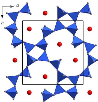
Structurally it belongs to feldspathoids - tectosilicates char-acterized with Al-Si framework structure. Voids within framework are partly filled with K atoms, Figure 1.
A
Figure 1. Polyhedral representation of one pseudolayer in
leucite KAlSi2O6 structure. Blue tetrahedra represent SiO4
102 A. KREMENOVIĆ et al.: Tailoring of K0.8Al0.7Fe0.15Si2.25O6 Leucite Based …
Croat. Chem. Acta2016, 89(1), 101–104 DOI: 10.5562/cca2860
Leucite crystalizes as euhedral pseudocubic crystals in tetragonal I41/a space group. Characteristic unit cell
pa-rameters are: a = 13.056 Å, c = 13.751 Å (a: c = 1:1.053). Inside the unit cell there are 16 asymmetric units (Z=16). Common impurities in natural leucites are: Ti, Fe, Mg, Ca, Ba, Na, Rb, and Cs. Impurities concentrations are rather small in natural leucites. However, leucite structures exist even after complete exchange of Al with Fe.[7]
The optical properties of the leucite glass-ceramic make it one of the most appropriate materials for the fab-rication of dental restorations. The leucite has almost the same refractive index as the glass. Therefore, the translu-cency is never hindered by the crystallization of the leucite in the glass. Leucite based glass-ceramics has ability to match the colour of the natural tooth. Addition of other chemical elements, like iron, could slightly change the
colour of the dental ceramic to desirable hue. Another advantage of the leucite based glass-ceramic materials in dental industry is that due to low thermal expansion coef-ficient its stability during fusion is remarkable.[6]
In order to tailor potassium based ceramic materials with good properties for dental industry we have synthe-sized them by mechanochemical homogenization and an-nealing of K2O-SiO2-Al2O3-Fe2O3 mixtures. Our main goal
was to synthesize leucite in which 10 % of Al is exchanged with Fe and to characterize it by X-ray Powder Diffraction (XRPD) and Scanning Electron Microscopy coupled with En-ergy Dispersive X-ray spectroscopy (SEM/EDX).
EXPERIMENTAL DETAILS
Sample Preparation
The starting compounds were SiO2, Al2O3, K2CO3 and Fe2O3.
They were mixed in appropriate molar ratio according to the stoichiometric formula KAl0.9Fe0.1Si2O6.
Mechanochem-ical treatment was performed during one hour in a plane-tary ball mill (Fritsch Pulverisette 5) equipped with tungsten carbide bowls (250 ml in volume) and balls (10 mm in diam-eter). The mass of the powder was 10 g and the balls-to-powder mass ratio was 20:1. The milling was done in air atmosphere without any additives. The angular velocity of the supporting disc and vial was 32.2 and 40.3 rad s–1,
re-spectively. The intensity of milling corresponded to an ac-celeration of about 10 times the gravitational acac-celeration. The milling vessels were opened for removing of the CO2
which evaporate during milling. After milling the obtained powders were pressed in pellets under pressure of 50 MPa and sintered at temperature of 1100 °C for 24 hours. Than specimens were milled again but not opened for removing of the CO2. At the end specimens were pressed
in pellets under pressure of 50 MPa and sintered at 1100 °C for 24 hours.
Experimental Techniques and Methods
For the collection of the XRPD data a D8 Advance (Bruker, Germany) X-ray powder diffractometer was used. The dif-fractometer was equipped with a Cu-tube and a Xe-filed proportional counter. The divergence and receiving slits were 1 ° and 0.1 mm, respectively. The scanning range was 4–90 ° in 2θ, with a step of 0.03 ° and a scanning time of 22 s per step. The determination of the structural parameters of the K0.8Al0.7Fe0.15Si2.25O6 was carried out using the
Rietveld method implemented in the FullProf program package.[8]
SEM images were recorded using a MIRA3 FE-SEM microscope (TESCAN, Czech Republic) equipped with an EDX detector (Oxford Instruments, UK).
Table 1. Experimental details for XRPD data collection.
Estimated standard deviations are in parenthesis.
Crystal data
Chemical formula K0.8Al0.7Fe0.15Si2.25O6
Mr 217.74
Crystal system, space group Tetragonal, I41/a
Temperature / K 295
a, c / Å 13.1334(3), 13.7343(4)
V / Å3 2368.99(9)
Z 16
Radiation type Cu Kα1, Cu Kα2 radiation,
λ = 1.540562, 1.544390 Å
Specimen shape Irregular
Data collection
Diffractometer Brucker D8 Advance Specimen mounting packed powder pellet Data collection mode Reflection
Scan method Step
2θ values 2θmin = 4.000 °, 2θmax = 90.010 °, 2θstep = 0.030 °
Refinement
Profile function TCH pseudo-Voigt
Background function linear extrapolation between points; 15 points were determined by visual estimation and refined R factors and
goodness of fit RRp = 11.00, Bragg = 6.11, Rwp = 14.10, χ2 = 2.15 Rexp = 9.64, No. of data points 2868
No. of parameters 45 No. of restraints 10
Computing details: Data collection: D8 Software;[11] program used to refine
A. KREMENOVIĆ et al.: Tailoring of K0.8Al0.7Fe0.15Si2.25O6 Leucite Based … 103
DOI: 10.5562/cca2860 Croat. Chem. Acta2016, 89(1), 101–104
RESULTS AND DISCUSSION
Collected XRPD pattern corresponds to reference leucite KAlSi2O6.[9] Few low intensity peaks in the pattern belongto hematite.[10] Quantitative phase analysis indicated that
only 4(1) % of hematite is in the specimen. An amorphous phase could not be recognized by XPRD analysis. Therefore, it is reasonable to assume that part of Fe is incorporated into leucite structure. Obtained unit cell parameters for leucite (a = 13.1334(3) Å and c = 13.7343(4) Å; a: c = 1: 1.046) are different from the reference (a = 13.09(1) Å and
c = 13.75(1) Å; a : c = 1:1.050) indicating that Fe partly ex-changes Al during synthesis procedure. Vacancy creation during mechanochemial treatment is possible. That could be another reason for the unit cell difference compared to the reference material. Moreover, the Rietvled refinement results (Figure 2, Tables 1–3) show that about 5 % of Al is exchanged with Fe and that ca. 20 % of vacancies exist in the position of K atoms. Chemical formula of synthesized compound recalculated from site occupation parameter values is K0.8Al0.7Fe0.15Si2.25O6, Table 2. However, estimated
standard deviations of site occupation parameters are rel-atively high suggesting that they are not reliable for recal-culation of leucite chemical formula. Ionic radii for Si4+, Al3+
and Fe3+ are 0.26, 0.39, and 0.49 Å respectively. Therefore,
interatomic distance values are more reliable than site oc-cupation parameters indicating that T1 atomic site is mostly
occupied with Si while T2 and T3 are occupied with Si, Al and Fe.
Atomic site T3 contains more Al and Fe than other two T sites,
as given in Table 3. Interatomic distances, as well as overall temperature parameters, are in good agreement with lit-erature data.[9] Obtained accuracy parameter values are
reas-onable, indicating reliable refinement, as shown in Table 1.
Morphology of as-prepared sample was determined by SEM analyses. Leucite, K0.8Al0.7Fe0.15Si2.25O6, occurs in
microsphe-rical forms, approximately 0.5–1 µm in size, Figure 3a. Semi-quantitative chemical composition of the synthesized compound calculated from the EDX results, Figures 3b and 3c, K0.8Al0.7Fe0.15Si2.25O6 is in good agreement with XRPD
results. Moreover, elemental mapping shows that all constituent elements are uniformly distributed over the as-prepared sample confirming its homogeneity.
Table 3. Selected interatomic distances (expressed in Å) for
K0.8Al0.7Fe0.15Si2.25O6. Estimated standard deviations are in
parenthesis.
connection interatomic distance connection interatomic distance
T1-O1 1.628(4) K- O1 3.22(1)
T1-O1 1.611(4) K- O2 3.07(1)
T1-O2 1.637(9) K- O3 3.06(2)
T1-O4 1.63(1) K- O4 3.04(2)
<T1-O> 1.626 K- O5 3.00(1)
T2-O2 1.665(8) K- O6 3.00(2)
T2-O3 1.64(1) <K- O> 3.065
T2-O4 1.66(1)
T2-O5 1.643(5) <T2-O> 1.652
T3-O3 1.69(1)
T3-O5 1.665(6)
T3-O6 1.633(2)
T3-O6 1.691(3) <T3-O> 1.670
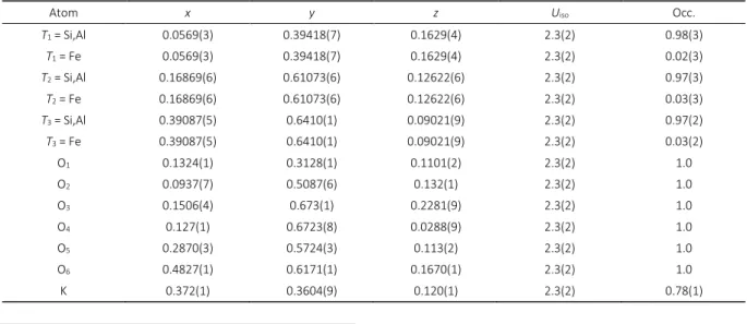
Table 2. Fractional atomic coordinates x, y and z, isotropic displacement parameters Uiso / Å2 and site occupation parameters
Occ. for K0.8Al0.7Fe0.15Si2.25O6. Estimated standard deviations are in parenthesis.
Atom x y z Uiso Occ.
T1 = Si,Al 0.0569(3) 0.39418(7) 0.1629(4) 2.3(2) 0.98(3)
T1 = Fe 0.0569(3) 0.39418(7) 0.1629(4) 2.3(2) 0.02(3)
T2 = Si,Al 0.16869(6) 0.61073(6) 0.12622(6) 2.3(2) 0.97(3)
T2 = Fe 0.16869(6) 0.61073(6) 0.12622(6) 2.3(2) 0.03(3)
T3 = Si,Al 0.39087(5) 0.6410(1) 0.09021(9) 2.3(2) 0.97(2)
T3 = Fe 0.39087(5) 0.6410(1) 0.09021(9) 2.3(2) 0.03(2)
O1 0.1324(1) 0.3128(1) 0.1101(2) 2.3(2) 1.0
O2 0.0937(7) 0.5087(6) 0.132(1) 2.3(2) 1.0
O3 0.1506(4) 0.673(1) 0.2281(9) 2.3(2) 1.0
O4 0.127(1) 0.6723(8) 0.0288(9) 2.3(2) 1.0
O5 0.2870(3) 0.5724(3) 0.113(2) 2.3(2) 1.0
O6 0.4827(1) 0.6171(1) 0.1670(1) 2.3(2) 1.0
104 A. KREMENOVIĆ et al.: Tailoring of K0.8Al0.7Fe0.15Si2.25O6 Leucite Based …
Croat. Chem. Acta2016, 89(1), 101–104 DOI: 10.5562/cca2860
All obtained results, which are in good agreement, suggest that synthesis was quite successful. In the future, a synthesis should be slightly changed in order to obtain pure leucite enriched with Fe, i.e. without hematite.
CONCLUSIONS
Potassium based ceramic material with promising proper-ties for dental industry was tailored by mechanochemical homogenization and annealing of K2O-SiO2-Al2O3-Fe2O3
mixtures. Chemical and mineral compositions, as well as crystal structures, were investigated by X-ray Powder Dif-fraction (XRPD) and Scanning Electron Microscopy coupled with Energy Dispersive X-ray spectroscopy (SEM/EDX). Ob-tained results, which are in good agreement, suggest that synthesis was quite successful. In the future, a synthesis should be slightly changed in order to obtain pure leucite enriched with Fe, i.e. specimen without hematite.
Acknowledgment. The Serbian Ministry of Science has
finan-cially supported this work through grant Nos. 176016, 45015. The Swiss National Science Foundation has finan-cially supported this work under contract No. SCOPES IZ73Z0_127961. M.F. and J.B. thankVEGA (2/0097/13) and APVV (14-0103) for support of their work.
REFERENCES
[1] I. G. Talmy, D. A. Haught, E. J. Wuchina, Proceedings of Sixth International SAMPE Electronics Conference, USA, 1992, 687.
[2] W. B. Im, Y. Kim, D. Y. Yeon, Chem. Mater.2006, 18, 1190. [3] A. Kremenović, Ph. Colomban, B. Piriou, D. Massiot,
P. Florian, J. Phys. Chem. Solids2003, 64, 2253. [4] A. Kremenović, P. Norby, R. Dimitrijević, V. Dondur
Phase Transitions1999, 68, 587.
[5] A. Kremenović, P. Norby, R. Dimitrijević, V. Dondur
Phase Transitions 2004, 77, 955.
[6] E. El-Meliegy, R. van North, Glasses and Glass Ceramics for Medical Applications, Springer, London, 2012, p.p. 167–192.
[7] R. A. Lange, I. S. E. Carmichael, J. F. Stebbins, Am. Mineral.1986, 71, 937.
[8] J. Rodriguez-Carvajal, FULLPROF. Abstracts of the Satellite Meeting on Powder Diffraction of the XVth Congress of the IUCr, Toulouse, France, 1990, p. 127. [9] F. Mazzi, E. Galt, G. Gottardi, Am. Mineral.1976, 61, 108. [10] R. L. Blake, R. E. Hessevick, T. Zoltai, L. W. Finger, Am.
Mineral.1966, 51, 123.
[11] Bruker D8 Software, Bruker AXS Inc., Madison, Wisconsin, USA, 2005.
[12] K. Brandenburg, H. Putz, DIAMOND, Crystal Impact GbR, Bonn, Germany, 2005.
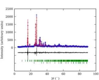
Figure 2. Final Rietvled plot for K0.8Al0.7Fe0.15Si2.25O6 leucite
based ceramic material. Blue crosses denote observed step intensities; the red line represents the corresponding calculated values. The difference curve between observed and calculated values is given at the bottom (black line). Vertical green bars represent diffraction line positions; upper bars correspond to K0.8Al0.7Fe0.15Si2.25O6 leucite, lower
to hematite.
Figure 3. Scanning electron micrograph (a), EDX analysis (b)
and elemental mapping (c) for K0.8Al0.7Fe0.15Si2.25O6 leucite