Received on 2 April 2008; revised 18 September 2008.
Address for correspondence: Dr. Marcus Vinícius T. Fadel. Rua João Evangelista Espíndola 123, Curitiba, PR - Brasil. Zip Code: 82520-070. Phone number: (041)30263456. Fax number: (041)30263456. E-mail: [email protected].
The Brazilian Journal of Infectious Diseases 2008;12(5):416-422. © 2008 by The Brazilian Journal of Infectious Diseases and Contexto Publishing. All rights reserved.
Inadequate Timing Between Corticosteroid and Antibiotics Applications Increases Mortality Due to Sepsis
Marcus Vinícius Telles Fadel1, João Carlos Repka2, Cláudio Leinig Pereira da Cunha1 and Maria Terezinha C. Leão1 1Federal University of Paraná; 2Laboratory of Experimental Microbiology of the Faculdade Evangélica de Medicina do Paraná; Curitiba, PR, Brazil
This study tested the hypothesis that the use of corticosteroids prior to antibiotics can lower the mortality rate in severe infections by S. aureus or Gram-negative bacilli, using an animal model. This study was a prospective and controlled study, placed in a university laboratory. Seven hundred and sixty mice distributed into three groups (Staphylococcus aureus, Escherichia coli and Klebsiella pneumoniae infected). The interventions in each group were: I) infection control (intra-peritoneal); II) treatment solely with antibiotics (teicoplanin or amikacin); III) antibiotics administered prior to the corticosteroid (methylprednisolone); IV) antibiotics administered after the corticosteroid. Mortality in the E. coli group, subgroup I: 100%; subgroup II: 55% (p<0.001); subgroup III: 62.5% (p=0.2488, compared to subgroup II); subgroup IV: 20% (p<0.01 compared to subgroups II and III). Mortality in the K. pneumoniae group: subgroup I: 100%; subgroup II: 72.5% (p<0.01); subgroup III: 80% (p=0.215 compared to subgroup II); subgroup IV: 45% (p<0.01 compared to subgroups II and III). Mortality in the S. aureus group: subgroup I: 82.5%; II: 42.5% (p<0.001); subgroup III: 77.5% (p=0.2877 compared to subgroup I); subgroup IV: 32.5% (p=0.1792 compared to subgroup II). The use of corticosteroids prior to antibiotics lowered the mortality rate caused by Gram-negative bacteria and did not affect the mortality caused by S. aureus. When used after starting treatment with antibiotics, the corticosteroid was not superior to the use of antibiotics alone in the case of the Gram-negative bacteria, and was not significantly different from non-treatment of the infection, in the case of S. aureus.
Key Words: Adrenal cortex hormones, antibiotics, sepsis, survival, mice, animals, laboratory, severe infection, peritonitis, shock, endotoxin.
The current line of thought regarding aggression to the host during severe infections is that the primary determinant is not the activity of the microorganisms themselves, but an uncontrolled response on the part of the host, culminating in a reaction of self-aggression and death. In fact, evidence continues to accumulate implicating numerous biochemical participants included in the collective “septic cascade”; which if not controlled ultimately lead to multiple organ failure [1-4]. The use of suitable anti-microbial medicines, even if they eliminate the microorganisms that triggered the septic process, often will not be enough to prevent the host’s worsening and dying [5].
Several pro-inflammatory cytokines take part in this cascade, among them tumor necrosis factor-alpha (TNF-alpha) seems to be the most important and is the first to appear. TNF-alpha triggers activation of several other mediators, culminating in the metabolic, hemostatic and hemodynamic features of septic shock [6-10]. Although the lipopolysaccharide (LPS) produced by Gram-negative bacilli (GNB) is the most potent stimulus for the production of TNF-alpha, this cytokine is also produced during infections by Gram-positive bacteria (the main sensitizing factors seem to be the peptidoglycans and theicoic acid, which are components of the bacterial cell wall, as well as exotoxins), viruses, fungi and protozoans [11,12].
In the case of Gram-negative bacteria, the production and secretion of TNF-alpha is conditioned by the presentation of the bacterial lipopolysaccharide linked to a specific protein, the LPS binding protein [13,14], to receptor CD 14 of macrophages.
As long as the cell wall remains intact and the sensitizing portion of the LPS, called Lipid A, is hidden within it, the host will not produce an immunological reaction against the bacteria [15]. The toxic effects of Lipid A are only observed in situations in which the cell wall ruptures, i.e., during bacterial lysis [16], or during the processes of bacterial growth and multiplication, in which the LPS is also released into circulation [8]. The use of antibiotics can induce an exaggerated increase in the amount of LPS in the circulation during infections [17-19]; this allows us to make the assumption that rupture of the outer bacterial covering (induced by antibiotics) is associated with release of sensitizing factors, such as the LPS, triggering an unrestrained response on the part of the host [20]. Our objective was to determine if it was possible, in severe infections, to initiate corticosteroid therapy (inhibiting the macrophage response prior to the release of LPS) before starting antibiotics, and thereby inhibit the otherwise exaggerated response of the host.
activation factor), protection of the endothelium against free-oxygen radicals; inhibition of the function of polymorphonuclear leukocytes; and stabilization of the vascular response to vasopressors [23-25].
Material and Methods
The care and handling of the animals included in this study were in accordance with the Ethical Principles of Animal Experimentation - International Council for Laboratory Animal Science-1990 USA, and was approved by the animal use review committees of all three institutions involved in this study (Universidade Federal do Paraná, Faculdade Evangélica de Medicina do Paraná and Instituto de Tecnologia do Paraná). All moribund animals were euthanized after the experiment.
Seven hundred and sixty male Swiss-albino mice,, non consanguineous, lineage CF1 (Carawarth Farm 1), 45 days old, weighing between 18 and 22g, were obtained from the animal-rearing facility of the Institute of Technology of Parana. The mice were placed in groups of 10 in disinfected polypropylene boxes containing sterilized wood shavings and provided with water and food (Nuvital) ad libitum. The animals
were kept in an acclimatized environment (18-22ºC), with 12 hour light / dark cycles. The boxes were exchanged every 48 hours, providing fresh wood shavings and water [26]. The animals were divided into three groups: Escherichia coli
(ATCC25922, sensitive to amikacin), Klebsiella pneumoniae
(isolated from patients, sensitive to amikacin) and
Staphylococcus aureus (ATCC 25923, not enterotoxigenic and
sensitive to teicoplanin). All three groups were studied simultaneously, and each group was subdivided into seven subgroups according to treatment with antibiotics and/or corticosteroids, as well as the corresponding controls:
- subgroup Control A: 40 mice that received only antibiotics,
intramuscularly(antibiotic control subgroup);
- subgroup Control B: a subgroup of 40 mice was considered
common to all the other three groups, serving as a reference for the control of the diluent solution.
- subgroup Control C: 40 mice were treated only with corticosteroids,
intraperitoneally (corticosteroid control subgroup);
- subgroup I: 40 mice were treated with the bacterial inoculum
alone, intraperitoneally. (Infection control subgroup); - subgroup II: 40 mice were treated with the bacterial inoculum;
two hours later, the first dose of antibiotic was given; - subgroup III: 40 mice were treated with the bacterial inoculum,
two hours later the first dose of antibiotic was given, and four hours later they were treated with the corticosteroid;
- subgroup IV: 40 mice were injected with the bacterial
inoculum, one hour later the corticosteroid was given, and 2.5 hours after the bacterial inoculum, they were treated with the first dose of antibiotic.
Total number of animals: 760 mice (instead of 840, because the diluent used in the diluent control subgroup -subgroup
Control B- was the same for all three groups, thus only 40 mice were used (not 120) for this purpose).
Preparation of the Bacterial Inocula
The strains were re-suspended in soy-casein agar, incubated for 48 hours at 35ºC, and then utilized. After inoculation, the concentrations of the inoculum samples were checked by determining colony-forming units/mL in soy-casein agar [27]. The samples were diluted in decimal dilutions (1/10, 1/100, 1/1,000 and 1/10,000); 0.1 mL of each dilution was placed in a Petri dish and homogenized with a Drigalsky’s loop. These were incubated at 35ºC for 24 hours; the number of colonies was converted into colony-forming units /mL [28, 29].
Inoculation of Mice
The animals were inoculated in the left inferior abdominal quadrant with 1 mL of the bacterial suspension; the same procedure was followed when the corticosteroid was inoculated, but then in a volume of 0.2 mL, carrying 30mg/kg of methylprednisolone [Solumedrol, Pharmacia & Upjohn, São Paulo-SP, Brazil] in a single dose. For the intramuscular administration of antibiotics, in the left posterior limb, 1 mL tuberculin-type syringes were used. An analysis after the inoculations confirmed the adequacy and the homogeneity of the inocula administered to each animal.
Monitoring
Each group was monitored for 10 days. During the first four days, mortality was assessed twice daily, then once daily thereafter.
An analysis made after the inoculations confirmed the adequacy and the homogeneity of the inocula administered to each animal; no significant differences were found compared to the samples collected prior to inoculation.
Preparation and Administration of the Drugs
The antibiotics selected were teicoplanin [Targocid, Hoechst-Marion-Roussel, São Paulo-SP, Brazil] for S.
aureus and amikacin [Novamin, Bristol-Myers-Squibb, São
Paulo- SP, Brazil] for E. coli and K. pneumoniae. The
sensitivity of the bacteria was previously confirmed in vitro
for both antibiotics. The teicoplanin solution was administered in a load dose of 6 mg/kg and a maintenance dose of 6mg/kg/day, divided into doses of 3mg/kg in volumes of 0.1mL, given intramuscularly every 12 hours. Each antibiotic was administered during the first four days of the experiment, and then discontinued. The amikacin solution was administered at a dose of 15mg/kg/day, divided into doses of 7.5mg/kg, in volumes of 0.1mL, given intramuscularly every 12 hours. This antibiotic was used during the first four days of the experiment and then discontinued.
Statistical Analysis
Results
Gram-Negative Bacilli
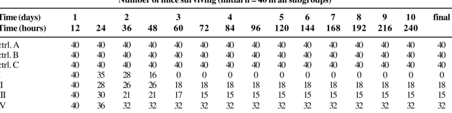
Survival Tables 1 and 2 summarize the outcomes with E. coli and K. pneumoniae, respectively. In all subgroups, the
initial number of mice was 40. The control subgroups of antibiotic, corticosteroid and diluent yielded no mortality. During the first 12 hours of observation, the mortality in all subgroups was null.
After the third day there were no changes in the mortality rates in any subgroup. The final mortality in subgroup I (infection control subgroup) was 100%.
When the antibiotic was used alone(subgroup II; amikacin 15mg/kg/day for four days), the mortality rate decreased significantly (p<0.00001, when compared to the infection control subgroup) and 18 mice survived (a mortality of 55%). The use of a corticosteroid prior to amikacin (subgroup IV) reduced the mortality even more: 32 of the initial 40 mice survived (mortality of 20%), giving a better result than amikacin alone (p<0.001). However, when the corticosteroid was administered after the antibiotic (subgroup III), the final result was inferior to its use prior to the antibiotic (p<0.001), and there was no significant difference compared to subgroup II (antibiotic alone, p=0.2488).
At the 24th hour of the experiment, subgroup II had a higher mortality rate when compared to the non-treated subgroup (p<0.05).
Mortality caused by K. pneumoniae (Table 2) was more
pronounced than that induced by E. coli (comparison between
both subgroups IV: p<0.01; comparison between both subgroups III: p<0.05; for subgroups II this difference in mortality caused by E. coli and K. pneumoniae was not quite
significant: p=0.051). In the infection control subgroup, the final observed mortality was also 100%. When the infection was treated with the antimicrobial alone, the mortality decreased to 72.5% (p<0.001). An additional protective effect was seen when the corticosteroid was used prior to the antibiotic (subgroup IV); in this subgroup, the mortality (45%) was significantly lower than in subgroup II (p<0.01). On the other hand, when the corticosteroid was administered after the antibiotic (subgroup III), the final result was inferior compared to the administration of the corticosteroid prior to the antibiotic (subgroup IV, p<0.01), and there was no significant difference when compared to the results of the subgroup which received amikacin alone (p=0.215), which also resulted in a higher mortality within the first 24 hours (37.5%) when compared to the non-treated subgroup (5%, p<0.001).
Table 1. Survival in the E. coli group.
Table 2. Survival in the K. pneumoniae group.
Number of mice surviving (initial n = 40 in all subgroups)
Time (days) 1 2 3 4 5 6 7 8 9 10 final
Time (hours) 12 24 36 48 60 72 84 96 120 144 168 192 216 240
ctrl. A 40 40 40 40 40 40 40 40 40 40 40 40 40 40 40
ctrl. B 40 40 40 40 40 40 40 40 40 40 40 40 40 40 40
ctrl. C 40 40 40 40 40 40 40 40 40 40 40 40 40 40 40
I 40 35 28 16 0 0 0 0 0 0 0 0 0 0 0
II 40 28 26 26 18 18 18 18 18 18 18 18 18 18 18
III 40 30 21 21 17 15 15 15 15 15 15 15 15 15 15
IV 40 36 32 32 32 32 32 32 32 32 32 32 32 32 32
ctrl. A = antibiotic control; ctrl. B = diluent control; ctrl. C = corticosteroid control; subgroup I: infection control; subgroup II: mice treated only with antibiotics; subgroup III: mice treated with antibiotics prior to the corticosteroid; subgroup IV: mice treated with the corticosteroid prior to antibiotics.
Number of mice surviving in the different treatment groups (initial n = 40 in all subgroups)
Time (days) 1 2 3 4 5 6 7 8 9 10 final
Time(hours) 12 24 36 48 60 72 84 96 120 144 168 192 216 240
ctrl. A 40 40 40 40 40 40 40 40 40 40 40 40 40 40 40
ctrl. B 40 40 40 40 40 40 40 40 40 40 40 40 40 40 40
ctrl. C 40 40 40 40 40 40 40 40 40 40 40 40 40 40 40
I 40 38 21 9 0 0 0 0 0 0 0 0 0 0 0
II 40 25 18 11 11 11 11 11 11 11 11 11 11 11 11
III 40 30 23 21 21 10 8 8 8 8 8 8 8 8 8
IV 40 31 26 22 22 22 22 22 22 22 22 22 22 22 22
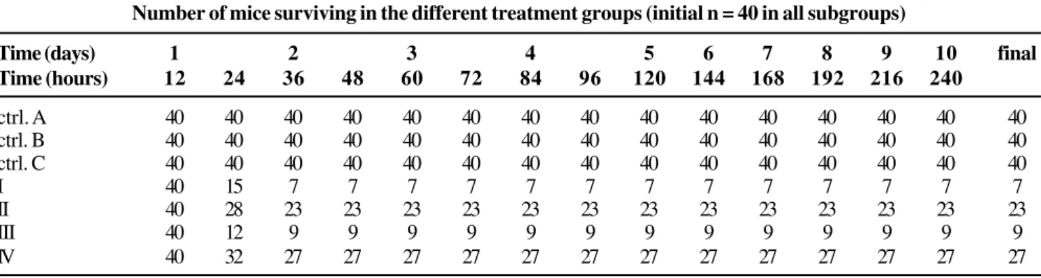
Infection with S. aureus
Table 3 shows the results of the experiment with S. aureus.
The final mortality of control subgroups of antibiotic, diluent and corticoid was zero. During the first 12 hours of observation, the mortality in all subgroups was zero.
Relevant differences in the results regarding Gram-negative bacteria were observed: at 24 hours, the mortality was higher in subgroup I than in subgroup II (62.5 and 30% respectively, p<0.001).
Although the final mortality in subgroup I (infection control) in the S. aureus group was lower than in the GNB
groups, mortality in the first 24 hours for this same subgroup was considerably higher in the S. aureus experiment than in
the E. coli or K. pneumoniae experiments (p<0.0001).
The difference in mortality between subgroups II and III (mortality rates of 42.5 and 32.5%, respectively) was not significant (p=0.1792); nevertheless, the mortality in these two subgroups was significantly lower than the mortality observed in the same subgroups in the experiments with Gram-negative bacilli.
In subgroup III, mortality was higher than in the subgroups that used only the antibiotic or the antibiotic after the corticosteroid (p<0.0001).
In subgroup I (infection control), mortality (82.5%) was significantly higher than in subgroups II and IV (p<0.00001). The incidence of mortality did not change after 48 hours.
Discussion
We chose those three kinds of bacteria to include the most important mechanisms or models of sepsis in clinical practice, that is, Gram-negative bacilli (encapsulated and unencapsulated) and S. aureus.
The subdivision of the three groups (S. aureus, E. coli
and K. pneumoniae) into seven subgroups allowed us to
include all the possible controls. A subgroup treated only with corticosteroid was not included because this would only be used to treat endotoxic shock [31]. Given the effectiveness of corticosteroids for the treatment of endotoxic shock, we understood the importance of excluding the possibility of a large amount of pre-formed endotoxin in the inoculum (from
bacterial growth and multiplication) [32] in the bacterial solution. Without this precaution, any eventual beneficial effect of methylprednisolone in our study would only be due to its action against the effects of the endotoxin (consequently, a possible higher mortality in the subgroups not treated with this drug could be attributed to the effects of a large volume of injected endotoxin). The precaution of washing the bacteria was taken for this reason, and the bacterial suspension was injected into the mice immediately thereafter; this procedure minimizes the amount of endotoxin in the inoculum. The inoculation was also done immediately to prevent changes in the characteristics of the inoculum. To confirm that the inocula had not changed, colony forming counts per mL of the suspension were calculated in in soy-casein agar.
The choice of antibiotics, amikacin for GNBs and teicoplanin for S. aureus, was made based on information
available in the literature and on their use in humans. We administered the antibiotics to the animals using the same doses, routes and dosing intervals recommended for human treatments; these two drugs were selected because 1) the dosing intervals are longer (12/12h), which make the technicians’ work easier; and 2) both can be administered intramuscularly (adequate and repeated IV administration of drugs in mice is quite difficult). The antimicrobials were used for four days and then discontinued; this procedure did not affect the evolution of disease in any group, as no further deaths occurred in the subgroups where antibiotics were used alone after the third day (in the S. aureus group, after the
second day).
Methylprednisolone was chosen among all corticosteroids because there was data concerning optimal dosages in mice [32]. An intraperitoneal route was chosen, instead of intramuscular, because it provides better and quicker absorption.
It could be argued that the large surface of the peritoneum facilitates rapid mobilization of bacteria into the circulation, making this model closer to one of endotoxic shock. But if this was true, then the survival rates would have been similar for the subgroups that received the corticosteroid prior to the antibiotic (subgroups IV) and those that received it after the
Table 3. Survival in the group S. aureus.
Number of mice surviving in the different treatment groups (initial n = 40 in all subgroups)
Time (days) 1 2 3 4 5 6 7 8 9 10 final
Time (hours) 12 24 36 48 60 72 84 96 120 144 168 192 216 240
ctrl. A 40 40 40 40 40 40 40 40 40 40 40 40 40 40 40
ctrl. B 40 40 40 40 40 40 40 40 40 40 40 40 40 40 40
ctrl. C 40 40 40 40 40 40 40 40 40 40 40 40 40 40 40
I 40 15 7 7 7 7 7 7 7 7 7 7 7 7 7
II 40 28 23 23 23 23 23 23 23 23 23 23 23 23 23
III 40 12 9 9 9 9 9 9 9 9 9 9 9 9 9
IV 40 32 27 27 27 27 27 27 27 27 27 27 27 27 27
antibiotic (subgroups III), because corticosteroids alone can abort endotoxic shock. Consequently, even if a human sepsis model is not fully mimicked by experimental models, we believe that our study design was adequate to test the benefits of corticosteroids given early and prior to antibiotics.
Analysis was simplified by the large differences found. Though the mortality rates were higher with K. pneumoniae
when compared with E. coli (we attributed this fact to the
pathogenic characteristics of K. pneumoniae, due mainly to
the protective capsule), we found similar trends in the results found with these two species; for this reason we discuss them together, separately from the results obtained with S. aureus.
The severity of infection with GNB was clear from the high mortality (100%) of the infection control subgroups (subgroups I). The final mortality in the subgroups treated with corticosteroid prior to the antibiotic (subgroups IV) was considerably lower when compared to the subgroups treated only with antibiotics (subgroups II) (p = 0.0009 for the E. coli
group and 0.0073 for the K. pneumoniae group). However,
when the administration sequence was inverted and antibiotics started prior to corticosteroid treatment (subgroups III), these effects were not apparent and mortality was not significantly different from that found in subgroups where only amikacin was administered.
Mortality due to GNB during the first 24 hours was lower in subgroup I than in subgroup II (p<0.05 in the E. coli group
and p<0.001 in the K. pneumoniae group). Although a
superficial analysis might conclude that this finding is paradoxical, it actually reinforces the previously-discussed postulates: this mortality, initially higher in the subgroup treated with antibiotics can be understood as secondary to the sudden release of large amounts of LPS, due to bacterial lysis, which was induced by the antibiotic. In the subgroup treated with methylprednisolone prior to amikacin, the steroid probably inhibited the macrophages, so that subsequent reactions were blocked, resulting in lower mortality rates in this subgroup throughout the study (Tables 1 and 2). In the
K. pneumoniae-infection group, whenever the corticosteroid
was administered after the antibiotic (subgroup III), mortality increased enormously after the withdrawal of the antibiotics, suggesting that the use of corticosteroids after beginning antibiotics can be counterproductive; possibly it would be helpful if antibiotics were continued for a longer period.
Results regarding S. aureus. The reaction in the S. aureus
group was quite different from that with GNB. Though the differences of mortality between subgroups II or IV and subgroup I were significant (p<0.00001), the small advantage observed in the survival of subgroup IV over subgroup II (27 survivors against 23) was not quite significant (p=0.0603).
Contrary to what was found with the GNB, the S. aureus
group did not reproduce the initial difference in mortality (during the first 24 hours) favorable to subgroup I (infection control) in comparison with subgroup II (treated only with antibiotics): the difference was quite significant in favor of
this latter subgroup (p = 0.0023, Table 3). Nevertheless, in the subgroup treated with antibiotics prior to the corticosteroid (subgroup III), the mortality in 24 hours was higher than in all the other infected and treated subgroups (p< 0.001) and not significantly different from subgroup I (p=0.2388). Possibly, in the case of S. aureus, the corticosteroid given after the
antibiotic only exerts its immune-depressant effects, thus accelerating the death of the animals and overcoming the effects of the antibiotic, leading to a kind of response that is equivalent to non-treatment of infection.
The mortality rate in the infection control subgroup during the first 24 hours was much higher in the S. aureus group than
in the equivalent subgroups of the other bacteria. The final rate of mortality in this subgroup was not 100%, as occurred with the GNB, but it was high enough (82.5%) to demonstrate the severity of the infection.
These differences reinforce the hypothesis that the pathogenic mechanisms of S. aureus are considerably different
from those of GNB; the response of the host apparently follows different pathways in such a way that it is not affected by the corticosteroids. Similar findings, using antibodies against TNF–alpha instead of corticosteroids, and infection by other Gram-positive bacteria (Streptococcus pyogenes), were
reported by Wayte [33].
These differences between GNB and S. aureus infection
reinforce the hypothesis that the evolution of sepsis at the biomolecular level is quite different for positive and Gram-negative bacteria. Possibly, in the case of S. aureus, there are
one or more parallel chain-reactions, besides that of TNF-alpha, which are not responsive to the blockages exerted by corticosteroids.
action before the antibiotic had caused the rupture of a large number of bacteria. High doses corticosteroids would have inhibited the macrophages before these cells had contact with large amounts of lipopolysaccharide, thus preventing triggering of the septic cascade.
Our opinion is that corticosteroids should be used early and prior to the use of antibiotics, in order to achieve the best results.
Study Limitations
1) We did not check the minimum inhibitory concentrations (MIC) of the antibiotics used for each microorganism; even though the good survival rates for infected mice treated only with antibiotics suggests an adequate MIC, it would have been useful to check this item. However, an antibiogram by Kirby-Bauer’s method was provided.
2) Unfortunately endotoxins were not measured in our study, so that our conclusions regarding endotoxin release rest on the logic of the vast literature concerning this issue.
3) We did not proceed to a post-mortem study.
4) Finally, TNF-alpha and other mediators were not measured; such measurements might have reinforced our conclusions, but we did not have sufficient funds for such analyses.
Conclusions
1) The use of methylprednisolone early and prior to antibiotics reduced the mortality in this model of severe experimental infection by E. coli or K. pneumoniae.
2) The response of S. aureus to application of
methylprednisolone prior to the antibiotic did not coincide with that observed in the Gram-negative bacilli experiments, failing to promote a significant reduction in mortality;
3) The use of methylprednisolone after the antibiotic had already been started was not better than the use of the antibiotic alone against infection with Gram-negative bacilli, and it increased the mortality rate in the case of S. aureus infection.
This is a primary observation in an experimental model; clearly, these conclusions should not be translated to humans. However, a potential clinical implication of this work would be to use corticosteroids in patients not yet managed with antibiotics who arrive in the emergency room with a disease that provokes high mortality rates, such as, for example, acute severe community-acquired pneumonia. More preliminary studies must be done with animal models before attempting this in humans.
Acknowledgements
We thank CAPES for financial support and Professor Paulo R.B. Guimarães for the statistical work.
References
1. Cannon J.G, Tompkins R.G., Gelfand J.A., et al. Circulating Interleukin-1 and Tumor Necrosis Factor alpha in Septic Shock and Experimental Endotoxin Fever. J Infect Dis 1990;161:79-84.
2. Okusawa S., Gelfand A.J., Ikejima T., et al. Interleukin-1 Induces a Shock-Like State in Rabbits. J Clin Invest 81:1162-72. 3. Charbonneau P., Suisse A. Le Syndrome de Défaillance
Multiviscérale Rev Prat 40:2329-36.
4. Lamy M., Deby-Dupont G. Is Sepsis a Mediator-Inhibitor Mismatch? Intensive Care Med 1995;21:S250-7.
5. Wenzel R.P. Anti-Endotoxin Monoclonal Antibodies: a Second Look. N Engl J Med 1992;326:1151-3.
6. Thijs L.J., Hack C.E. Time Course of Cytokine Levels in Sepsis. Intensive Care Med 1995;21:S258-63.
7. Tracey K.J., Cerami A. Tumor Necrosis Factor: an Updated Review of its Biology. Crit Care Med 1993;21:S423-35.
8. Giroir B.P. Mediators Of Septic Shock: New Approaches For Interrupting The Endogenous Inflammatory Cascade. Crit Care Med 1993;21:780-9.
9. Tracey K.J., Beutler B., Lowry S.F., et al: Shock and Tissue Injury Induced by Recombinant Human Cachectin. Science 1986;234:470-4.
10. Strieter R.M., Kunkel S.L., Bone R.C. Role of Tumor Necrosis Factor - alpha in Disease States and Inflammation. Crit Care Med 1993;21:S447-63.
11. Beutler B, Grau G: TNF-alpha in the Pathogenesis of Infectious Diseases. Crit Care Med 1993;21:S423-35.
12. Bone R.C. Gram-Positive Organisms and Sepsis. Arch Intern Med 1994;154:26-34.
13. Schumann R.R., Leong S.R., Flaggs G.W., et al. Structure and Function of Lipopolyssacaride Binding Protein. Science 1990;249:1429-31.
14. Wright S.D., Ramos R.A., Tobias P.S., et al. CD 14 a Receptor for Complexes of Lipopolyssacaride (LPS) and LPS Binding Protein. Science 1990;249:1431-3.
15. Lepine G. Bacterial Structure. In: Medical Microbiology 2nd ed. Murray P.R., Kobayashi G.S., Pfaller M.A., et al. (Eds). St Louis, Mosby 1994, pp1-16.
16. Salyers A.A., Whitt D.D. Virulence Factors that Damage the Host. In: Bacterial Pathogenesis a Molecular Approach. Washington DC: ASM Press, 1994, pp 47-61.
17. Dofferhoff ASM, Buys J: Effects of Antibiotics on Endotoxin Release. In: Yearbook of Intensive Care and Emergency Medicine. Vincent J-L. Brussels, Springer, 1995, pp 465-472.
18. Shenep J.L., Flynn P.M., Barrett F.F., et al: Serial Quantitation of Endotoxemia and Bacteremia During Therapy for Gram-Negative Bacterial Sepsis. J Infect Dis 1988;157:565-8. 19. Shenep J.L., Morgan K.A. Kinetics of Endotoxin Release During
Antibiotic Therapy for Experimental Gram-Negative Bacterial Sepsis J Infect Dis 1984;150:380-8.
20. Hopkin B.D.A. Frapper Fort ou Doucement: a Gram-Negative Dilemma. Lancet 1978;2:1193-4.
21. Christman J.W., Holden E.P., Blackwell T.S. Strategies for Blocking the Systemic Effects of Cytokines in the Sepsis Syndrome. Crit Care Med 1995;23:955-63.
22. Eidelman L.A., Pizov R., Sprung C.L. New Therapeutic Approaches in Sepsis: a Critical Review. Intensive Care Med 1995;21:S269-72.
24. Barber A.E., Coyle S.M., Marano M.A., et al. Glucocorticoid Therapy Alters Hormonal and Cytokine Responses to Endotoxin in Man. J Immunol 1993;150:1999-2006. 25. Boumpas D.T., Paliogiani F., Anastassiou E.D., et al.
Glucocorticosteroid Action on the Immune System: Molecular and Cellular Aspects. Clin Exp Rheumatol 1991;9:413-23. 26. Romero S., Fuenzalida S. Manual de Manejo y Experimentación
Animal. Instituto de Salud Publica de Chile. Santiago Chile 1991. 27. Kanemann E.W., Allen S.D., Dowel J.R., et al: Diagnóstico Microbiológico 2nd ed. São Paulo, Editorial Panamericana 1993. 28. Marchal N., Bourdon J.L., Richard C.L. Les Millieux de Culture Pour l’Isolement et l’Identification Biochimique des Bactéries. Paris: Soin Éditeurs 1987.
29. Pessoa G.V., Silva M. Proposição de um Meio Simplificado para Identificação de Enterobactérias. Revista do Instituto Adolfo Lutz 1975;24:29-35.
30. Balows A., Hausler J.R.W.J., Herrmann K., et al. Manual of Clinical Microbiology 5th ed. American Society for Microbiology USA 1995. 31. Pitcairn M., Schuler J., Erve P.R., Schumer W., et al. Glucocorticoid and Antibiotic Effect on Experimental Gram-Negative Bacteremic Shock. Arch Surg 1975;110:1012-5.
32. Greisman S.E. Experimental Gram-Negative Bacterial Sepsis: Optimal Methylprednisolone Requirements for Prevention of Mortality not Preventable by Antibiotics Alone. Proc Soc Exp Biol Med 1982;170:436-42.
33. Wayte J., Silva A.T., Krausz T., et al. Observations on the Role of Tumor Necrosis Factor in a Murine Model of Shock Due to Streptococcus pyogenes. Crit Care Med 1993;21:1207-12. 34. Jansen N.J.G., Van Oeveren W., Hoiting B.H., Wildevuur C.H.
Methylprednisolone Prophylaxis Protects Against Endotoxin -Induced Death in Rabbits. Inflammation 1991;15:91-101. 35. Loggie J.M.H., Privitera P.J., Sugarman S. Effects of Hydrocortisone
on Survival in Neonatal Beagles given Endotoxin. Proc Soc Exp Biol Med 1968;128:326-9.
36. Schumer W. Steroids in the Treatment of Clinical Septic Shock. Annals of Surgery 1976;184;3333-341.
37. Bone R.C., Fisher C.J., Clemmer T.P., et al. A Controlled Clinical Trial of High-Dose Methylprednisolone in the Treatment of Severe Sepsis and Septic Shock N Engl J Med 1987;317:653-8. 38. Sprung CL, Caralis VP, Marcial EH, et al: The Effects of High-Dose Corticosteroids in Patients with Septic Shock. N Engl J Med 1984; 311:1137-43.
39. The Veterans Administration Systemic Sepsis Cooperative Study Group: Effect of High-Dose Glucocorticoid Therapy on Mortality in Patients with Clinical Signs of Systemic Sepsis. N Engl J Med 1987;317:659-65.
40. Lefering R., Neugebauer E.A.M. Steroid Controversy in Sepsis and Septic Shock: a Meta-Analysis. Crit Care Med 1995 ;23:1294-1303.
41. Neugebauer E., Dietrich A., Bouillon B., et al. Glucokortikoide beim Polytrauma und bei Sepsis- Immer noch ein Thema? Klin Wochenschr 1991;69(SupplXXVI)211-23.
42. Opal S.M. Clinical Trials of Novel Therapeutic Agents: Why did They Fail? In: Yearbook of Intensive Care and Emergency Medicine. Vincent J-L. Brussels, Springer, 1995, pp 425-36. 43. Bollaert P.E., Charpentier C., Levy B., et al. Reversal of late
septic shock with supraphysiological doses of hydrocortisone. Crit Care Med 1998;26:645-50.
44. Annane D., Sebille V., Charpentier C., et al. Effect of treatment with low doses of hydrocortisone and fludrocortisone on mortality in patients with septic shock. JAMA 2002;21;288(7):862-71.
45. Annane D. Resurrection of steroids for sepsis resuscitation. Minerva Anestesiol 2002;68(4):127-31.
46. Annane D., Cavaillon J.M. Corticosteroids in sepsis: from bench to bedside? Shock 2003;20(3):197-207.