ORIGINAL
ARTICLE
DMFT index and oral mucosal lesions associated with HIV
infection: cross-sectional study in Porto Velho, Amazonian
Region – Brazil
Authors
Rodrigo Queiroz Aleixo1 Alexandre Prado Scherma2 Gustav Guimarães1 José Roberto Cortelli3 Sheila Cavalca Cortelli3
1Master’s Degree in Dentistry, Unitau – Professor of Faculdade São Lucas.
2Post-Graduation in Dentistry – Assistant Professor of Instituto Básico de Biociências, Unitau.
3Post-Graduation in Dentistry – Assistant Professor of Preventive Dentistry and Cariology, and Periodontoly, Unitau.
Submitted on: 08/02/2009 Approved on: 11/04/2009
Correspondence to:
Rodrigo Queiroz Aleixo Rua Beethoven, 4604 - Nova Esperança Porto Velho – RO – Brazil CEP: 76822-200 Phone: +55-69-92316955 E-mail: rodrigoaleixo@ yahoo.com.br
We declare no confl ict of interest.
ABSTRACT
We evaluated the DMFT (decayed, missing and fi lled teeth) index and the prevalence of candidia-sis, linear gingival erythema, oral hairy leukoplakia, herpes simplex, aphthous ulcers, Kaposi’s sar-coma and lymphoma, as well as the association with TCD4 count, viral load (VL) and antiretroviral therapy (ART) in 140 HIV-infected adult individuals. A standardized examination to determine the DMFT index and the presence of oral lesions was conducted. Demographic data, TCD4 count and use of ART were obtained from medical records. A high number of decayed teeth detected among patients undergoing ART resulted in a mean DMFT of 16.9 teeth. It was observed that 24.2% of the individuals had at least one oral lesion. Candidiasis was the most frequent lesion and was associated with the TCD4 count. Oral hairy leukoplakia was associated with an increased VL. Regular use of ART was inversely associated with the occurrence of lesions. Overall, the studied population showed low prevalence of oral lesions and high DMFT index. The use of ART seems to reduce the occurrence of these lesions. Higher TCD4 count and a lower VL were associated with an improved oral health status in HIV+ individuals.
Keywords: oral mucosal lesions, DMFT index, tooth decay, HIV infection.
[Braz J Infect Dis 2010;14(5):449-456]©Elsevier Editora Ltda.
INTRODUCTION
Between 1980 and 2007, 474,237 cases of ac-quired immunodefi ciency syndrome (AIDS) were notifi ed in Brazil, with the Southeast region being the one presenting the highest number of cases (289,074). The Northern re-gion had 16,103 cases, with 1,862 in the state of Rondônia. Additionally, during a similar pe-riod of time (1980 – 2006), 192,709 deaths due to AIDS were notifi ed in Brazil, with 549 in the state of Rondônia.1
The initial infection by the human immu-nodefi ciency virus (HIV) is accompanied by unspecifi c symptoms, such as fever and ma-laise. Subsequently, the lymphadenopathy pro-ceeds to a chronic and asymptomatic phase, as viral replication is, partially controlled by the immune system. Duration of this stage var-ies to factors such as viral phenotype, host’s immune response, and use of antiretroviral therapy (ART). If it is not controlled by medi-cation, viral replication continues and other symptoms, such as diarrhea, weight loss and oral
candidiasis, appear and full-blown AIDS is es-tablished, which consists of a large number of acquired or reactivated diseases due to immu-nosuppression. Opportunistic diseases caused by viruses, bacteria, protozoa, and fungi and, together with some related neoplasias,2
indi-cate a severely immunocompromised state.3
Around 1% of HIV-infected individuals, so called non-progressors, can maintain nor-mal TCD4 lymphocyte levels and low viral load (VL) for periods as long as 10 to 20 years.4
Specifi cally, in the oral environment the lesions associated with HIV infection include candi-diasis, herpes simplex, oral hairy leukoplakia (OHL), cytomegalovirus (CMV) infection, varicella-zoster virus infection, papilloma vi-rus infection, linear gingival erythema (LGE), gingivitis and necrotizing ulcerative periodon-titis, Kaposi’s sarcoma, and aphthous ulcers, which are acknowledged as important markers of AIDS clinical stages.5-9
immuno-suppression increases, so does the incidence of caries.10,11
Phelan et al.12 observed a higher incidence of caries and a
lower number of teeth present in the oral cavity of HIV-in-fected adult women. However, in Brazil, the DMFT data in HIV-infected populations are scarce. Awareness of the mag-nitude of this problem in Brazil may allow for comparison with other countries and might highlight the need to imple-ment or intensify preventive programs aimed at this group of individuals and at professionals working in this area. Such actions could guarantee a better quality of life through ad-equate care for the ones in need.
The treatment of the HIV infection/AIDS aims at lim-iting viral replication and, consequently, preventing and controlling opportunistic infections. To monitor disease progression, TCD4 cell count and VL13 must be monitored
throughout treatment. TCD4 counts < 200 cells/mm3
indi-cate higher probability of developing opportunistic infec-tions, whereas VL > 50,000 to 100,000 copies/mL can result in a rapidly compromised immune system.3 In Brazil, ART
is recommended for all symptomatic individuals infected by HIV, and asymptomatic individuals with a TCD4 count < 350 cells/mm3.3,14
Therefore, the objective of the present cross-sectional study was to assess the DMFT index in association with oral mucosal lesions HIV-infected individuals living in the city of Porto Velho, state of Rondônia, Brazil. Moreover, a pos-sible association between the oral health status and TCD4 count, VL quantifi cation, and ART use was investigated.
METHODS
Sample calculation
Of 970 medical records of individuals registered at the Refer-ence Outpatient Service (ROS), receiving care at Policlínica Municipal Dr. Rafael Vaz e Silva, a sample of 130 partici-pants was chosen, to provide a power of 80% and an α error of 0.05. For the sample calculation, we considered the mean prevalence observed in the literature of oral candidiasis, lin-ear gingival erythema, oral hairy leukoplakia, aphthous ul-cers, Kaposi’s sarcoma, herpes simplex and lymphoma.
Inclusion and exclusion criteria
The convenience sample consisted of patients that came to Policlínica for medical, dental or psychological care, or to collect ART medication during the period of August 2007 to January 2008. HIV-infected men and women, aged 18 years or older, registered and followed at the ROS were evaluated. Only individuals that agreed to participate in the study and signed the Free and Informed Consent Form were kept in the study (protocol of approval by the Eth-ics Committee in Research #121/07). Former smokers who had quit smoking less than three years before enrollment, and individuals that did not follow the outpatient clinic treatment were excluded.
Data collection
Participants underwent an oral assessment by a single ex-aminer at the dental office of the clinic, under artificial light, according to the appropriate biosafety guidelines and with the help of a clinical dental mirror #5 (Duflex, SS White, Rio de Janeiro, RJ, Brazil), a dental probe #5 (Duflex, SS White, Rio de Janeiro, RJ, Brazil), a blunt-tip probe (Duflex, SS White, Rio de Janeiro, RJ, Brasil), a wooden spatula and sterilized gauze pads.
The examiner was trained to identify the following oral mucosal lesions: candidiasis (erythematous, pseudo-membranous and angular cheilitis), linear gingival ery-thema, oral hairy leukoplakia, aphthous ulcers, herpes simplex, Kaposi’s sarcoma and lymphoma, which were photographed (Sony DSC-H2, San Diego, CA, USA). Following a standardized oral exam, a single examiner assessed all present teeth, except for the third molars, in order to determine the DMFT (decayed, missing and filled teeth) index, according to the criteria established by the World Health Organization (WHO).15 Each tooth
was classified as Decayed: D (decayed teeth and filled teeth with caries or temporary filling material), Missing – subdivided in two categories; Extracted: E (teeth lost due to caries or other reasons) or Extraction indicated: Ei (extensive tooth decay that prevented tooth restoration procedures), or Filled: F (teeth that had been filled with permanent filling material and with no caries). Accord-ing to the Brazilian Ministry of Health,16 an examiner is
adequately trained when his intra-examiner index reach-es a reproducibility rate of 97.5%. The information on age, gender, laboratory data, smoking habit, and time of ART use were collected from medical records. TCD4 lym-phocyte counts and VL quantifications obtained up to three months before or after the oral clinical assessment were considered in the study, as the VL tends to stabilize with the beginning of the humoral response.17
As the use of ART depends on symptomatology and the TCD4 cell count,14 patients that had used ART regularly for
at least twelve months were considered users. All data were entered in a form especially created for the present study.
Participants were advised on the importance of follow-up and regular use of the antiretroviral therapy, as well as the importance of oral hygiene. Those who presented with oral mucosal lesions received adequate treatment. Those who needed dental treatment were scheduled to receive it. Only cases that needed endodontic treatment and/or pros-thesis were referred to the Dental Clinic of Faculdade São Lucas and the Dental Specialization Centers – DSCs.
Statistical analysis
software, and the level of significance was set at 5% (p < 0.05). The normality of data was initially evaluated by Kolmogorov-Smirnov test (Lilliefors Test).
The Chi-square test was used for the comparative analyses regarding the occurrence of oral mucosal le-sions, as well as their presence according to smoking status. The Mann-Whitney test was used for the intra-group analysis of the association between these lesions and TCD4 counts, VL quantification and use of ART, whereas ANOVA was used to verify the differences be-tween the number of decayed teeth and TCD4 counts, VL quantification and use of ART.
The individual DMFT was obtained by adding the teeth included in any of criteria (D, E, Ei or F) and the mean DMFT was obtained by calculating the sim-ple arithmetic mean of the DMFT index of all partici-pants.
RESULTS
To reach a sample size of 140 eligible participants (39 ± 10.7 years), 238 individuals were invited to participate. Two participants were excluded as they did not meet the age criterion. A high number of subjects refused to par-ticipate (96) due to the need to undergo the intra-oral clinical assessment.
The study sample consisted of 66 men (40.8 ± 11.5 years), 74 women (37.5 ± 9.8 years); 37 were smokers. There were 2.9% participants in the age-range between 14 and 19 years, 15% between 20 and 29 years, 37.9%
between 30 and 39 years, 25.0% between 40 and 49 years, and 19.2% were older than 50 years.
The mean TCD4 count was of 380 cells/mm3. Of the
140 participants, 58% had intermediate counts of TCD4 (between 200-500 cells/mm3), 23% presented more than
500 cells/mm3, 14% presented 50-200 cells/mm3 and
only 5% had fewer than 50 lymphocytes/ mm3.
Regarding ART use, 68% of the individuals reported using a combination of three antiretroviral drugs for a mean time of 45.4 months (± 37.5 months).
Fifty-three percent of individuals had undetectable HIV VL, 23% had < 10,000 copies/mL, 16% had between 10,000 and 100,000 copies/mL, and only 8% of the par-ticipants had more than 100,000 copies/mL.
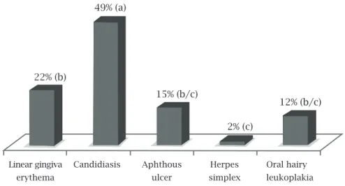
Out of the total, 75.8% (106/140) disclosed no oral mucosal lesion. In the remaining 34 patients only one oral lesion was present in 82.4% (28/34). whereas 14,7% (5/34) had two lesions, and only one patient (2.9%) had simultaneously three different lesions (linear gingival erythema, candidiasis and aphthous ulcer). The most frequently identified oral mucosal lesion was candidiasis (20 patients) (Figure 1), with erythematous candidiasis being observed in 16 cases, followed by angular cheilitis in three cases, and pseudomembranous candidiasis in two patients. There were no cases of Kaposi’s sarcoma or lymphoma. Additionally, aphthous ulcers was present in six patients, herpes simplex in one, and oral hairy leu-koplakia in five patients. Smoking, reported by 37 par-ticipants, was not associated with the occurrence of oral mucosal lesions (p = 0.99).
Figure 1: Oral lesion types. Different small letters between parenthesis indicate significant statistical difference (Chi-square test, p < 0.05) between the occurrence of lesions.
Oral mucosal lesion occurrence
22% (b)
49% (a)
15% (b/c)
2% (c)
12% (b/c)
Linear gingiva Candidiasis Aphthous Herpes Oral hairy
The presence of lesions was inversely proportional to the TCD4 count, i.e., the higher the number of cells, the lower the occurrence of lesions (Figure 2). As for the VL, as its lev-els increased, so did the occurrence of lesions.
There was a trend towards the association of an increas-ing number of oral mucosal lesions and beincreas-ing without ART, albeit without statistical signifi cance.
Among the 64 individuals who had an undetect-able VL, 96.8% (62) was on regular use of ART, and 82.8% (53) had a TCD4 count > 200 cells/mm3. Only
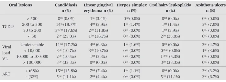
Table 1. Presence of oral lesions according to TCD4 count, viral load (VL) and use of ART
Oral lesions Candidiasis Linear gingival Herpes simplex Oral hairy leukoplakia Aphthous ulcers n (%) erythema n (%) n (%) n (%) n (%)
> 500 0(b) (0.0%) 1(a) (3.4%) 0(a) (0.0%) 0(a) (0.0%) 0(a) (0.0%) 200 to 500 14(a)(19.7%) 4(a) (5.9%) 1(a) (1.4%) 1(a) (1.4%) 5(a) (7.0%) TCD4+
50 to 200 3(a/b) (17.6%) 2(a) (11.8%) 0(a) (0.0%) 1(a) (5.9%) 0(a) (0.0%) < 50 2(a) (25.0%) 1(a) (16.7%) 0(a) (0.0%) 2(a) (25.0%) 0(a) (0.0%)
Viral Undetectable 11(a) (17.2%) 4(a) (6.3%) 1(a) (1.6%) 0(b) (0.0%) 3(a) (4.7%)
load < 10,000 3(a) (10.7%) 3(a) (10.7%) 0(a) (0.0%) 0(b/c) (0.0%) 1(a) (3.6%) VL 10,000 to 100,000 2(a) (10.5%) 1(a) (5.3%) 0(a) (0.0%) 1(a/b) (5.3%) 0(a) (0.0%)
> 100,000 3(a) (33.3%) 0(a) (0.0%) 0(a) (0.0%) 3(a) (33.3%) 0(a) (0.0%)
ART + (68%) 15
(a) (15.8%) 7(a) (7.4%) 1(a) (1.1%) 0(a) (0.0%) 3(a) (3.2%)
- (32%) 5(a) (11.1%) 2(a) (4.4%) 0(a) (0.0%) 5(b) (11.1%) 3(a) (6.7%)
Different small letters in the same column indicate significant statistical difference (p < 0.05) between the subgroups of TCD4, viral load and ART use.
+, user; -, non-user.
one patient with undetectable VL had TCD4 count < 50 cells/mm3.
The group with TCD4 count > 500 cells/mm3 showed
a lower prevalence of candidiasis, when compared to the group with TCD4 count between 200-500 cells/mm3
(p = 0.0238) and to the group with TCD4 count < 50 cells/mm3 (p = 0.0254). Oral hairy leukoplakia was more prevalent among patients with VL > 100,000 copies/mL than among those with undetectable VL and with < 10,000 copies/mL (p < 0.05). These data are shown in Table 1.
Figure 2: Percentage of individuals with a positive diagnosis for at least one of the oral mucosal lesions, evaluated
comparatively to those that did not exhibit any lesions at the moment of the intra-oral assessment. * Statistically significant difference at the intra-group analysis (p < 0.05; Mann-Whitney).
Presence of oral mucosal lesions in relation to TCD4 count (A) and viral load (B)
lesion no lesion lesion no lesion
A
B
96.6% *
3.4%
26.8% 73.2%
* 64.7%
* 66.7%
15.2% 84.8%
*
60.7% *
39.3% 33.3%
* 35.3%
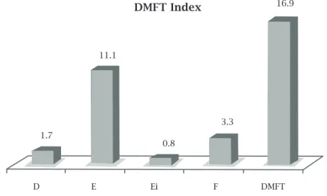
Figure 3 shows the means of decayed – D, extracted – E, extraction indicated – Ei and fi lled teeth – F (restored), as well as the general mean DMFT index. There was no sta-tistically signifi cant difference when the DMFT index was correlated with TCD4 cell counts (ANOVA), or when each
Figure 3: Mean DMFT Index: D, decayed; Missing: E, extracted; Ei, extraction indicated; F, filled (restored).
Table 2. Association between the DMFT index and the use of ART
Decayed (D) Extracted (E) Extraction indicated (Ei) Filled (F) DMFT n (Mean) n (Mean) n (Mean) n (Mean) n (Mean)
ART + (n = 95) 120 (1.3)
(a) 1094 (11.6)(a) 79 (0.8)(a) 343 (3.6)(a) 1636(17.4)(a)
- (n = 45) 123 (2.7)(b) 454 (9.9)(a) 37 (0.8)(a) 117 (2.5)(a) 731 (15.9)(a)
Different small letters in the same column indicate significant statistical difference (p < 0.05) between the participants who were users (+) or non-users (-) of ART.
DISCUSSION
In the state of Rondônia, Amazonian region, Brazil, there are only six healthcare centers providing specialized care for patients with HIV/AIDS, and two of them are located in the capital city, Porto Velho. Of these, only ROS offers dental care, which was the setting of the present study. The main objective of the present study was to determine the DMFT index and the prevalence of oral mucosal le-sions in HIV-infected patients and its association with TCD4 count, viral load (VL) quantification, and antiret-roviral therapy (ART) use.
criterion (D, E, Ei and F) were compared among the indi-viduals with different VL counts (ANOVA).
However, higher number of decayed teeth – D was ob-served among patients who were not on ART (p = 0.004; t-Test) (ANOVA) (Table 2).
These data represents important information on which to base the planning of more effective actions to copy with the studied health problem. The results of the present study confi rmed two trends of the AIDS epidemics: the aging of the infected population due to higher survival rates9,18,19 and
the feminization.20,21
Availability of ART at the Brazilian Public Healthcare Sys-tem (SUS) was established by Federal Law # 9313, of December 1996, and guaranteed the broad and free distribution of ART to the Brazilian population.22 According to Hammer et al.,23
com-bination ART therapy is more effi cient than monotherapy, as it results in an increase in TCD4 count and concomitant decrease
1.7
11.1
0.8
3.3
16.9
D E Ei F DMFT
in VL, which, in turn, results in a decrease in the rate of oral mu-cosal lesions.24-26 This low prevalence of lesions was confi rmed
by the present study. Moreover, the combination therapy has shown to be effective in improving/restoring the quality of life and increasing survival rates of infected patients.27
The type of candidiasis most frequently observed was the erythematous type, in accordance with Noce et al.,9 but in
disagreement with other authors, such as Souza et al.,11
Mat-tos, Santos and Ferreira,28 Ranganathan and Hemelatha,29
and Ranganathan et al.,30 who reported that
pseudomebra-nous candidiasis was the most common type. These authors also mentioned the presence of angular cheilitis as a frequent fungal infection, a manifestation also observed in this study, albeit with a low incidence (2%).
Linear gingival erythema represented 6% of the cases, a value that was similar to that of several studies involving Bra-zilian populations7,17,28,31 (Fabro et al.7 – 2%; Grando et al.17
– 5.9%; Mattos, Santos and Ferreira28 – 4.3%; and Ferreira et al.31 – 2.5%).
Aphthous ulcers represented 4% of the cases in the present study. The literature presents an assorted prevalence of these alterations. Ulcers are reported by studies carried out in Brazil as ranging from 0.9%32 up to 23.5%,5 with most of studies
showing a prevalence of approximately 3%.
Oral hairy leukoplakia (OHL) has also been reported with prevalences varying from 2.1% through 6%.9,11,17,30,33
It is noteworthy that several authors described OHL as a mark-er of HIV infection progression.13,34,35 Thus, the relatively low
frequency of this lesion in the population of the present study suggests an indicator of HIV control as a result of combina-tion ART. Patton et al.36 reported that HIV-infected patients
who present OHL only among those with VL > 20,000 copies/ mL, regardless of the TCD4 count. In contrast, there were fi ve patients with OHL identifi ed in the present study.
Kaposi’s Sarcoma and lymphoma were not seen in this study. According to Girotto et al.,37 the decrease in the prevalence of
these lesions is associated with the use of combination ART. In Porto Velho, since the ROS started treating patients, there have never been any report of lymphoma in the oral cavity.
Candiani et al.38 reported a signifi cant decrease in
oppor-tunistic infections, hospitalizations and death in HIV-infected patients following the introduction of highly-active antiret-roviral therapy (HAART), a combination of three antiretro-viral drugs. Birnbaum et al.,24 Greenwood, Zakrzewska and
Robinson,26 Ferreira et al.,31 and Coogan, Greenspan and
Challacombe39 reported changes in the prevalence of oral
mucosal lesions following the introduction of ART, including the reduction of the incidence of candidiasis, OHL and KS. A low prevalence of KS was also associated with the use of ART, as reported by Greenspan et al.25 In the present study,
68% of the patients used combination ART for a mean pe-riod of 45 months (SD: 37.5 months). In approximately 20% (95) of the patients on regular use of ART, some type of oral
mucosal lesion was identifi ed, whereas in the group with-out ART therapy, approximately 31% (45) presented lesions. Greenspan et al.40 reported that after the start of HAART
ther-apy, a signifi cant decrease in the quantifi cation of VL occurs, as well as an increase in TCD4 count. The results obtained in the present study show a decrease in the prevalence of lesions in the oral mucosa among those on HAART, thus suggesting that, in fact, the combined HAART maintains or restores the immune response and, additionally, decreases the VL, result-ing in a better quality of life and longer survival rates.
According to Mattos, Santos and Ferreira,28 most of the
patients with oral mucosal lesions had TCD4 counts be-tween 200 and 399 cells/mm3, whereas in patients with VL
up to 10,000 copies/mL fewer lesions were identifi ed. Birn-baum et al.24 and Patton et al.36 showed that the increase in
VL is also characterized by the increase in the prevalence of lesions in the oral mucosa.
Similarly, in the present study, patients with VL values > 10,000 copies/mL had higher prevalence of lesions. Miziara, Lima and Cortina35 demonstrated that patients with
candidiasis or OHL had lower TCD4 lymphocyte counts and higher VL values, when compared to patients that did not express these manifestations clinically. A study carried out by Ferreira et al.31 demonstrated a higher prevalence of
lesions in the oral mucosal lesions in patients with TCD4 count < 200 cells/mm3 and a higher prevalence of these
le-sions in patients with high VL values. In agreement, in the present study, the increase in the TCD4 count was associated with the decrease in the occurrence of oral mucosal lesions.
In Brazil, few studies have reported their experience on den-tal caries and HIV infection, and most of them were carried out with HIV-infected children.10,12,32,41 The differences between the
prevalence and the incidence of caries in individuals with and without HIV infection have yet to be elucidated. However, by rec-ognizing that dental caries is an infectious disease, and knowing that several side effects of ART therapy can contribute to an inad-equate oral hygiene, as well as to an inappropriate diet, the HIV infection seems to function as a risk factor for caries.
Bretz et al.41 observed a high prevalence of caries (mean
of 42.9 surfaces) in an HIV-infected population from Hou-ston, Texas. A study carried out by Phelan et al.12 showed a
comparison of prevalence of caries between the HIV-infected and uninfected women. These authors observed higher rate of coronal and radicular caries, in addition to a lower number of permanent teeth in the oral cavity of HIV-infected women, concluding that the occurrence of caries was higher in indi-viduals with decreased TCD4 count. In Brazil, Silveira and Rangel32 observed a high prevalence of caries with a mean
DMFT = 23 (D = 12; E = 7.5; Ei =1.5; F = 2) in 123 adult patients of both sexes.
teeth with extraction indicated was low (1.7 and 0.8, respec-tively) and the number of extracted teeth was high (11.1). It can be observed that these numbers are far from the targets proposed by the WHO for the year 2010, such as: the pres-ence of 20 or more teeth in the oral cavity for 96% of the in-dividuals at the age range of 35 to 44 years and no lost teeth at the age of 18 years.42 In the present study, at the age range
of 35 to 44 years, 49 individuals were assessed. Of these, 23 individuals (47%) had fewer than 20 teeth in the oral cavity, that is, less than half of the target number proposed by the WHO; there were only two participants who were 18 years old, and both had at least two missing teeth.
The SB Brazil epidemiological surveillance, carried out by the Ministry of Health in 2002, indicated that, at the age range 35 to 44 years, the mean of missing teeth was of 14.77 for the Northern region, whereas in the present study, the mean was of 12.12 for HIV-infected individuals. Regarding the decayed and fi lled teeth criteria, the results demonstrat-ed better oral health status for HIV-infectdemonstrat-ed patients (D = 1.41; F = 4.0), when compared to the general population of the Northern region (D = 2,97; F = 1.89).43 Therefore, the
comparison of these data suggest a better access to dental care services for the studied population, confi rming the im-portance of the surgeon-dentist in reference services provid-ing care for HIV/AIDS patients.
The results from the present study refl ect the importance of adequate care given to HIV/AIDS patients, as the correct management provides a better quality of life to the patient, which was demonstrated here through the low prevalence of oral mucosal lesions. It should also be pointed out the impor-tance of creating/implementing oral rehabilitation programs (dental prosthesis), considering the large number of missing teeth, as well as the creation of more reference centers with multiprofessional teams, so that a new effective management and follow-up of HIV-infected patients can be accomplished.
CONCLUSIONS
The studied population, which consisted primarily of indi-viduals with intermediate TCD4 counts and undetectable VL, showed low prevalence of oral mucosal lesions and a high DMFT index, with the high number of extracted teeth contributing the most for this high index.
The most frequently observed lesion was candidiasis, which was related to TCD4 count, whereas the increase in the VL was associated with the occurrence of OHL. The DMFT index was not associated with VL, TCD4 count or ART use. Only the criterion Decayed tooth, when analyzed separately, was higher in the group that was not on ART use.
Individuals with higher counts of TCD4 lymphocytes and lower counts of VL tended to present fewer oral mucosal lesions associated with HIV infection. Additionally, the use of antiretroviral therapy showed an association with lower prevalence of lesions, especially OHL.
REFERENCES
1. Brasil. Ministério da Saúde. Secretaria de Vigilância em Saúde. Programa Nacional de DST E AIDS. Boletim Epidemiológico Aids e DST 2007; IV(1):48 [cited 2008 Apr 19]. Available from <http://www.aids.gov.br>.
2. Mims C, Dockrell HM, Goering RV, et al. Microbiologia médi-ca. Rio de Janeiro: Elsevier, 2005. p. 284-95, p. 464-72. 3. Lewi DS, Turcato Jr G, Castelo Filho A, Diaz RS. Síndrome da
Imunodefi ciência Adquirida (Aids). In: Salomão R, Pignatari AC, editors. Guias de medicina ambulatorial e hospitalar-Infectologia Unifesp/Escola Paulista de Medicina. Barueri SP: Manole, 2004. p. 125-34.
4. Marques AR, Masur H. História natural da infecção pelo HIV.
In: Focaccia R, editor. Veronesi: tratado de infectologia. 3ª edi-ção. São Paulo: Atheneu, 2005. p. 143-7.
5. Ferreira RI, Valença Neto AAP, Vianna DC, et al. Estudo da prevalência de alterações bucais em pacientes HIV+ do Hos-pital Universitário Prof. Edgard Santos – Salvador, BA. Rev Fac Odontol Univ Fed Bahia 1999; 18:22-7.
6. Volkweis MR, Rocha RS da, Leonardo LLN, Wagner, JCB. Lesões bucais manifestadas em pacientes aidéticos e tuberculosos, re-lacionadas com a contagem celular cd4+/cd8+. PGR-Pós-Grad Rev Fac Odontol São José dos Campos 2001; 4(3):74-82. 7. Fabro SML, Rath IBS, Grando LJ, et al. Estudo das manifestações
estomatológicas em pacientes infectados pelo HIV, atendidos no Hospital Nereu Ramos – Florianópolis – SC, Brasil. RPG Rev Pos Grad 2002; 9(1):12-9.
8. Silverman S Jr, Glick M. Doenças associadas com o vírus da Imunodefi ciência Humana. In: Silverman S Jr, Eversole LR, Truelove EL, editors. Fundamentos de Medicina Oral. Rio de Janeiro: Editora Guanabara Koogan, 2004. p. 128-43.
9. Noce CW, Pinheiro RS, Souza ACB, et al. Prevalência de lesões orais na infecção por HIV em adultos e crianças. Rev Bras Od-ontol 2006; 63(1-2):126-9.
10. Castro GF, Souza IPR, Oliveira RHS, et al. Prevalência de cárie e sua correlação com a classifi cação clínica e imunológica em crianças infectadas pelo HIV. Pesqui Odontol Bras 2001; 15(2):91-7.
11. Souza LB, Pereira Pinto L, Medeiros AMC, et al. Manifestações orais em pacientes com Aids em uma população brasileira. Pesqui Odontol Bras 2000; 14(1):79-85.
12. Phelan JA, Mulligan R, Nelson E et al. Dental caries in HIV-seropositive women. J Dent Res 2004; 83(11):869-73.
13. Patton L, Shugars DC. Immunologic and viral markers of HIV-1 disease progression: implications for dentistry. JADA 1999; 130(9):1313-22.
14. Brasil. Ministério da Saúde. Secretaria de vigilância em saúde. Programa Nacional de DST e Aids. Recomendações para tera-pia antiretroviral em adultos e adolescentes infectados pelo HIV: 2005/2006. 6th ed. Brasília: Ministério da Saúde, 2007. 15. World Health Organization. Assessment Forms. In: Oral
Health Surveys Basic Methods. 4th ed. Geneva, 1997. p. 21-50.
16. Brasil. Ministério da Saúde. Projeto SB Brasil – Condições de saúde bucal da população brasileira. Manual do Examinador. Brasília: Ministério da Saúde, 2001.
17. Grando LJ, Yurgel LS, Machado DC, et al. Manifestações esto-matológicas, contagem de linfócitos T-CD4+ e carga viral de
crianças brasileiras e norte-americanas infectadas pelo HIV. Pesqui Odontol Bras 2002; 16(1):18-25.
19. Stoff DM, Khalsa JH, Monjan A, Portegies P. Introduction: HIV/AIDS and Aging. AIDS 2004; 18(S1):S1-2.
20. Fonseca MGP, Bastos FI. Twenty-fi ve years of the AIDS epi-demic in Brazil: principal epidemiological fi ndings, 1980-2005. Cad. Saúde Pública 2007; 23(3):S333-44.
21. Souto BGA. HIV/AIDS in the small cities: a Brazilian epidemi-ology study. Rev. Cubana Med Trop 2004; 56(2):91-3. 22. Noce CW, Silva Júnior A, Ferreira SMS. Panorama mundial da
epidemia pelo HIV/Aids: Aspectos sociais e lesões bucais. DST – J Bras Doenças Sex Transm 2005; 17(4):301-5.
23. Hammer SM, Katzenstein DA, Hughes MD et al. A trial com-paring nucleoside monotherapy with combination therapy in HIV-infected adults with CD4 cell counts from 200 to 500 per cubic millimeter. N Engl J Med 1996; 335(15):1081-90. 24. Birnbaum W, Hodgson TA, Reichart PA, et al. Prognostic
sig-nifi cance of HIV-associated oral lesions and their relation to therapy. Oral Diseases 2002; 8(S2):110-4.
25. Greenspan D, Canchola AJ, MacPhail LA, Cheikh B, Greenspan JS. Effect of highly active antiretroviral therapy on frequency of oral warts. Lancet 2001; 357(9266):1411-2.
26. Greenwood I, Zakrzewska JM, Robinson PG. Changes in the prevalence of HIV-associated mucosal disease at a dedicated clinic over 7 years. Oral Diseases 2002; 8(2):90-4.
27. Barbosa MTS, Struchiner CJ. Impacto da terapia antirretrovi-ral na magnitude da epidemia do HIV-Aids no Brasil: diversos cenários. Cad Saúde Pública 2003; 19(2):535-41.
28. Mattos SL, Santos VR, Ferreira EF. Prevalência das lesões de mucosa bucal em pacientes HIV-positivos da unidade de referência especializada em doenças infecciosas e parasitárias especiais – URE-DIPE (Belém-Pará). Rev Bras Patol Oral 2004; 3(1):7-16.
29. Ranganathan K, Hemelatha R. Oral lesion in HIV infection in developing countries: an overview. Adv Dent Res 2006; 19(1):63-8.
30. Ranganathan K, Umadevi M, Saraswathi TR, Kumarasamy N, Solomon S, Johnson N. Oral lesions and conditions associated with human immunodefi ciency virus infection in 1000 South-Indian patients. Ann Acad Med Singapore 2004; 33(S):37-42. 31. Ferreira S, Noce CW, Silva Jr A et al. Prevalence of Oral
Mani-festations of HIV Infection in Rio de Janeiro, Brazil from 1988 to 2004. Aids Patient Care and STDs 2007; 21(10):724-31.
32. Silveira FM, Rangel M. Perfi l Biopsicossocial de pacientes do programa de atenção à saúde bucal. Pesq Bras Odontoped Clín Integr 2004; 4(3):221-6.
33. Hamza OJM, Matee MIN, Simon ENM et al. Oral manifesta-tions of HIV infection in children and adults receiving highly active antiretroviral therapy [HAART] In: Dar Es Salaam, Tan-zania. BMC Oral Health 2006; 6(12):1-9.
34. Pithan SA, Schardosim LR, Figueiredo MAZ. Manejo clínico da leucoplasia oral. Rev Bras Patol Oral 2003; 2(3):40-5. 35. Miziara ID, Lima AS, Cortina RAC. Candidíase oral e
leuco-plasia pilosa como marcadores de progressão da infecção pelo HIV em pacientes brasileiros. Rev Bras Otorrinolaringol 2004; 70(3):310-4.
36. Patton LL, McKaig RG, Eron JJ, Lawrencw HP, Strauss RP. Oral hairy leukoplakia and oral candidiasis as predictors of HIV vi-ral load. AIDS 1999; 13(15):2174-6.
37. Girotto GC, Coutinho Neto M, Moreira NLM et al. Sarcoma de Kaposi: novas perspectivas. J Bras Med 2004; 87(4):59-70. 38. Candiani TMS, Pinto J, Cardoso CAA et al. Impact of highly
active antiretroviral therapy (HAART) on the incidence of op-portunistic infections, hospitalizations and mortality among children and adolescents living with HIV/AIDS in Belo Hori-zonte, Minas Gerais State, Brazil. Cad Saúde Pública 2007; 23(Suppl 3):S414-23.
39. Coogan MM, Greenspan J, Challacombe SJ. Oral lesions in infection with human immunodefi ciency virus. Bull World Health Organ 2005; 83(9):700-6.
40. Greenspan D, Gange SJ, Phelan JA et al. Incidence of Oral Le-sions in HIV-1-infected Women: Reduction with HAART. J Dent Res 2004; 83(2):145-50.
41. Bretz WA, Flaitz C, Moretti A, et al. Medication usage and dental caries outcome-related variables in HIV/AIDS patients. Aids patient care and STDs 2000; 14(10):549-54.
42. Organização Mundial da Saúde. Organização Pan-Americana da Saúde (OPAS/OMS) – Brasil. Saúde Bucal [cited 2008 May 1st]. Available from <http://www.opas.org.br/sistema/fotos/
bucal.pdf>.