Cyto kine accum ulatio n in o ste itis
fibro sa o f re nal o ste o dystro phy
1Programa Avançado de Biologia Celular Aplicado à Medicine (PABCAM),
Hospital Universitário Clementino Fraga Filho, and 2Departamento de Histologia
e Embriologia, Universidade Federal do Rio de Janeiro, Rio de Janeiro, RJ, Brasil Departamentos de 3Patologia e 4Medicina Clínica,
Universidade Federal Fluminense, Rio de Janeiro, RJ, Brasil
5Serviço de Hematologia, Universidade Estadual do Rio de Janeiro, Rio de Janeiro,
RJ, Brasil
6Veterans Administration Medical Center and the Department of Medicine,
University of Washington, Seattle, WA, USA M.E.L. Duarte1,2,
E.F. Carvalho3,
E.A.S. Cruz4,
S.B.G. Lucena5
and D.L. Andress6
Abstract
Bone marrow fibrosis occurs in association with a number of patho-logical states. Despite the extensive fibrosis that sometimes character-izes renal osteodystrophy, little is known about the factors that contri-bute to marrow accumulation of fibrous tissue. Because circulating cytokines are elevated in uremia, possibly in response to elevated parathyroid hormone levels, we have examined bone biopsies from 21 patients with end-stage renal disease and secondary hyperparathyroid-ism. Bone sections were stained with antibodies to human interleukin-1a (IL-1a), IL-6, IL-11, tumor necrosis factor-a (TNF-a) and trans-forming growth factor-ß (TGF-ß) using an undecalcified plastic em-bedding method. Intense staining for IL-1a, IL-6, TNF-a and TGF-ß was evident within the fibrotic tissue of the bone marrow while minimal IL-11 was detected. The extent of cytokine deposition corre-sponded to the severity of fibrosis, suggesting their possible involve-ment in the local regulation of the fibrotic response. Because immu-noreactive TGF-ß and IL-6 were also detected in osteoblasts and osteocytes, we conclude that selective cytokine accumulation may have a role in modulating bone and marrow cell function in parathy-roid-mediated uremic bone disease.
Co rre spo nde nce
M.E.L. Duarte PABCAM, UFRJ Hospital Universitário Clementino Fraga Filho Av. Brigadeiro Trompowsky, s/n 4º andar
21941-970 Rio de Janeiro, RJ Brasil
Fax: + 55-21-2610-4593 E-mail: eugenia@ urbi.com.br
Received July 27, 2001 Accepted September 17, 2001
Ke y wo rds
·Cytokines ·O steitis fibrosa ·Renal osteodystrophy ·Chronic renal failure ·Dialysis
·Secondary
hyperparathyroidism
Intro ductio n
Bone marrow fibrosis occurs in patho-logical states that characterize idiopathic myelofibrosis (1,2) and hyperparathyroid bone disease (3,4). While recent investiga-tions have identified important constituents within fibrotic marrow in patients who have certain hematologic malignancies (2,5-7), little is known about the pathogenesis of marrow fibrosis that occurs in patients with renal osteodystrophy. Specifically, questions
regarding fibrosis composition, its genesis and the potential role of parathyroid hor-mone (PTH) in the fibrous replacement of the marrow space remain to be answered.
in regulating normal bone metabolism (8,9) and circulating levels of specific cytokines are elevated in renal failure (10,11). Cyto-kines specifically function as translators in the mixed population of cells that reside in the bone marrow and are particularly impor-tant in modulating the bone resorptive phase of the remodeling cycle (12). Thus, the find-ing that PTH can stimulate selective cy-tokine synthesis (13) suggests that the hyper-parathyroidism of renal failure may be a significant stimulus for cytokine accumula-tion in renal osteodystrophy.
The purpose of the present study was to determine whether specific cytokines are lo-calized to the bone marrow stroma in dialy-sis patients with osteitis fibrosa.
Mate rial and Me tho ds
Patie nts
The study group consisted of 21 dialysis patients who had been previously submitted to an iliac crest bone biopsy for the investi-gation and treatment of high turnover bone disease. All these patients had clinical find-ings of bone disease such as unexplained bone and/or muscular pain and/or fractures and high (6-35-fold increase) plasma PTH levels. Written consent was obtained from all patients prior to the biopsy procedure.
Bo ne bio psie s
The bone specimens were fixed in 70% ethanol and then dehydrated in graded etha-nol prior to embedding in methyl-methacry-late (MMA). All patients were diagnosed with varying degrees of hyperparathyroid-ism and bone marrow fibrosis and graded as mild (N = 7), moderate (N = 7) and severe (N = 7) osteitis fibrosa.
Embe dding
The infiltrated specimens were
embed-ded in fresh MMA, dibutyl phthalate (3:1) and 2.5% benzoyl peroxide solution at 38ºC overnight. Five-micrometer sections were cut using a Leica RM 2155 microtome equipped with a disposable carbide steel knife. Adja-cent sections were stained with Goldner tri-chrome (14) in order to establish the inten-sity of marrow fibrosis. Sections were then mounted on the slide with Haupts gelatin and deplasticized in fresh acetone for 12 h at room temperature.
Im m uno histo lo gy
Sections were stained using a Vectastain Elite ABC kit (Vector Laboratories, Burlin-game, CA, USA) following the manufac-turers recommendations. The immunohis-tochemical procedure for plastic embedded bone samples has been previously described (15). Briefly, the hydrated bone sections were treated with 3% hydrogen peroxide in 70% methanol in order to block the endogenous peroxidase. The sections were then incu-bated with the primary antibody for 12 h at 4ºC after the blockage of nonspecific sites with 20% normal serum from the same spe-cies of the secondary antibody.
Antibo die s
Peroxidase-conjugated avidin-biotin complex was al-lowed to react with the secondary antibody and the complexes were visualized after the addition of diaminobenzidine. Sections were rinsed in distilled water and counterstained with Mayer hematoxylin.
Re sults
Table 1 shows the clinical and biochemi-cal characteristics of the patients with chronic renal failure and secondary hyperparathy-roidism. Bone histology was defined by his-tomorphometric criteria previously described (3). Hyperparathyroid bone changes were mild in 7 patients, moderate in 7, and severe in 7, as indicated by increased rates of bone formation and increased numbers of osteo-blasts and osteoclasts lining the bone sur-faces (3). Serum PTH and alkaline phos-phatase were highest in the group with the most severe form of osteitis fibrosa.
Table 2 characterizes the cellular and extracellular location of immunoreactive cytokine accumulation. For all cytokines tested, except IL-11, abundant staining was present diffusely throughout most areas of fibrotic tissue. Mineralized bone and unmin-eralized osteoid did not stain for any of the cytokines.
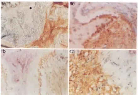
Figure 1 demonstrates the relative abun-dance of IL-1a (a), IL-6 (b), TGF-ß (c) and TNF-a (d) in the bone samples. Areas of non-fibrous marrow stroma were consistently negative stained for all tested cytokines (Fig-ure 1a) and served also as internal negative control for the immunostaining procedure. Higher power micrographs showed positive osteoblast staining for IL-6 (Figure 1b) and TGF-ß (Figure 1c) but not for TNF-a (Figure 1d). Osteocytes also stained positive for TGF-ß.
While highly specific immunostaining was obtained in the fibrous marrow tissue of patients with osteitis fibrosa using each of the antisera for IL-1a, IL-6, IL-11, TNF-a and TGF-ß, the removal of any one of the
Table 1. Clinical and biochemical features of patients w ith osteitis fibrosa.
Bone histology*
M ild M oderate Severe
(N = 7) (N = 7) (N = 7)
Age 44 ± 6 47 ± 7 46 ± 3
Gender (M /F) 3/4 3/4 3/4
Time on dialysis (months) 101 ± 16 74 ± 9 115 ± 3 Serum
Calcium (mg% ) 9 ± 0.4 9 ± 0.5 9.5 ± 0.5
PTH (pg/ml) 465 ± 103 1107 ± 63 2510 ± 335
Alkaline phosphatase (U/l) 213 ± 46 659 ± 100 2399 ± 361
Data are reported as mean ± SEM . *All patients had increased rates of bone formation
and mild, moderate or severe amounts of marrow fibrosis. PTH = parathyroid hor-mone.
Figure 1. Immunohistochemical localization of cytokines in marrow stroma and in bone cells in osteitis fibrosa. a, Distinct IL-1a accumulation in marrow fibrous tissue. Note sharp transition zone betw een heavily positive fibrous marrow stroma and the negative stain for IL-1a in non-fibrous marrow . M ineralized bone (asterisk) and unmineralized osteoid matrix are negatively stained. b, c, Active, plump osteoblasts immediately adjacent to the osteoid seam expressing immunopositivity for IL-6 (b) and TGF-ß (c). d, High magnification of the transition zone betw een heavily positive fibrous marrow stroma and the negative stain for TNF-a of bone cells and non-fibrous marrow . M agnification 4X (a) and 40X (b-d).
Table 2. Bone localization of immunoreactive cytokines in patients w ith osteitis fibrosa.
Cytokines M arrow stroma Osteoblasts Osteocytes
Fibrotic Non-fibrotic
IL-1a +++ 0 0 0
IL-6 +++ 0 ++ 0
IL-11 + 0 0 0
TNF-a +++ 0 0 0
TGF-ß +++ 0 +++ ++
immunostaining steps resulted in negative immunostaining of the fibrous marrow. Also, the application of nonimmune sera from ani-mals which produced the same type of sec-ondary antibody failed to stain the bone marrow fibrous stroma or cellular elements.
D iscussio n
We have shown that the bone marrow space of patients with secondary hyperpara-thyroidism shows significant accumulations of IL-1a, IL-6, TNF-a and TGF-ß within discrete areas of fibrosis. The protein stain-ing was usually homogeneous for each cy-tokine, though occasionally there were areas of fibrosis with apparently higher concentra-tions of one or more cytokines. Except for IL-11, all of the cytokines tested were rela-tively equal in their expression at the protein level and, in general, the greater the fibrosis the more intense was the cytokine staining. Thus, patients with the highest circulating PTH levels had the largest amount of fibrosis and the most marrow space that was positive for cytokine accumulation.
We did not attempt to determine which cell type was responsible for cytokine pro-duction. As such, any of the marrow stromal elements could be responsible since macro-phages, fibroblasts, megakaryocytes and en-dothelial cells are all capable of secreting one or more cytokines (16). Regardless of the cell type responsible, it is clear that much of the immunoreactivity is localized to the extracellular matrix of the fibrotic areas, suggesting that these cytokines are able to bind certain extracellular matrix proteins. Thus, cytokine accumulation may represent a feed forward phenomenon where cells are stimulated to produce cytokines that stimulate cellular proliferation and matrix proteins which then bind and retain the bioactive cytokines. Alternatively, the el-evated circulating levels of IL-6, IL-1a and TNF-a, which are present in dialysis pa-tients (10,11) may result in a trapping
phenomenon within the marrow fibrosis that leads to their accumulation. Because of the descriptive nature of our observation we cannot determine whether there is a causal relationship between cytokine expression and fibrotic response.
One major difference in cytokine expres-sion was noted with respect to cellular local-ization within bone. Cytoplasmic staining within osteoblasts lining the bone surface was clearly positive for IL-6 and TGF-ß but not for the others. This response was more apparent in those patients whose serum PTH levels were the highest, suggesting that PTH may have had a stimulatory role. Consistent with this notion are several in vitro studies
showing that PTH stimulates IL-6 (13,17) and TGF-ß (18) synthesis in cultured osteo-blasts. Interestingly, osteocytes within the mineralized matrix stained positive for TGF-ß only. This is the first report that demon-strates osteocyte immunoreactivity to TGF-ß and suggests that this cytokine may have a role in mediating the osteocytic regulation of osteoblast and osteoclast activity (19).
cy-tokine accumulation would be increased bone turnover.
We have shown that the high turnover bone lesion of uremic hyperparathyroidism is characterized by abnormal cytokine accu-mulation within marrow fibrosis. Our find-ing that TGF-ß and IL-6 are prominent within cells of the osteoblast lineage suggests that
they may have a direct role in stimulating bone turnover. The results of this study also provide insight into the utilization of immu-nohistochemical staining of plastic embed-ded bone sections as a way to investigate mechanisms of bone remodeling in normal and pathologic conditions.
Re fe re nce s
1. Hasselbach H (1990). Idiopathic myelofi-brosis: A review . European Journal of Haematology,45: 65-92.
2. Reilly JT (1992). Pathogenesis of idio-pathic myelofibrosis: Role of grow th fac-tors. Journal of Clinical Pathology, 45: 461-464.
3. Andress DL & Sherrard DJ (1997). Renal osteodystrophy of chronic renal failure. In: Schrier RW & Gottschalk CW (Editors), Diseases of the Kidney. Little Brow n & Company, Boston, M A, USA.
4. Hruska KA & Teitelbaum SL (1995). Renal osteodystrophy. New England Journal of M edicine, 333: 166-174.
5. Johnston JB, Dalal BI, Israels SJ, Oh S, M cM illan E, Begleiter A, M ichaud G, Israels LG & Greenberg AH (1995). Depo-sition of transforming grow th factor ß in the marrow in myelofibrosis, and the in-tracellular localization and secretion of TGF-ß by leukemic cells. American Jour-nal of Clinical Pathology, 103: 574-582. 6. Lisse I, Hasselbach H & Junker P (1991).
Bone marrow stroma in idiopathic my-elofibrosis and other haematological dis-eases. An immunohistochemical study. Acta Pathologica, M icrobiologica et Im-munologica Scandinavica, 99: 171-178. 7. M artyré M C, M adelenat H, Bryckaert M C,
Laine-Bidron C & Calvo F (1991). In-creased intraplatelet levels of platelet-de-rived grow th factor and transform ing grow th factor-ß in patients w ith myelofi-brosis w ith myeloid metaplasia. British Journal of Haematology, 77: 80-86. 8. M anolagas SC (1998). The role of IL-6
type cytokines and their receptors in bone. Annals of the New York Academy of Sciences, 840: 194-204.
9. Canalis E, M cCarthy TL & Centrella M (1991). Grow th factors and cytokines in bone cell metabolism. Annual Review of M edicine, 42: 17-24.
10. Herbelin A, Urena P, Nguyen AT, Zingraff J & Descamps-Latscha B (1991). Elevated circulating levels of interleukin-6 in pa-tients w ith chronic renal failure. Kidney International, 39: 954-960.
11. Herbelin A, Nguyen AT, Zingraff J, Urena P & Descamps-Latscha B (1990). Influ-ence of uremia and hemodialysis on circu-lating interleukin-1 and tumor necrosis factor a. Kidney International, 37: 116-125.
12. Yoneda T (1993). Cytokines in bone: local translators in cell-to-cell communications. In: Noda M (Editor), Cellular and M olecu-lar Biology of Bone. Academic Press, San Diego, CA, USA.
13. Greenfield EM , Shaw SM , Gornic SA & Banks M A (1995). Adenyl cyclase and in-terleukin 6 are dow nstream effectors of parathyroid hormone resulting in stimula-tion of bone resorpstimula-tion. Journal of Clinical Investigation, 96: 1238-1244.
14. Villanueva AR & M ehr LA (1977). M odifi-cations of the Goldner and Gomori-one-step trichrome stain for plastic embedded thin sections of bone. American Journal of M edical Technology, 43: 536-538. 15. Lucena SB, Duarte M EL & Fonseca EC
(1997). Plastic embedded undercalcified bone biopsies: an immunohistochemical method for routine study of bone marrow extracellular matrix. Journal of Histotech-nology,20: 253-257.
16. Groopman JE, M olina JM & Scadden DT (1989). Hematopoietic grow th factors. Bi-ology and clinical applications. New
Eng-land Journal of M edicine, 321: 1449-1459. 17. Feyen JHM , Elford P, Di Padova FE & Trechsel U (1989). Interleukin-6 is pro-duced by bone and modulated by parathy-roid hormone. Journal of Bone and M in-eral Research, 4: 633-638.
18. Oursler M J, Cortese C, Keeting P, Ander-son M A, Bonde SK, Riggs BL & Spelsberg TC (1991). M odulation of transforming grow th factor-ß production in normal hu-man osteoblast-like cells by 17 ß-estradiol and parathyroid hormone. Endocrinology, 129: 3313-3320.
19. Nijw eide PJ, Burger EH, Nulend JK & Van der Plas A (1996). The osteocyte. In: Bilezikian JP, Raisz LG & Rodan GA (Edi-tors), Principles of Bone Biology. Academ-ic Press, Boston, M A, USA.
20. Centrella M , Horow itz M C, Wozney JM & M cCarthy TL (1994). Transforming grow th factor-ß gene family members and bone. Endocrine Review s, 15: 27-39.
21. Littlew ood AJ, Russel J, Harvey GR, Hughes DE, Russell RGGG & Gow en M (1991). The modulation of the expression of IL-6 and its receptor in human osteo-blasts in vitro. Endocrinology, 129: 1513-1520.
22. M anolagas SC & Jilka RL (1995). Bone marrow , cytokines, and bone remodeling. Emerging insights into the pathophysiol-ogy of osteoporosis. New England Jour-nal of M edicine, 332: 305-311.