Effects of 5-aza-2
9
deoxycytidine on
RECK
gene expression and tumor invasion in
salivary adenoid cystic carcinoma
X.Q. Zhou
1,2,3*, S.Y. Huang
3*, D.S. Zhang
1,3*, S.Z. Zhang
3, W.G. Li
3, Z.W. Chen
3and H.W. Wu
31Department of Oral and Maxillofacial Surgery, School of Stomatology, Shandong University, Jinan, China 2Department of Oral and Maxillofacial Surgery, The First People’s Hospital of Jining, Shandong, China 3Department of Oral and Maxillofacial Surgery, Shandong Provincial Hospital, Affiliated to Shandong University, Jinan, China
Abstract
Reversion-inducing cysteine-rich protein with kazal motifs (RECK), a novel tumor suppressor gene that negatively regulates matrix metalloproteinases (MMPs), is expressed in various normal human tissues but downregulated in several types of human tumors. The molecular mechanism for this downregulation and its biological significance in salivary adenoid cystic carcinoma (SACC) are unclear. In the present study, we investigated the effects of a DNA methyltransferase (DNMT) inhibitor, 5-aza-29deoxycytidine (5-aza-dC), on the methylation status of theRECKgene and tumor invasion in SACC cell lines. Methylation-specific PCR (MSP), Western blot analysis, and quantitative real-time PCR were used to investigate the methylation status of theRECKgene and expression of RECK mRNA and protein in SACC cell lines. The invasive ability of SACC cells was examined by the Transwell migration assay. Promoter methylation was only found in the ACC-M cell line. Treatment of ACC-M cells with 5-aza-dC partially reversed the hypermethylation status of theRECKgene and significantly enhanced the expression of mRNA and protein, and 5-aza-dC significantly suppressed ACC-M cell invasive ability. Our findings showed that 5-aza-dC inhibited cancer cell invasion through the reversal ofRECKgene hypermethylation, which might be a promising chemotherapy approach in SACC treatment.
Key words: RECK; Salivary adenoid cystic carcinoma; 5-aza-29deoxycytidine; Methylation; Transwell assay
Introduction
Salivary adenoid cystic carcinoma (SACC) is one of the most common subtypes of malignant tumors. It accounts for only 1% of all head and neck malignancies, but it is well known for both distant metastasis and perineural invasion (1,2). Despite aggressive surgery, long-term survival is only 39.6% after 15 years (3). The underlying molecular mechanisms of carcinogenesis are still unclear.
The reversion-inducing cysteine-rich protein with kazal motifs (RECK) gene was identified by screening an expression library for cDNAs that could reverse the round morphology associated with v-Ki-ras-transformation of 3T3 cells to the nontransformed flat morphology (4). It is thought to be a novel matrix metalloproteinase (MMP) inhibitor that can inhibit tumor angiogenesis, invasion, and metastasis.RECK is expressed in various normal human tissues, but downregulated in several types of human cancers, including pancreatic (5), colorectal (6), breast (7), and hepatocellular carcinoma (8). In a previous study (9), we
found thatRECKexpression was significantly lower in SACC than in normal tissues, but the mechanism was not clear.
DNA methylation changes are frequently found in human cancers (10,11). Hypomethylation of oncogenes can result in aberrant activation, and hypermethylation of suppressor genes can lead to silencing. Several methylation-regulated candidate genes have been identified in SACC, including auprabasin (SBSN) (12), aquaporin 1 (AQP1) (13), phos-phatase and tensin homolog deleted on chromosome 10 (PTEN) (14), cyclin-dependent kinase inhibitors (15), RAS-associated domain family protein 1A (RASSF1) (16), and death-associated protein kinase (DAPK) (17), but the methylation ofRECKin SACC has not yet been reported.
In the present study, we examined the methylation status of theRECKgene and determined the effects of 5-aza-2’deoxycytidine (5-aza-dC) treatment on its methylation status and expression level in human adenoid cystic carcinoma cell lines. The overall aim was the development
Correspondence: Dong-sheng Zhang:,ds63zhang@sdu.edu.cn.. *These authors contributed equally to this study.
of new strategies for cancer prevention and therapeutic interventions.
Material and Methods
Cell lines and cell cultures
Two human salivary adenoid cystic carcinoma cell lines, ACC-2 and ACC-M, were used. Both cell lines were kindly provided by Professor Wantao Chen (Department of Oral and Maxillofacial Surgery, Ninth People’s Hospital, College of Stomatology, Shanghai Jiao Tong University, Shanghai, China). The cells were cultured in Dulbecco’s modified Eagle’s medium (DMEM) with 10% fetal bovine serum (FBS; Hyclone, USA) and antibiotics in a humidi-fied 376C incubator containing 5% CO2.
Cell viability assay
Briefly, cells were seeded on 96-well plates and treated with 5-aza-dC in triplicate. After 24, 72, or 144 h incubation, the medium was replaced with fresh medium containing 0.5 mg/mL 3-(4,5-dimethylthiazol-2-yl)-2,5-diphenyltetra-zolium bromide (MTT). After 4 h, the supernatants were removed and the resulting MTT formazan was solubilized in DMSO and measured spectrophotometrically at 570 nm.
5-aza-dC treatment
We treated the SACC cell lines in triplicate with 5-aza-dC (Sigma, USA) to determine dose- and time-dependent changes as described previously (18). Briefly cells were diluted to a low density (56105/100 mm dish) 24 h before
5-aza-dC treatment. Stock solutions of 5-aza-dC were dissolved in DMSO. Cells were treated with 1, 5, and 10mM 5-aza-dC for 144 h, or 10mM 5-aza-dC for 24, 72, or 144 h. Baseline expression was established in mock treatment of cells with the same volume of DMSO in triplicate.
Bisulfite modification and methylation-specific PCR (MSP)
Genomic DNA was isolated and modified using the CpGenomeTMDirect Prep Bisulfite Modification kit (Millipore, USA) according to the manufacturer’s instructions. MSP reactions were performed using 26Taq PCR MasterMix
(Tiangen, China) in 25 mL volumes under the following conditions: 946C for 3 min, then 30 cycles at 946C for 30 s, 556C for 30 s, 726C for 60 s, and 5 min at 726C. The PCR product lengths for methylated and unmethylated RECK are 195 and 199 bp. Universal Methylated DNA (Millipore) and normal human blood DNA were used as positive controls for the methylated and unmethylated conditions. Water blanks were used as a negative control. Positive and negative controls were used in each round of MSP. All assays were performed in triplicate. For each DNA sample, primer sets for methylated (M) and unmethylated (U) DNA were used for analysis (Table 1). The PCR products were separated on 2% agarose gels and visualized by ethidium bromide
RNA extraction and quantitative real-time PCR Total RNA was isolated from cultured cells using a Trizol reagent kit (TaKaRa, China) according to the manufacturer’s protocol. The quantity and quality of the RNA samples were measured by spectrophotometry and electrophoresis. cDNA was synthesized from 1mg of total RNA using PrimeScriptTM RT reagent kits with gDNA Eraser (TaKaRa). Quantitative real-time PCR reactions were performed using SYBR1 Premix Ex TaqTM (TaKaRa) in 20 mL volumes under the following conditions: 956C for 30 s, followed by 40 cycles of 956C for 5 s and 606C for 20 s. Glyceraldehyde-3-phosphate dehydrogenase (GAPDH) was used as an internal control to check the efficiency of cDNA synthesis and PCR amplification. The RECK and GAPDH primers are shown in Table 2. The 2-DDCTmethod was used for analysis
of quantitative real-time PCR data, and DDCT(threshold
cycle)=(CT RECK, treated group––CT GAPDH, treated
group)––(CT RECK gene, untreated group––CT GAPDH,
untreated group). The change in RECK mRNA expression was calculated as 2-DDCT. All reactions were performed in triplicate with water controls.
Western blot analysis
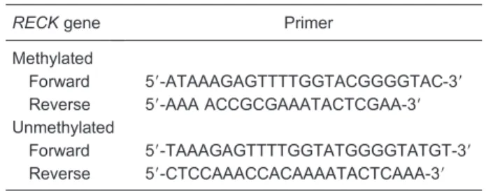
Total cellular proteins were extracted, and were sepa-rated by 10% sodium dodecyl sulfate polyacrylamide gel electrophoresis (SDS-PAGE) as described previously (19). Proteins were transferred from gels to polyvinylidene fluoride membranes followed by incubation with the primary anti-RECK and anti-GAPDH antibodies. GAPDH was used as a loading control on each membrane. Enhanced chem-iluminescence reagents were used to detect the proteins on the membranes. Anti-RECK antibody was purchased from Abcam (USA, at 1:1000 dilution) and anti-GAPDH antibody Table 1. RECK primer sequences for methylation-specific PCR.
RECKgene Primer Methylated
Forward 59-ATAAAGAGTTTTGGTACGGGGTAC-39
Reverse 59-AAA ACCGCGAAATACTCGAA-39 Unmethylated
Forward 59-TAAAGAGTTTTGGTATGGGGTATGT-39 Reverse 59-CTCCAAACCACAAAATACTCAAA-39
Table 2. Primer sequences for quantitative real-time PCR.
Gene Primer
RECK
Forward 59-TGCAAGCAGGCATCTTCAAA-39
Reverse 59-ACCGAGCCCATTTCATTTCTG-39
GAPDH
was obtained from Santa Cruz Biotechnology (USA, at 1:2500 dilution).
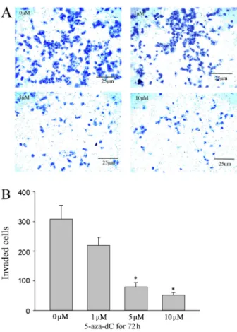
In vitroinvasion assay
The invasiveness of cells exposed to 5-aza-dC was assayed using Matrigel (BD, USA) coated 8 mm pore size filter inserts in 24-well plates (Sigma-Aldrich, USA). Cells exposed to 5-aza-dC (1, 5, 10mM) or DMSO for 72 h were collected and 16105cells in 200mL of medium were placed in the upper part of the Transwell unit and allowed to invade for 24 h. The lower part of the Transwell unit was filled with 500mL medium containing 10% FBS. After incubation, noninvasive cells on the upper part of the membrane were removed with a cotton swab. Invasive cells on the bottom surface of the membrane were fixed in formaldehyde for 20 min, stained with crystal violet for 5 min, and observed by light microscopy at 1006magnification. The number of
cells in 5 randomly selected fields were counted; all assays were performed in triplicate.
Statistical analysis
SPSS (Statistic Package for Social Sciences) 13.0 for Windows (SPSS Inc., USA) was used to analyze data. Differences between groups were assessed by ANOVA
with Dunnett’spost hoc test. Statistical significance was defined as P,0.05.
Results
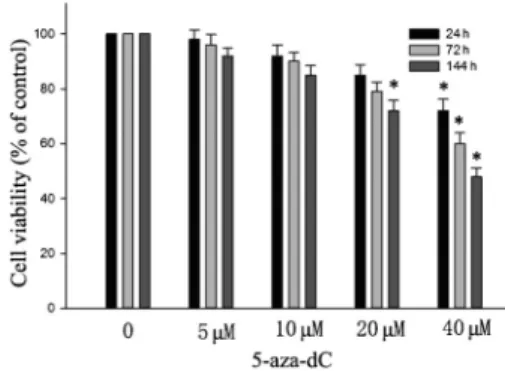
Cytotoxic effect of 5-aza-dC in ACC-M cells
The cytotoxic effect of 5-aza-dC in human adenoid cystic carcinoma ACC-M cells is shown in Figure 1. It is demonstrated that treatment with 40mM for 24 h, or 20mM 5-aza-dC for 144 h significantly decreased the viability of ACC-M cells. Treatment with 5-aza-dC at doses of 10mM or less for 144 h did not cause cytotoxicity of ACC-M cells.
Methylation status ofRECKgene in ACC-2 and ACC-M cell lines
MSP assays showed that the ACC-M cell lines contained both unmethylated and methylated promoters, but that ACC-2 had only a strongly unmethylated promoter (Figure 2A). Normal human blood DNA (NBD) was used as a positive control for unmethylated status and universal methylated DNA (UMD) as a positive control for methylated status, H2O was a negative control.
Reversal of hypermethylation status in ACC-M cells by 5-aza-dC
The time- and dose-dependent effects of 5-aza-dC in ACC-M cell lines were investigated in cells treated with 1, 5, or 10mM of 5-aza-dC for 144 h. Methylation-specificRECK gene bands still existed but had a very weak appearance; however, the unmethylation-specific bands appeared to be enhanced. After treating the cells with 10mM of 5-aza-dC for 24, 72, or 144 h, unmethylation-specific bands of the RECKgene became increasingly intensive, and methyla-tion-specific bands of theRECKgene became increasingly weaker (Figure 2B).
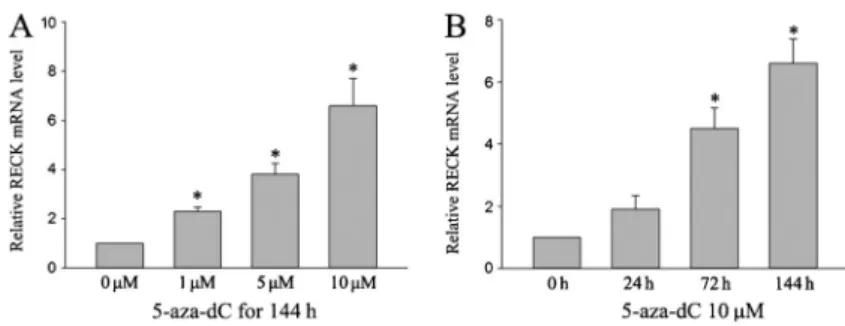
5-aza-dC enhanced RECK mRNA and protein expression in ACC-M cell lines
To confirm the effect of 5-aza-dC on expression of RECK mRNA, real-time quantitative PCR was performed in ACC-M cell lines after treatment with different concentrations of Figure 1.Effect of 5-aza-29deoxycytidine (5-aza-dC) on viability
of ACC-M cells. Cells were treated with various concentrations of 5-aza-dC for 24, 72, and 144 h. Cell viability is reported as means±SD of 3 independent experiments. *P,0.05 compared with control (ANOVA).
Figure 2.A, Methylation status of theRECKgene in ACC-2 and ACC-M cell lines. M: methylation-specific band; U: unmethylation-methylation-specific band; NBD: normal blood DNA as positive control for unmethylated status; UMD: universal methylated DNA as positive control for methylated status; H2O: negative control.B, Alterations of
5-aza-dC for different times (Figure 3). The results showed that the relative amounts of RECK mRNA expressed increased in a dose- and time-dependent manner with significant effects at 1, 5, and 10mM (P,0.05) and at 72 and 144 h (P,0.05).
As shown in Figure 4, RECK protein expression was low in ACC-M cells. After treatment with different doses of 5-Aza-dC for 144 h, or with 10mM of 5-Aza-dC for different times, RECK protein expression increased. These findings suggest that inhibition of RECK protein expression may be associated with RECK promoter methylation.
DNA methyltransferase (DNMT) inhibitor 5-aza-dC reduced the invasive ability of human ACC-M cells
As shown in Figure 5, treatment with 1, 5, or 10mM 5-aza-dC significantly suppressed the invasive ability of ACC-M cells. We conclude that restoration of RECK expression by 5-aza-dC is important for the inhibition of cell invasion by ACC-M cells.
Discussion
In the present study, we demonstrated that 5-aza-dC enhanced expression of RECK mRNA and protein by reversal of the hypermethylation status of the RECK promoter in a time- and dose-dependent manner, and could inhibit cancer cell invasion in ACC-M cell lines.
RECK is an important MMP inhibitor and is involved in the regulation of various physiological and pathological processes. Many studies have demonstrated that RECK
mRNA and protein are frequently highly expressed in both human tissue and untransformed cells, but are lost or not detectable in most tumor cells. Several hypotheses have been advanced to explain the mechanism of low RECK expression in tumor tissue. Epigenetic mechanisms have been shown to play an important role in the regulation of gene expression. Hsu et al. (20) reported that the oncogene HER-2/neu represses RECK by inducing the binding of Sp1 and Sp3 proteins as well as histone deacetylase (HDAC) to the Sp1-binding site. Sasahara et al. (21) reported that the HDAC inhibitor trichostatin A inhibited the interaction between HDAC and Sp1, which prevented binding to the Sp1 site, thereby restoring RECK expression.
DNA methylation of the RECK promoter and histone acetylation/deacetylation have been studied to uncover the underlying mechanisms of RECK expression (22). DNA methylation, a crucial epigenetic alteration, is associated with the silencing of tumor suppressor genes in several cancers. Cho et al. (23) found that downregulation of RECK mRNA and protein expression in colon tumor tissues significantly correlated with methylation of the RECK promoter. Chang et al. (24) suggested that down-regulation of the metastasis suppressor RECK is caused by promoter methylation in non-small-cell lung cancer. In our previous research, we found RECK expression in SACC was significantly lower than in normal tissues, but the underlying mechanism by which RECK was downregulated in tumors has never been reported. In our present research, the methylation status of the RECK promoter assayed by MSP in ACC-M cell lines, characterized by frequent Figure 3. Relative mRNA expression levels of
RECKgene by quantitative real-time PCR in ACC-M cell lines. 5-aza-29deoxycytidine (5-aza-dC) enhanced RECK mRNA expression in a dose-and time-dependent manner. All expression levels are shown relative to the untreated sample. Data are reported as means±SD (n=3). *P,0.05 compared with control (ANOVA).
pulmonary metastasis and screened in ACC-2 cells, was both unmethylated and methylated, but ACC-2 had only unmethylated bands. The present study thus suggests that a decrease in or deficiency of RECK expression in ACC-M cells may be caused by the methylation of CpG islands in the RECK promoter region.
59-Azacytidine has been approved by the FDA for the treatment of myelodysplastic syndrome (25), which, as a classic DNMT inhibitor, was reported to restore RECK mRNA and protein expression by demethylation of the RECK promoter. Epigallocatechin-3-gallate (EGCG) is the major polyphenol and possibly the key active ingredient in green tea. Previous studies (26-29) have shown that EGCG inhibited DNMT and blocked the hypermethylation of newly synthesized DNA strands, resulting in the reversal of the hypermethylation and re-expression of silenced genes with reduced side effects and toxicity. This study demonstrated that almost no RECK protein was expressed in ACC-M cells (isolated from advanced lung metastases),
which are characterized by high pulmonary metastasis and are isolated from ACC-2 lines. After ACC-M cells were treated with 5-aza-dC, the expression of RECK mRNA and protein was increased in a dose- and time-dependent manner. The invasiveness of ACC-M cells was significantly reduced by 5-aza-dC treatment.
Degradation of extracellular matrix (ECM) is an important step during cell invasion and is usually mediated by extracellular proteases, such as MMPs. The inhibition of MMP-2 is a potential means for the prevention of the metastasis of cancer cells (30,31). RECK acts as an inhibitor of MMPs and can inhibit tumor angiogenesis, invasion and metastasis (32). The expression of RECK in SACC is lower than that in normal tissue counterparts, and in our previous research decreased RECK was correlated with a poor prognosis (9). Enhancement of RECK ex-pression may suppress cancer cell invasion (33). In the present study, 5-aza-dC had a significant effect on the invasive capability of ACC-M that was associated with increasing expression of RECK.
The mitogen-activated protein kinase (MAPK) pathway is known to participate in various signaling cascades that play an important regulatory role in cell growth, apoptosis, differentiation, and metastasis (34). Inhibition of the MAPK pathway may potentially prevent angiogenesis, prolifera-tion, invasion, and metastasis in a wide range of tumors (35,36). Metastasis is also regulated by the phosphati-dylinositide-3 kinase/protein kinase B (PI3K/Akt) signaling pathway, which is involved in many cellular processes, including survival, adhesion, and metastasis (37,38). The inhibition of the MAPK and PI3K/Akt pathways may potentially prevent cancer cell proliferation, invasion, and metastasis. Yan (39) reported that tomatidine inhibits the invasion of A549 cells by reducing the expression of MMPs, and also inhibits extracellular signal-regulated (ERK) and Akt signaling pathways and nuclear factor kappa light-chain enhancer of activated B cells (NF-kB) activity. Although the effect of 5-aza-dC on suppressing invasiveness in ACC-M cells was demonstrated, the signal pathway remains unclear. In future studies, we will investigate the signal pathway to better understand the metastasis of SACC.
In conclusion, our findings are the first to show that 5-aza-dC can inhibit cancer cell invasion through reversal of RECK gene hypermethylation, which could serve as a promising chemotherapeutic strategy for SACC treatment. Additional studies are needed to find a more tolerable and effective drug, and to uncover the mechanism underlying RECK hypermethylation in SACC.
Acknowledgments
Research supported by the Natural Science Foundation of Shandong Province (#ZR2012HQ022). Figure 5.5-aza-29deoxycytidine (5-aza-dC) inhibited the invasion
ability of ACC-M in a dose-dependent manner. Cells were incubated with DMSO or different doses of 5-aza-dC for 72 h. After incubation for 24 h, invaded cell numbers were counted as described previously (1006magnification). *P,0.05 compared with control
References
1. Barrett AW, Speight PM. Perineural invasion in adenoid cystic carcinoma of the salivary glands: a valid prognostic indicator?
Oral Oncol2009; 45: 936-940, doi: 10.1016/j.oraloncology. 2009.07.001.
2. Spiro RH, Huvos AG, Strong EW. Adenoid cystic carcinoma of salivary origin. A clinicopathologic study of 242 cases.Am J Surg1974; 128: 512-520, doi: 10.1016/0002-9610(74)90265-7. 3. Fordice J, Kershaw C, El-Naggar A, Goepfert H. Adenoid cystic carcinoma of the head and neck: predictors of morbidity and mortality.Arch Otolaryngol Head Neck Surg
1999; 125: 149-152, doi: 10.1001/archotol.125.2.149. 4. Takahashi C, Sheng Z, Horan TP, Kitayama H, Maki M,
Hitomi K, et al. Regulation of matrix metalloproteinase-9 and inhibition of tumor invasion by the membrane-anchored glycoprotein RECK. Proc Natl Acad Sci U S A1998; 95: 13221-13226, doi: 10.1073/pnas.95.22.13221.
5. Masui T, Doi R, Koshiba T, Fujimoto K, Tsuji S, Nakajima S, et al. RECK expression in pancreatic cancer: its correlation with lower invasiveness and better prognosis.Clin Cancer Res2003; 9: 1779-1784.
6. Takemoto N, Tada M, Hida Y, Asano T, Cheng S, Kuramae T, et al. Low expression of reversion-inducing cysteine-rich protein with Kazal motifs (RECK) indicates a shorter survival after resection in patients with adenocarcinoma of the lung.
Lung Cancer2007; 58: 376-383, doi: 10.1016/j.lungcan.2007. 07.004.
7. Figueira RC, Gomes LR, Neto JS, Silva FC, Silva ID, Sogayar MC. Correlation between MMPs and their inhibitors in breast cancer tumor tissue specimens and in cell lines with different metastatic potential.BMC Cancer2009; 9: 20, doi: 10.1186/1471-2407-9-20.
8. Zhang C, Ling Y, Zhang C, Xu Y, Gao L, Li R, et al. The silencing of RECK gene is associated with promoter hypermethylation and poor survival in hepatocellular carci-noma.Int J Biol Sci2012; 8: 451-458, doi: 10.7150/ijbs.4038. 9. Zhou X, Huang S, Jiang L, Zhang S, Li W, Chen Z, et al. Expression of RECK and MMP-2 in salivary adenoid cystic carcinoma: Correlation with tumor progression and patient prognosis.Oncol Lett2014; 7: 1549-1555.
10. Ehrlich M. DNA hypomethylation in cancer cells.Epigenomics
2009; 1: 239-259, doi: 10.2217/epi.09.33.
11. Herman JG, Baylin SB. Gene silencing in cancer in associa-tion with promoter hypermethylaassocia-tion.N Engl J Med2003; 349: 2042-2054, doi: 10.1056/NEJMra023075.
12. Shao C, Tan M, Bishop JA, Liu J, Bai W, Gaykalova DA, et al. Suprabasin is hypomethylated and associated with metastasis in salivary adenoid cystic carcinoma.PLoS One
2012; 7: e48582, doi: 10.1371/journal.pone.0048582. 13. Shao C, Sun W, Tan M, Glazer CA, Bhan S, Zhong X, et al.
Integrated, genome-wide screening for hypomethylated onco-genes in salivary gland adenoid cystic carcinoma.Clin Cancer Res2011; 17: 4320-4330, doi: 10.1158/1078-0432.CCR-10-2992.
14. Fan X, Chen B, Xu J, Zhang H, Deng F, Xiang X. Methylation status of the PTEN gene in adenoid cystic carcinoma cells.
Mol Med Rep2010; 3: 775-779.
15. Daa T, Kashima K, Kondo Y, Yada N, Suzuki M, Yokoyama S. Aberrant methylation in promoter regions of cyclin-dependent
salivary gland.APMIS2008; 116: 21-26, doi: 10.1111/j.1600-0463.2008.00773.x.
16. Williams MD, Chakravarti N, Kies MS, Maruya S, Myers JN, Haviland JC, et al. Implications of methylation patterns of cancer genes in salivary gland tumors.Clin Cancer Res2006; 12: 7353-7358, doi: 10.1158/1078-0432.CCR-06-1272. 17. Li J, El-Naggar A, Mao L. Promoter methylation of
p16INK4a, RASSF1A, and DAPK is frequent in salivary adenoid cystic carcinoma. Cancer 2005; 104: 771-776, doi: 10.1002/cncr.21215.
18. Yamashita K, Upadhyay S, Osada M, Hoque MO, Xiao Y, Mori M, et al. Pharmacologic unmasking of epigenetically silenced tumor suppressor genes in esophageal squamous cell carcinoma.Cancer Cell2002; 2: 485-495, doi: 10.1016/ S1535-6108(02)00215-5.
19. Liu LT, Chang HC, Chiang LC, Hung WC. Histone deacetylase inhibitor up-regulates RECK to inhibit MMP-2 activation and cancer cell invasion.Cancer Res2003; 63: 3069-3072.
20. Hsu MC, Chang HC, Hung WC. HER-2/neu represses the metastasis suppressor RECK via ERK and Sp transcription factors to promote cell invasion.J Biol Chem 2006; 281: 4718-4725, doi: 10.1074/jbc.M510937200.
21. Sasahara RM, Takahashi C, Noda M. Involvement of the Sp1 site in ras-mediated downregulation of the RECK metastasis suppressor gene.Biochem Biophys Res Commun1999; 264: 668-675, doi: 10.1006/bbrc.1999.1552.
22. Sasahara RM, Brochado SM, Takahashi C, Oh J, Maria-Engler SS, Granjeiro JM, et al. Transcriptional control of the RECK metastasis/angiogenesis suppressor gene. Cancer Detect Prev2002; 26: 435-443, doi: 10.1016/S0361-090X(02) 00123-X.
23. Cho CY, Wang JH, Chang HC, Chang CK, Hung WC. Epigenetic inactivation of the metastasis suppressor RECK enhances invasion of human colon cancer cells. J Cell Physiol2007; 213: 65-69, doi: 10.1002/jcp.21089. 24. Chang HC, Cho CY, Hung WC. Downregulation of RECK by
promoter methylation correlates with lymph node metasta-sis in non-small cell lung cancer.Cancer Sci2007; 98: 169-173, doi: 10.1111/j.1349-7006.2006.00367.x.
25. Das PM, Singal R. DNA methylation and cancer. J Clin Oncol2004; 22: 4632-4642, doi: 10.1200/JCO.2004.07.151. 26. Stresemann C, Brueckner B, Musch T, Stopper H, Lyko F. Functional diversity of DNA methyltransferase inhibitors in human cancer cell lines.Cancer Res2006; 66: 2794-2800, doi: 10.1158/0008-5472.CAN-05-2821.
27. Hellebrekers DM, Griffioen AW, van Engeland M. Dual targeting of epigenetic therapy in cancer.Biochim Biophys Acta2007; 1775: 76-91.
28. Shaw RJ, Hall GL, Lowe D, Bowers NL, Liloglou T, Field JK, et al. CpG island methylation phenotype (CIMP) in oral cancer: associated with a marked inflammatory response and less aggressive tumour biology.Oral Oncol2007; 43: 878-886, doi: 10.1016/j.oraloncology.2006.10.006. 29. Kato K, Long NK, Makita H, Toida M, Yamashita T,
30. Bodey B, Bodey B Jr, Groger AM, Siegel SE, Kaiser HE. Invasion and metastasis: the expression and significance of matrix metalloproteinases in carcinomas of the lung.In Vivo
2001; 15: 175-180.
31. Hrabec E, Strek M, Nowak D, Greger J, Suwalski M, Hrabec Z. Activity of type IV collagenases (MMP-2 and MMP-9) in primary pulmonary carcinomas: a quantitative analysis. J Cancer Res Clin Oncol2002; 128: 197-204, doi: 10.1007/ s00432-001-0320-3.
32. Clark JC, Thomas DM, Choong PF, Dass CR. RECK - a newly discovered inhibitor of metastasis with prognostic significance in multiple forms of cancer.Cancer Metastasis Rev2007; 26: 675-683, doi: 10.1007/s10555-007-9093-8. 33. Rabien A, Ergun B, Erbersdobler A, Jung K, Stephan C.
RECK overexpression decreases invasive potential in pros-tate cancer cells.Prostate2012; 72: 948-954, doi: 10.1002/ pros.21498.
34. Chan-Hui PY, Weaver R. Human mitogen-activated protein kinase kinase kinase mediates the stress-induced activation of mitogen-activated protein kinase cascades. Biochem J
1998; 336 (Part 3): 599-609.
35. Chen PN, Hsieh YS, Chiou HL, Chu SC. Silibinin inhibits cell invasion through inactivation of both PI3K-Akt and MAPK signaling pathways.Chem Biol Interact2005; 156: 141-150, doi: 10.1016/j.cbi.2005.08.005.
36. Chien CS, Shen KH, Huang JS, Ko SC, Shih YW. Antimeta-static potential of fisetin involves inactivation of the PI3K/Akt and JNK signaling pathways with downregulation of MMP-2/9 expressions in prostate cancer PC-3 cells.Mol Cell Biochem
2010; 333: 169-180, doi: 10.1007/s11010-009-0217-z. 37. Kim D, Kim S, Koh H, Yoon SO, Chung AS, Cho KS, et al.
Akt/PKB promotes cancer cell invasion via increased motility and metalloproteinase production.FASEB J2001; 15: 1953-1962, doi: 10.1096/fj.01-0198com.
38. Shukla S, MacLennan GT, Hartman DJ, Fu P, Resnick MI, Gupta S. Activation of PI3K-Akt signaling pathway promotes prostate cancer cell invasion.Int J Cancer2007; 121: 1424-1432, doi: 10.1002/ijc.22862.