ABSTRACT
ORIGINAL AR
Marcelo Lopes de Lima
Ubirajara Ferreira
Elba Cristina Sá Camargo Etchebehere
Allan de Oliveira Santos
Nelson Rodrigues Netto Júnior
Edwaldo Eduardo Camargo
dimercaptosuccinic acid: direct
comparison with the radioactivity
of nephrectomy specimens
Division of Nuclear Medicine, Department of Radiology, and Division
of Urology, Department of Surgery, Universidade Estadual de Campinas
(Unicamp), Campinas, São Paulo, Brazil
CONTEXT AND OBJECTIVE: Studies using radio-nuclides are the most appropriate method for estimating renal function. Dimercaptosuccinic acid chelate labeled with technetium-99m (99m
Tc-DMSA) is the radiopharmaceutical of choice for high-resolution imaging of the renal cortex and estimation of the functional renal mass. The aim of this study was to evaluate a simplifi ed method for determining the absolute renal uptake (ARU) of 99mTc-DMSA prior to nephrectomy, using the
radioactivity counts of nephrectomy specimens as the gold standard.
DESIGN AND SETTING: Prospective study at the Division of Nuclear Medicine, Department of Ra-diology, Universidade Estadual de Campinas. METHODS: Seventeen patients (12 females; range 22-82 years old; mean age 50.8 years old) underwent nephrectomy for various reasons. Renal scintigraphy was performed three to four hours after intravenous administration of a mean dose of 188.7 MBq (5.1 mCi) of 99mTc-DMSA,
which was done six to 24 hours before surgery. The in vivo renal uptake of 99mTc-DMSA was
determined using the radioactivity of the syringe before the injection (measured using a dose calibrator) and the images of the syringe and kidneys, obtained from a scintillation camera. After surgery, the reference value for renal uptake of 99mTc-DMSA was determined by measuring the
radioactivity of the nephrectomy specimen using the same dose calibrator.
RESULTS: The ARU measurements were very similar to those obtained using the reference method, as determined by linear regression (r-squared = 0.96).
CONCLUSION: ARU estimation using the pro-posed method before nephrectomy seems to be accurate and feasible for routine use. KEY WORDS: 99mTc-DMSA. Scintigraphy. Ne-phrectomy. Kidney cortex. Nuclear medicine.
INTRODUCTION
Studies using radionuclides are the most appropriate method for estimating renal function, to add functional information to anatomical studies such as ultrasound and X-ray methods. Dimercaptosuccinic acid chelate labeled with technetium-99m (99mTc-DMSA) was introduced in the early 1970s1,2 and, ever since, it has been the radio-pharmaceutical of choice for high-resolution imaging of the renal cortex and estimation of the functional renal mass.3
Several studies have demonstrated excel-lent correlation between 99mTc-DMSA uptake and creatinine clearance,4 para-aminohippuric acid (PAH) clearance5 and renal technetium-99m diethylenetriamine pentaacetic acid (99mTc-DTPA) accumulation.6,7
Different techniques to measure the ab-solute uptake of 99mTc-DMSA by the kidneys have been described using planar scintigraphy and single photon emission computed tomog-raphy (SPECT), but the methods remain too complex for routine use.8,9 Moreover, none of these techniques for in vivo quantifi ca-tion of isolated renal 99mTc-DMSAuptake has been compared with ex vivo determination of renal uptake.
OBJECTIVE
The aim of this study was to evaluate a simplifi ed method for determining the abso-lute renal uptake (ARU) of 99mTc-DMSA, using the radioactive counts from nephrec-tomy specimens as the reference.
METHODS
Type of study
This was a prospective study conducted in a tertiary public institution.
Patients
Seventeen patients were studied: twelve females and fi ve males, ranging from 22 to 82 years of age (mean 50.8 years). All of these patients had been selected for nephrectomy by means of open or laparoscopic surgery, because of chronic pyelonephritis (eleven patients) or neoplasia (six patients) (Table 1). All patients signed a consent form that had been approved by the Institution’s Ethics Committee.
The weight (in kg) and height (in cm) of all patients were measured for subsequent use in the Tonnensen equations for kidney depth estimation10 (see image processing).
Radiopharmaceutical preparation
The radiopharmaceutical was reconsti-tuted in accordance with the manufacturer’s instructions (CIS Bio International DMSA agent, Gif-sur-Yvette, France; IPEN Mo-lybdenum generator [Mo-99-Tc99m], São Paulo, Brazil).
The labeling effi ciency was determined by means of thin layer chromatography silica gel (TLC-SG) kits (Merck, Darmstadt, Ger-many). The syringes with the doses of 99m Tc-DMSA were measured in a dose calibrator (Capintec CRC-15R, Ramsey, New Jersey, United States). The doses ranged from 173.9 to 207.2 MBq (4.7 to 5.6 mCi), with a mean dose of 188.7 MBq (5.1 mCi). After intrave-nous injection, the residual radioactivity of the syringes was also measured. The radioactivity administered to the patients was calculated by subtracting this residual radioactivity from the syringe radioactivity before injection.
Syringe images
adminis-tration to the patients, to determine the effi-ciency of the scintillation camera detector. The images were acquired for 120 seconds (matrix 256 x 256, zoom 1.8) in a single-head scintil-lation camera equipped with a high-resolution collimator (SP4 HR Elscint-General Electric, Haifa, Israel).
To keep the radioactivity measured in the syringes within the linear range of the detector, the syringes were placed inside a lead cylinder during acquisition. This lead cylinder was developed by the Institution’s Physics Division and consisted of a cylindrical polyvinyl chloride (PVC) tube of 3.8 cm in diameter by 20.3 cm in length inside a lead sheath of 0.1 cm in thickness. The attenuation correction factor was obtained experimentally, as follows.
Determination of the lead device attenuation correction factor
Radioactivity counts were acquired ten times for each of five samples of techne-tium-99m with different radioactivity levels (18.5 MBq; 37 MBq; 74 MBq; 148 MBq; 222 MBq) with the syringes inside the lead cyl-inder, using the same scintillation camera and acquisition parameters. All acquisitions were then repeated without the lead cylinder.
After decay and background corrections, the mean count for each technetium-99m sample was determined. The data obtained with and without the lead device were plotted on two graphs and the attenuation correction factor for the cylinder was then obtained by dividing the two angular coefficients, resulting in the factor 2.8087.
Patient images
Renal scintigraphy was performed three to four hours after administering the radiophar-maceutical, and six to 24 hours prior to surgery. Posterior view images in the supine position (matrix 256 x 256; zoom 1.8) were acquired with 800,000 counts, using the same camera as used for syringe imaging (Figure 1A). Tracer extravasation to soft tissue during injection did not occur in any of the patients, as demonstrated by imaging each patient’s injection site.
The same couch and position used to acquire the patient images were also used to acquire the syringe image, in order to stan-dardize the attenuation factor.
Image processing
Determination of renal counts per second
Using the isocontour method (30%), regions of interest (ROIs) were drawn over
Table 1. Characteristics of the patients who underwent nephrectomy Patient Gender Age Histopathology
1 Female 56 Chronic pyelonephritis
2 Female 66 Chronic pyelonephritis
3 Female 28 Renal angiomyolipoma
4 Male 82 Urothelial carcinoma of the kidney pelvis and atrophy of the renal parenchyma
5 Male 51 Hydronephrosis and chronic pyelonephritis
6 Male 45 Chronic pyelonephritis
7 Female 54 Chronic pyelonephritis and hydronephrosis
8 Female 60 Renal cell carcinoma
9 Female 45 Chronic pyelonephritis and atrophy of the parenchyma
10 Female 38 Chronic pyelonephritis and hydronephrosis
11 Female 76 Renal cell carcinoma
12 Female 29 Hydronephrosis and atrophy of the parenchyma
13 Female 33 Hydronephrosis, xanthogranulomatosis and chronic pyelonephritis
14 Female 34 Chronic pyelonephritis and hydronephrosis
15 Male 67 Transitional cells carcinoma of the pelvis and ureter
16 Female 22 Ureteral duplication, with severe atrophy of the lower pole; hydronephrosis and hydroureter of the upper unit
17 Male 77 Renal cell carcinoma
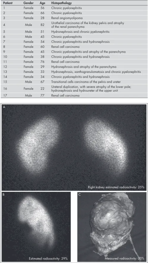
Figure 1. Estimation of absolute renal uptake with technetium-99m dimercaptosuccinic acid (99mTc-DMSA) in a 28-year-old patient with a right kidney tumor diagnosed by
computed tomography (a: in vivo image, posterior view; b: ex vivo image, posterior view; c: nephrectomy specimen, anterior view).
Right kidney estimated radioactivity: 25%
Estimated radioactivity: 29% Measured radioactivity: 30%
A
the kidneys that would be removed. All ROIs were visually checked for accuracy. In four cases, the ROI was not properly drawn by the isocontour method, and a new ROI was manually drawn. The number of counts per second in the kidneys was determined. Background corrections were performed us-ing ROIs around the kidneys. All values were corrected for radioactive decay.
Estimation of kidney depth:
The depth of the kidneys in centimeters was estimated by the Tonnensen equations,10 based on the weight (W) in kg and height (H) in cm of each patient, as follows:
Right kidney = 13.3 (W/H) + 0.7 Left kidney = 13.2 (W/H) + 0.7
Kidney attenuation correction and determination of the renal corrected counts per second:
Tissue attenuation corrections were per-formed by taking into consideration the at-tenuation of technetium-99m photons in water (0.15),11 which is the most similar analogy to hu-man tissue. The following equation was used:
Renal counts per second
e –(0.15) x (kidney depth) Renal corrected
counts per second=
Determination of syringe counts per second:
The count per second was obtained from the syringe images, with correction for
radioactive decay, and multiplication by the attenuation factor (2.8087).
Estimation of the ARU of
99m
Tc-DMSA using the images
The estimation of the ARU was based on the efficiency of the detector (counts/MBq x seconds), by dividing the syringe count per sec-ond before injection by the radioactivity of the same syringe measured by the dose calibrator.
Syringe count per second before injection
Radioactivity of the syringe before injection Efficiency of
the detector =
The ARU was then calculated using the images of the kidneys before nephrectomy:
ARU x 100
Renal corrected count per second/ efficiency
of the detector
Radioactivity adminis-tered to the patient
ARU using nephrectomy specimen images (ex vivo renal images) for evaluating the attenuation correction accuracy
Immediately after surgery, posterior view images of the nephrectomy specimens were obtained using the same scintillation camera as before, with the same acquisition parameters (Figure 1B). These images were not subjected
to soft tissue attenuation (except for the at-tenuation of the renal parenchyma itself ).
All ARU values were then recalcu-lated using these images and the equation described above. These values were compared with the values obtained from the in vivo im-ages and with the reference value.
Determination of the
reference value for the ARU of 99m
Tc-DMSA
Each nephrectomy specimen was placed in a plastic bag and fitted in the same dose calibrator chamber that had been used for radioactivity measurement on the syringes. After correcting all measurements for ra-dioactive decay, the reference value for the ARU was determined for each excised kidney (Figure 1C) as a percentage of the injected dose, by dividing the nephrectomy specimen radioactivity by the injected dose radioactivity and multiplying by 100%, as follows:
Reference value of ARU = (specimen ra-dioactivity/injected radioactivity) x 100%.
Statistical analysis
Linear regression analysis was used to compare the ARU calculated using in vivo and ex vivo images with the ARU from the refer-ence method. It was also applied to compare the ARU values measured with the in vivo and ex vivo images in order to evaluate the simi-larity of these values, which is related to the accuracy of the attenuation correction method applied to the in vivo images of kidneys.
Results
The labeling efficiency of the radiophar-maceutical ranged from 98.7% to 99.3%. The mean ARU values obtained with the in vivo and ex vivo images and with the refer-ence method were, respectively, 5.6%, 6.4% and 6.8%. The calculated ARU values for all patients are listed in Table 2.
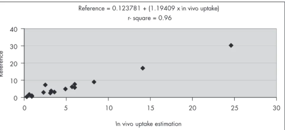
The linear regression analysis comparing the ARU values obtained with the in vivo and ex vivo images and the reference method result-ed in r-squared of 0.96 (in vivo image and the reference method, Figure 2) and 0.99 (ex vivo image and the reference method; Figure 3).
The attenuation correction method ap-plied to the in vivo images was found to be efficient, since the values obtained with the in vivo and ex vivo images were very similar, with r-squared of 0.95.
discussion
Determination of the functional capa-bility of an individual kidney is particularly
Table 2. Absolute renal uptake (ARU) of technetium-99m dimercaptosuccinic acid (99mTc-DMSA): comparison with the reference value
Patient ARU, in vivo image (%) ARU, ex vivo image (%) Reference (nephrectomy
specimen radioactivity) (%)
1 3.2 3.9 3.8
2 0.9 0.9 1.0
3 24.6 27.8 30.3
4 2.5 6.7 7.3
5 3.6 2.8 3.1
6 3.1 2.1 2.6
7 6.0 7.3 7.7
8 14.1 14.7 17.0
9 0.3 0.4 0.4
10 2.3 2.9 2.9
11 8.3 8.1 9.0
12 5.7 8.1 6.1
13 0.6 1.4 1.7
14 6.0 5.4 5.8
15 0.9 0.5 0.6
16 4.9 4.5 4.9
Figure 2. Linear regression analysis between the reference method and the in vivo
absolute renal uptake of technetium-99m dimercaptosuccinic acid (99mTc-DMSA).
Reference = 0.123781 + (1.19409 xin vivo uptake)
r- square = 0.96
0
0 5 10 15 20 25 30
10 20 30 40
In vivouptake estimation
Reference
Figure 3. Linear regression analysis between the reference method and the ex vivo
absolute renal uptake of technetium-99m dimercaptosuccinic acid (99mTc-DMSA).
Reference = -0.137464 + (1.08752 xex vivouptake)
0
0 5 10 15 20 25 30
5 10 15 20 25 30 35
Ex vivouptake estimation
Re
fe
re
nc
e
r- square = 0.99 important when nephrectomy is being
con-sidered. Many studies have evaluated different methods for quantifying 99mTc-DMSA up-take.8,9,12 However, few studies have specifically addressed the use of the ARU measurement to evaluate the kidneys for nephrectomy purpos-es.13,14 When surgery is considered, very precise measurement of the absolute renal function is important, since chronic renal failureafter nephrectomy is a possibility.13 In addition, we have observed that nephrectomy provides a good opportunity to check the efficiency of the method used for absolute renal uptake estimation using 99mTc-DMSA.
Goldraich et al.15 also determined the absolute renal uptake in 142 children with vesi-coureteral reflux based on the percentage of the injected dose of 99mTc-DMSA. Their technique was similar to that described by Raynaud16 using 197Hg-chlormerodrin (197HgCl
2), which was an innovative quantitative method at that time. Goldraich et al.15 reported a significant association between the degree of reflux nephropathy and the functional impairment measured by 99mTc-DMSA uptake. Their tech-nique needed a correction factor consisting of standard radioactivity for renal depth, calcu-lated according to a decreasing exponential curve that was obtained by plotting a series of measurements of the standard against increas-ing thicknesses of a Plexiglas® cover.
Morris et al.17 measured the absolute renal uptake of 99mTc-DMSA in 160 children, based on injected counts, by subtracting the count in the syringe after injection from the count before injection. The attenuation correction used the geometric mean count between the anterior and posterior positions. They considered the thickness of the patient’s intervening tissue at the center of each kidney, measured with the patients in the prone and supine positions. They determined the count in the syringe before injection by acquiring an image for only five seconds, in order to avoid pixel overflow in the computer. In the present study, we preferred to use a lead cylinder attenuator.
Groshar et al. proposed the use of a quantitative SPECT method to estimate the absolute renal function,12 based on the tech-nique of Iosilevsky et al.18 The latter is a very sophisticated method that avoids the need for renal depth determination. However, it is laborious and impractical, and depends on specific site standardizations, including the need to determine the threshold value for each institution. Groshar et al.12 did not use a gold standard test to confirm their final results.
In the present method, we performed attenuation correction by using the renal
depth estimated by the Tonnensen equa-tions.10 which are simple for routine use and have been used by several authors.19,20 These equations may underestimate renal depth, as previously described by Taylor et al.21 Never-theless, we were able to check the efficiency of this method by comparing the ARU measure-ments of the in vivo and ex vivo images, which turned out to be very similar, with excellent correlation (r-squared = 0.95).
The use of the cylindrical device made of lead and PVC was essential for keeping the radioactivity dose within the linear range of the scintillation camera detector, with cor-rection of the error due to dead time loss. A device of this nature can be easily developed in any institution.
In this study, we evaluated a simple meth-od for determining the ARU before surgery using a reliable gold standard, which consisted of direct measurement of renal radioactivity in the nephrectomy specimen. The ARU values
obtained were very similar to those of the ref-erence method, with r-squared of 0.96. Using simple parameters, namely the radioactivity and count in the syringe and the count in the kidney, this method is easy to introduce into routine clinical practice. In particular, measure-ment of the radioactivity and acquisition of an image of the syringe inside the lead device is necessary only once a day. Thus, the only parameters needed for each patient are the injected dose and the count in the kidney.
It is important to emphasize that the method proposed here was only performed on entopic kidneys that were being considered for nephrectomy, mostly with severely reduced function. Further studies are necessary in order to evaluate whether this method would be feasible for ectopic organs and kidneys with normal or mildly impaired function.
1. Lin TH, Khentigan A, Winchell HS. A 99mTc-chelate sub-stitute for organoradiomercurial renal agents. J Nucl Med. 1974;15(1):34-5.
2. Müller-Suur R, Magnusson G, Bois-Svensson I, Jansson B. Estimation of technetium 99m mercaptoacetyltriglycine plasma clearance by use of one single plasma sample. Eur J Nucl Med. 1991;18(1):28-31.
3. Taylor A. Radiopharmaceuticals for the measurement of ‘func-tional renal mass’. In: Blaufox MD, editor. Evaluation of renal function and disease with radionuclides: the upper urinary tract. Basel: Karger; 1989. p. 60-83.
4. Daly MJ, Jones W, Rudd TG, Tremann J. Differential renal func-tion using technetium-99m dimercaptosuccinic acid (DMSA): in vitro correlation. J Nucl Med. 1979;20(1):63-6. 5. Higashihara E, Tokuda H, Kishi H, et al.
Technetium-99m dimercaptosuccinic acid uptake in long-term cath-eterized kidney. Comparison with renal function. Urology. 1988;31(4):327-31.
6. Bingham JB, Maisey MN. An evaluation of the use of 99Tcm-dimercaptosuccinic acid (DMSA) as a static renal imaging agent. Br J Radiol. 1978;51(608):599-607.
7. Taylor A Jr. Quantitation of renal function with static imaging agents. Semin Nucl Med. 1982;12(4):330-44.
8. Kawamura J, Hosokawa S, Yoshida O. Renal function stud-ies using 99mTc-dimercaptosuccinic acid. Clin Nucl Med. 1979;4(1):39-46.
9. Kawamura J, Hosokawa S, Yoshida O, Fujita T, Ishii Y, Torizuka K. Validity of 99mTc dimercaptosuccinic acid renal uptake for an assessment for individual kidney function. J Urol. 1978;119(3):305-9.
10. Tonnensen KH, Munch O, Hald T, et al. Influence on the renogram of variation skin to kidney distance and the clinical importance hereof. In: Zum Winkel K, Blaufox MD, Funck-Brentano JL, editors. Radionuclides in Nephrology, Proceedings of the Third International Symposium on Radionuclides in Nephrology. Berlin: Thieme; 1974. p. 79-86.
11. Gates GF. Split renal function testing using Tc-99m DTPA. A rapid technique for determining differential glomerular filtra-tion. Clin Nucl Med. 1983;8(9):400-7.
12. Groshar D, Moskovitz B, Gorenberg M, et al. Quantitative SPECT of technetium-99m-DMSA uptake in the kidneys of normal children and in kidneys with vesicoureteral reflux: detection of unilateral kidney disease. J Nucl Med. 1994;35(3):445-9.
13. Mullerad M, Kastin A, Issaq E, Moskovitz B, Groshar D, Nativ O. The value of quantitative 99m technetium dimercaptosuc-cinic acid renal scintigraphy for predicting postoperative renal insufficiency in patients undergoing nephrectomy. J Urol. 2003;169(1):24-7.
14. Kawamura J, Itoh H, Okada Y, et al. Preoperative and postopera-tive cortical function of the kidney with staghorn calculi assessed by 99mtechnetium-dimercaptosuccinic acid renal scintigraphy. J Urol. 1983;130(3):430-3.
15. Goldraich NP, Goldraich IH, Anselmi OE, Ramos OL. Reflux nephropathy: the clinical picture in South Brazilian children. Contrib Nephrol. 1984;39:52-67.
16. Raynaud C. A technique for the quantitative measurement of the function of each kidney. Semin Nucl Med. 1974;4(1):51-60. 17. Morris SC, Chittenden SJ, Rivens I, Heary TA, Vanstone
C, Meller ST. Absolute 99Tcm-DMSA renal uptake in children: a study of 321 kidneys. Nucl Med Commun. 1995; 16(7):566-71.
18. Iosilevsky G, Israel O, Frenkel A, et al. A practical SPECT technique for quantitation of drug delivery to human tu-mors and organ absorbed radiation dose. Semin Nucl Med. 1989;19(1):33-46.
19. Gates GF. Glomerular filtration rate: estimation from fractional renal accumulation of 99mTc-DTPA (stannous). AJR Am J Roentgenol. 1982;138(3):565-70.
20. Chachati A, Meyers A, Godon JP, Rigo P. Rapid method for the measurement of differential renal function: validation. J Nucl Med. 1987;28(5):829-36.
21. Taylor A, Lewis C, Giacometti A, Hall EC, Barefield KP. Improved formulas for the estimation of renal depth in adults. J Nucl Med. 1993;34(10):1766-9.
22. Gordon I. Indications for 99mtechnetium dimercapto-succinic acid scan in children. J Urol. 1987;137(3):464-7. 23. Moorin R. 99mTc-DMSA absolute uptake: normal pediatric
values at 2-4 hours. J Nucl Med Technol. 2001;29(1):22-9. 24. Flower MA, Meller ST, Chittenden SJ, Fielding SL, Evans K,
Gor-don I. Absolute 99Tcm-DMSA renal uptake in children: optimum time to scan. Nucl Med Commun. 1995;16(7):572-4.
Sources of funding: Not declared
Conflicts of interest: Not declared
Date of first submission: April 27, 2007
Last received: May 8, 2008
Accepted: May 8, 2008
REFERENCES
By that time, it is postulated that the uptake will have reached a plateau.22 However, for ideal imaging characteristics, a time interval of 2-4 hours between injection and imaging is optimal, without any significant effect on ab-solute 99mTc-DMSA uptake.23 The difference between the values obtained four hours and six hours after injection is less than 6%.24
The fact that the in vivo images were acquired three to four hours after administer-ing the radiopharmaceutical, while the images of the nephrectomy specimens were acquired six to 24 hours later could represent a potential
error in the in vivo ARU values, compared with the specimen ARU values. Neverthe-less, we found a high correlation between the in vivo and specimen ARU values (r -squared = 0.96), which suggests that there was no significant change in the renal uptake over this interval of time, at least for this group of patients, whose kidneys mainly presented low functional capability.
Thus, in the present study, a simplified method was used to quantify kidneys that were scheduled for nephrectomy. The in vivo ARU was compared with the gold standard, i.e. the
“real” quantification of the functional capability of the resected kidney, which was determined as the percentage of the injected radioactivity present in the nephrectomy specimen.
CONCLUSIONS
AUTHOR INFORMATION Mariana da Cunha Lopes de Lima, MD. Attending physician, Division of Nuclear Medicine, Department of Radiology, Universidade Estadual de Campinas (Unicamp), Campinas, São Paulo, Brazil.
Celso Darío Ramos, MD, PhD. Professor, Division of Nuclear Medicine, Department of Radiology, Universidade Estadual de Campinas (Unicamp), Campinas, São Paulo, Brazil.
Sérgio Quirino Brunetto, PhD. Professor, Division of Nuclear Medicine, Department of Radiology, Universidade Estadual de Campinas (Unicamp), Campinas, São Paulo, Brazil.
Marcelo Lopes de Lima, MD, PhD. Attending physician, Division of Urology, Department of Surgery, Universidade Estadual de Campinas (Unicamp), Campinas, São Paulo, Brazil.
Ubirajara Ferreira, MD, PhD. Titular professor, Division of Urology, Department of Surgery, Universidade Estadual de Campinas (Unicamp), Campinas, São Paulo, Brazil.
Elba Cristina Sá Camargo Etchebehere, MD, PhD. Attending physician, Division of Nuclear Medicine, Department of Radiology, Universidade Estadual de Campinas (Unicamp), Campinas, São Paulo, Brazil.
Allan de Oliveira Santos, MD, PhD. Attending physician, Division of Nuclear Medicine, Department of Radiology, Universidade Estadual de Campinas (Unicamp), Campinas, São Paulo, Brazil.
Nelson Rodrigues Netto Júnior, MD, PhD. Titular professor, Division of Urology, Department of Surgery, Universidade Estadual de Campinas (Unicamp), Campinas, São Paulo, Brazil.
Edwaldo Eduardo Camargo, MD, PhD. Titular professor, Division of Nuclear Medicine, Department of Radiology, Universidade Estadual de Campinas (Unicamp), Campinas, São Paulo, Brazil.
Address for correspondence:
Mariana da Cunha Lopes de Lima Serviço de Medicina Nuclear
Av. Zeferino Vaz, s/no — Caixa Postal 6.142 Campinas (SP) — Brasil — CEP 13081-970 Tel. (+55 19) 3521-7801
Fax (+ 55 19) 3521-7821 E-mail: [email protected]
Copyright © 2008, Associação Paulista de Medicina
RESUMO
Estimativa da captação renal absoluta com ácido dimercaptosuccínico marcado com tecnécio-99m: comparação direta com a radioatividade medida em peças de nefrectomia
CONTEXTO E OBJETIVO: Os estudos com radionuclídeos são os mais adequados para se estimar a função renal. O ácido dimercaptosuccínico marcado com tecnécio-99m (DMSA-99mTc) é o radiofármaco de escolha
para imagens de alta resolução dos rins, permitindo, também, estimar massa de parênquima renal funcio-nante. O objetivo deste estudo foi avaliar um método mais simples para determinar-se a captação renal absoluta (CRA) de DMSA-99mTc antes de nefrectomias e validá-lo utilizando-se as contagens radioativas
das próprias peças de nefrectomia como padrão-ouro.
TIPO DE ESTUDO E LOCAL: Estudo prospectivo, desenvolvido no Serviço de Medicina Nuclear do Depar-tamento de Radiologia da Universidade Estadual de Campinas.
MÉTODOS: Foram estudados 17 pacientes (12 pacientes do sexo feminino, média de idade de 50,8 anos), selecionados para a realização de nefrectomia. A cintilografia renal foi realizada três a quatro horas após a administração venosa de 188,7 MBq de DMSA-99mTc, seis a 24 horas antes da cirurgia. A CRA in vivo
foi determinada utilizando-se a radioatividade da seringa antes da injeção (medida com um calibrador de dose) e as imagens da seringa e dos rins, obtidas em uma câmara de cintilação. Após a cirurgia, o valor de referência para a CRA foi determinado medindo-se a radioatividade da peça de nefrectomia com o mesmo calibrador de dose.
RESULTADOS: Os valores de CRA foram muito semelhantes àqueles obtidos com o método de referência, conforme foi demonstrado pela análise de regressão linear (r-quadrado = 0,96).
CONCLUSÃO: A estimativa da CRA com o método proposto antes de nefrectomiasparece ser acurado e aplicável ao uso rotineiro.