Determination of a cutof value for pelvic loor
distensibility using the Epi-no balloon to predict
perineal integrity in vaginal delivery: ROC curve analysis.
Prospective observational single cohort study
Determinação de um valor de ponto de corte para a extensibilidade do assoalho
pélvico pelo balão Epi-no para predizer integridade perineal no parto vaginal:
análise pela curva ROC. Estudo prospectivo observacional de coorte única
Miriam Raquel Diniz Zanetti
I, Carla Dellabarba Petricelli
II, Sandra Maria Alexandre
III, Aline Paschoal
IV, Edward Araujo Júnior
V,
Mary Uchiyama Nakamura
VHospital e Maternidade Amador Aguiar, Osasco, São Paulo, Brazil
ABSTRACT
CONTEXT AND OBJECTIVE: Several risk factors are involved in perineal lacerations during vaginal deliv-ery. However, little is known about the inluence of perineal distensibility as a protective factor. The aim here was to determine a cutof value for pelvic loor distensibility measured using the Epi-no balloon, which could be used as a predictive factor for perineal integrity in vaginal delivery.
DESIGN AND SETTING: Prospective observational single cohort study conducted in a maternity hospital.
METHODS: A convenience sample of 227 consecutive at-term parturients was used. All women had a single fetus in the vertex presentation, with up to 9.0 cm of dilation. The maximum dilation of the Epi-no balloon was measured using a tape measure after it had been inlated inside the vagina up to the parturi-ents’ maximum tolerance. The receiver operating characteristic (ROC) curve was used to obtain the Epi-no circumference measurement with best sensitivity and speciicity.
RESULTS: Among the 161 patients who were included in the study, 50.9% underwent episiotomy, 21.8% presented lacerations and 27.3% retained an intact perineum. Age > 25.9 years; number of pregnancies > 3.4; number of deliveries > 2.2 and circumference measured by Epi-no > 21.4 cm were all directly cor-related with an intact perineum. Circumference measurements using the Epi-no balloon that were greater than 20.8 cm showed sensitivity and speciicity of 70.5% and 66.7% (area under curve = 0.713), respec-tively, as a predictive factor for an intact perineum in vaginal delivery.
CONCLUSION: Circumferences greater than 20.8 cm achieved using the Epi-noballoon are a predictive factor for perineal integrity in parturients.
RESUMO
CONTEXTO E OBJETIVO: Diversos fatores de risco estão envolvidos nas lacerações do períneo durante o parto vaginal, contudo, pouco se sabe sobre a inluência da extensibilidade perineal como um fator protetor. O objetivo foi avaliar o ponto de corte da extensibilidade do assoalho pélvico medido pelo balão Epi-no, o qual poderia ser usado como fator preditor de integridade perineal no parto vaginal.
TIPO DE ESTUDO E LOCAL: Estudo prospectivo observacional de coorte única conduzido em maternidade.
MÉTODOS: Uma amostra de conveniência de 277 parturientes consecutivas no termo foi utilizada. Todas as mulheres tinham feto único com apresentação cefálica letida, com até 9,0 cm de dilatação. A máxima dilatação do balão Epi-no foi medida com ita métrica após a sua insulação dentro da vagina até a tole-rância máxima da parturiente. Uma curva característica de operação do receptor (ROC) foi utilizada para obter a medida da circunferência com a melhor sensibilidade e especiicidade.
RESULTADOS: Dentre as 161 pacientes que foram incluídas no estudo, 50,9% sofreram episiotomia, 21,8% lacerações e 27,3% tiveram o períneo intacto. Idade > 25,9 anos; número de gestações > 3,4; número de partos > 2,2; e medida do perímetro do Epi-no> 21,4 cm foram todos diretamente correlacionados com períneo intacto. Os valores do perímetro com o balão Epi-noque estavam acima de 20,8 cm mostraram sensibilidade e especiicidade de 70,5% e 66,7% (área sob a curva = 0,713), respectivamente, como fator preditor de períneo intacto no parto vaginal.
CONCLUSÃO: Circunferência medida pelo balão Epi-nomaior que 20,8 cm é fator preditor de integridade perineal em parturientes.
IPhD. Voluntary Physiotherapist, Pelvic Floor Unit,
Department of Obstetrics, Universidade Federal de São Paulo (Unifesp), São Paulo, Brazil.
IIMSc. Voluntary Physiotherapist, Pelvic Floor
Unit, Department of Obstetrics, Universidade Federal de São Paulo (Unifesp), São Paulo, Brazil.
IIIPhD. Adjunct Professor, Pelvic Floor Unit,
Department of Obstetrics, Universidade Federal de São Paulo (Unifesp), São Paulo, Brazil.
IVBSc. Postgraduate Student, Pelvic Floor Unit,
Department of Obstetrics, Universidade Federal de São Paulo (Unifesp), São Paulo, Brazil.
VPhD. Associate Professor, Pelvic Floor Unit,
Department of Obstetrics, Universidade Federal de São Paulo (Unifesp), São Paulo, Brazil.
KEY WORDS:
Physical therapy modalities. Pelvic loor.
Perineum. Labor stage, irst. Parturition.
PALAVRAS-CHAVE: Modalidades de isioterapia. Diafragma da pelve. Períneo.
INTRODUCTION
he pelvic loor muscles are a complex involving two layers of muscles. One layer involving the levator ani and puborectalis muscles is deeper and the other is more supericial and involves the perineum.1
Vaginal delivery has been considered to be an important pre-dictive factor for pelvic loor dysfunction, including urinary or fecal incontinence, genital prolapse and levator trauma.2 his is
due to the extensive stretching of the pelvic loor during delivery. Cesarean section reduces the risk of pelvic loor trauma but is not entirely protective.3
It has been proven that vaginal delivery increases the leva-tor hiatal dimensions, especially ater an avulsion injury.4 In a
prospective cohort study on 39 women who delivered vaginally, three-dimensional translabial ultrasound was performed dur-ing the postpartum period and was repeated two and six months ater delivery. Levator avulsion occurred in 39%, and vaginal delivery was correlated with higher maternal age, operative deliv-ery and worsened stress incontinence postpartum.5 In another
study, levator hiatal area > 25 cm in the Valsalva maneuver, mea-sured by three-dimensional ultrasound, was deined as abnormal distensibility or “ballooning” of the levator hiatus.6
he most severe obstetric perineal lesions occur when the sot tissue, muscle, fascia, adipose tissue, skin and mucosa are not suiciently extensible to permit fetal passage. However, these sot perineal tissues can distend, and the extent of the distension var-ies both between parturients and between pregnancvar-ies within an individual. Moreover, this distension can be reduced or increased during the course of the pregnancy by promoting shrinkage or stretching of the sot perineal tissues, respectively, using physio-therapeutic methods.7
Some risk factors for perineal trauma during vaginal deliv-ery have already been established, and these include advanced maternal age, “Caucasian and Asian” races, high maternal body mass index, operative vaginal deliveries, a prolonged expulsive period and high birth weight of the newborn.8-10 However, there
is a lack of studies on the importance of pelvic loor distensibil-ity and its relationship with birth trauma. Distensibildistensibil-ity of the perineum is very important during the second stage of labor, for preventing birth trauma, because of the high pressure imposed by the fetus head on the muscles of the pelvic loor.11
he Epi-noDelphine Plusvaginal dilator (Starnberg Medical, Tecsana GmbH, Munich, Germany) consists of an inlatable sil-icone balloon connected to a manometer via a rubber tube.12
Recently, Kubotani et al.13 compared perineal distensibility using
Epi-no in 23 singleton and 20 twin pregnancies. here was no diference in perineal distensibility between the two groups, but there was a positive correlation between perineal distensibility and abdominal circumference in twin pregnancies.
OBJECTIVE
Because of the absence of an instrument for objectively and quantitatively assessing the maximum degree of pelvic loor distensibility, we decided to use the Epi-no device as a method for measuring this biomechanical property. hus, the aim of this study was to determine a cutof value, in centimeters, for pelvic loor distensibility measured using the Epi-no balloon, which could be used as a predictive factor for muscle integrity in vaginal delivery.
METHODS
A prospective observational single cohort study was conducted at the Amador Aguiar Maternity Hospital (HMMAA), in Osasco, state of São Paulo, Brazil, between January and December 2009. he project was evaluated and approved by the Research Ethics Committee of Universidade Federal de São Paulo (Unifesp), under registration number 1283/08, and by the National Research Ethics Committee, under report number 676. HMMAA is the largest public maternity hospital in Osasco and provides care for low-risk pregnancies (70%) and high-risk pregnancies (30%), at a rate of 600 deliveries/month.
he study included 227 consecutive at-term single births in the cephalic presentation with up to 9.0 centimeters of dilatation and at a maximum station of zero, based on the American College of Obstetrics and Gynecologists classiication of fetal head sta-tion assessments.14 We included both primiparous and
multipa-rous parturients. Only collaborative parturients who wished to undergo the examination, who had not received anesthesia (e.g. rachidian, peridural or combined block) and whose fetus showed good vitality at the time of the assessment were included.
Patients irstly read and signed the informed consent form. If the patient was still a teenager, her mother needed to provide consent and sign for her. he participants then underwent pel-vic loor distensibility assessment (comprising pelpel-vic loor and perineum), which was measured as the circumference in centi-meters of the inlated balloon of the Epi-no device (Starnberg Medical, Tecsana GmbH, Munich, Germany). his was done upon admission to the delivery room. he Epi-no circumference measurements were made by a single examiner (MRDZ), who had had four years of experience of using the Epi-no balloon for perineal muscle training during pregnancy. To reduce the bias of individual tolerance, all parturients received information regard-ing the safety of this device through the assurance that its use does not increase the risk of vaginal infection.15
visible outside the vaginal introitus. his was the assurance that the balloon had reached not only the supericial layer of the pel-vic loor (perineum) but also the deepest layer (including the leva-tor ani muscle). he balloon was then gradually inlated until the tolerable limit, which was subjectively determined by the patient, was reached. All of the patient assessments were performed by the same examiner. Next, the balloon was slowly withdrawn while still fully inlated, the condom was removed and the largest circumfer-ence of the balloon was measured using a measuring tape.
he sample size was estimated such that suicient precision would be attained, i.e. a 95% conidence interval (CI) of width = 0.20, if the observed area under receiver operating characteristic (ROC) curve was greater than 0.60.16 For an the area under the
ROC curve of 0.713, we would need to assess 160 subjects to have a 95% CI width ≤ 0.20.
he perineal trauma was classiied based on third-degree laceration (when the extent of the lesion included the external anal sphincter totally or partially) and fourth-degree laceration (when the rectal mucosa was involved).17 he diagnosis of
peri-neal trauma was made both by doctors and by the midwifes who assisted the labor, but the repairs were made only by doctors.
Statistical analysis was performed using Statistical Package for the Social Sciences (SPSS) v.14 (SPSS Inc., Chicago, IL, USA) and Minitab v.13 (Minitab Inc., State College, PA, USA). he sample size used for our study provided a power of 82.7%. First of all, descriptive statistics were produced on all the variables stud-ied (age, number of gestations and deliveries, body mass index, pelvic loor muscle extensibility, newborn weight and newborn cephalic circumference). Next, univariate analysis was applied to determine which variables inluenced perineal outcomes. Student’s t test for analysis of continuous variables and the Mann-Whitney test were used when the data were not normally distrib-uted. Ater that, multivariate logistic regression was used, taking into consideration all the signiicant variables of the univariate
analysis at a signiicance level of 20%. Adjusted multivariate logistic regression was performed by means of a backward pro-cess. Appropriate odds ratios (OR) with 95% CI were calculated. Probability values < 0.05 were regarded as statistically signiicant.
RESULTS
Initially, we assessed 227 parturients, of whom 117 were nullipa-rous and 110 were multipanullipa-rous. White, mixed and black skin color corresponded to 45.8%, 44.9% and 8.8%, respectively. Following delivery, 66 patients (29.1% of the cohort) were excluded from the analysis: 57 (25.1%) because their delivery was via cesar-ean section, eight (3.5%) because they did not provide suicient medical data and one (0.44%) because the patient let the hospi-tal against medical advice. here was no use of forceps or vacuum extractor device for assisting in any parturient’s delivery.
he patients were not followed up ater delivery, because the hospital where this study was conducted is a public hospital that only provides delivery care, while puerperium follow-up is pro-vided at several primary healthcare units in the metropolitan region of São Paulo. Hence, proper follow-up for perineal trauma cases was impossible.
he 161 remaining parturients averaged 23.6 ± 5.1 years of age with an average body mass index of 27.6 ± 4.3 kg/m2.
he patients had an average Epi-no balloon maximum circum-ference of 19.9 ± 2.7 cm and gave birth to newborns that weighed 3,168 ± 428 g with a head circumference of 34.1 ± 1.5 cm.
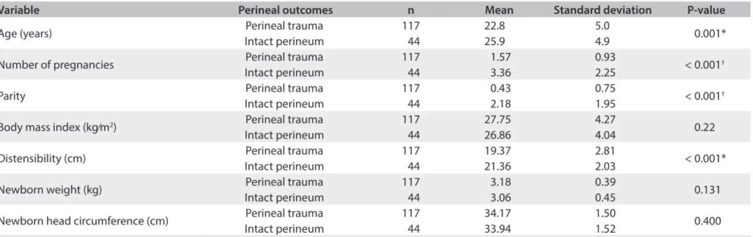
With regard to the perineal outcomes of the 161 patients who were included, 50.9% (n = 82 patients) received right mediolat-eral episiotomy, 21.8% (n = 35) sufered laceration and 27.3% (n = 44) maintained an intact perineum. he perineal outcomes were then analyzed based on variables including age, number of pregnancies, parity, body mass index, Epi-no balloon circumfer-ence, newborn weight and newborn head circumference. hese parameters are presented in Table 1.
Table 1. Univariate analysis from predictive factors for perineal integrity after vaginal delivery
Variable Perineal outcomes n Mean Standard deviation P-value
Age (years) Perineal trauma 117 22.8 5.0 0.001*
Intact perineum 44 25.9 4.9
Number of pregnancies Perineal trauma 117 1.57 0.93 < 0.001†
Intact perineum 44 3.36 2.25
Parity Perineal trauma 117 0.43 0.75 < 0.001†
Intact perineum 44 2.18 1.95
Body mass index (kg∕m2) Perineal trauma 117 27.75 4.27 0.22
Intact perineum 44 26.86 4.04
Distensibility (cm) Perineal trauma 117 19.37 2.81 < 0.001*
Intact perineum 44 21.36 2.03
Newborn weight (kg) Perineal trauma 117 3.18 0.39 0.131
Intact perineum 44 3.06 0.45
Newborn head circumference (cm) Perineal trauma 117 34.17 1.50 0.400
Intact perineum 44 33.94 1.52
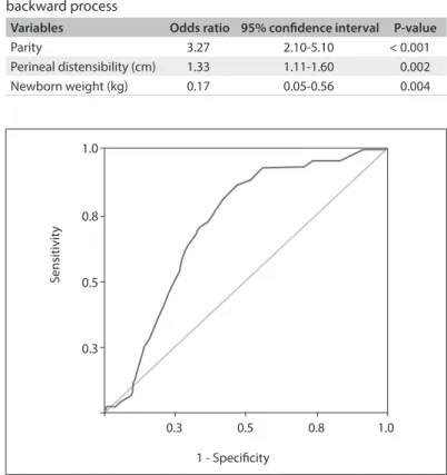
The results from adjusted multivariate logistic regression using the backward process are presented in Table 2. This shows that greater parity, higher distensibility (Epi-no balloon values) and lower newborn weight were predictive factors for perineal integrity.
he ROC curve was constructed, and this demonstrated that an Epi-no circumference measurement of 20.8 cm was the best cutof for perineal integrity ater vaginal delivery (area under curve = 0.713; sensitivity of 70.5% and speciicity of 66.7%) (Figure 1).
DISCUSSION
According to Astrand and Rodahl,18 muscle ibers have
biome-chanical properties such as excitability, contractility, distensi-bility and elasticity. Distensidistensi-bility and elasticity difer because the former property indicates the extent to which a iber can distend during a stretch stimulus, and the latter indicates how well the iber can return to its original length following the stretch stimulus.
To the best of our knowledge, no previous study has objec-tively investigated the maximum distensibility of the pelvic loor muscles. Shek and Dietz19 studied the inluence of levator ani
dis-tensibility on occurrences of levator avulsion ater vaginal delivery.
hey concluded that the levator avulsion could not be predicted antenatally, but they measured the pelvic loor distensibility using transperineal ultrasound when the women were doing a Valsalva maneuver. It is possible that, during this maneuver, the levator muscle does not achieve its maximum distensibility, because for this to occur, it would have to be stretched passively, i.e. the mus-cle would need to be relaxed while a movement elongating it and separating its origin from its insertion was being performed.20
However, our study did not have the objective of assessing leva-tor avulsion.
In the present study, an inlatable balloon was introduced into the vagina and was inlated to produce substantial sion of the pelvic loor muscles. A measure of muscle disten-sion can then be obtained by measuring the circumference of the fully inlated balloon. Although the device was not origi-nally designed for this purpose, this adaptation was necessary because no alternative method for measuring perineal distensi-bility is currently available.
One of the most frequent complaints from patients regard-ing vaginal birth is the fear of a perineal lesion (as occurs with episiotomy or laceration, for example) that could lead to sexual dysfunction ater delivery.21 In some Latin American countries,
including Brazil, the incidence of cesarean sections is as high as 80% in private care and leads to persistent concern within the healthcare system.22 Introduction of a test that could
pre-dict the likelihood that a lesion would not occur might allow the expectant mother to be more comfortable and “secure” in opt-ing for vaginal delivery. In this regard, predictive factors for pel-vic loor lesions during vaginal birth should be studied further. Although some factors predictive of lesions have been identi-ied, such as a prolonged expulsion period, a macrosomic fetus, advanced maternal age, ethnicity and high body mass index, pel-vic loor distensibility has received little attention.
Two previous studies investigated the use of the Epi-no bal-loon trainer to prepare the perineum for vaginal delivery and to reduce levator trauma. In the irst of these, Ruckhäberle et al.15
conducted a prospective, randomized study using Epi-no during pregnancy for perineal preparation (to increase muscle extensi-bility) prior to birth. A total of 135 primigravidae participated in the study and used the device for at least 15 minutes per day from the 37th gestational week onwards, for an average of 15
consecu-tive days; a control group of 135 primigravidae did not undergo any perineal preparation. Ater training, the study group had a mean circumference of 24.3 ± 4.4 cm and showed a tendency towards increased likelihood of having an intact perineum, in comparison with the control group (P = 0.05).
However, there are some possible caveats to the study by Ruckhäberle et al.15 First, the women were instructed to
per-form the stretching exercise with the Epi-no device at home Table 2. Final results from multivariate logistic regression using
backward process
Variables Odds ratio 95% conidence interval P-value
Parity 3.27 2.10-5.10 < 0.001
Perineal distensibility (cm) 1.33 1.11-1.60 0.002
Newborn weight (kg) 0.17 0.05-0.56 0.004
Figure 1. Receiver operating characteristic (ROC) curve for perineal
integrity assessment using the Epi-no circumference measurement.
Area under curve = 0.713; sensitivity of 70.5% and speciicity of 66.7%.
1 - Specificity
S
ensitivit
y
1.0
0.8
0.5
0.3
without any previous supervised training. Because using the Epi-no is not straightforward, this could lead to a bias in its application and in the subsequent results. In addition, the preg-nant women themselves were instructed to measure the max-imum balloon circumference, which could also introduce a bias in the results. Such considerations might explain the pri-mary diference between our results and theirs, in which they reported larger Epi-no circumference measurements. It is important to note that all of our measurements were conducted by the same examiner (MRDZ), which might have produced more reproducible data.
In the second of these investigations on the Epi-no trainer, Shek et al.23 conducted a randomized controlled trial to assess
if the pelvic trainer could reduce levator trauma. he authors selected 200 nulliparous women with singleton pregnancies, and these patients were divided into intervention and control groups. hese patients were examined by means of three-dimensional translabial ultrasound at 35-37 weeks and three months ater delivery. he patients in the intervention group were instructed to use the Epi no device from the 37th week onwards. A total of
156 women returned for the follow-up examination, of whom 78 had had vaginal deliveries. he risk of avulsion was halved in the intervention group (6% versus 13%; P = 0.19). he analysis on the treatment received revealed that the intervention group pre-sented nonsigniicant 42% and 30% reductions in levator avul-sion and microtrauma, respectively (P ≥ 0.22). he authors con-cluded that the Epi no balloon did not reduce the incidence of levator trauma.
One limitation of the present study is that it was diicult for a single examiner to operate the device without assistance. hus, two examiners were required (AP and CDP), in which the principal researcher introduced the balloon to the correct depth and held it in place while the second examiner inlated the balloon. In addi-tion, it would be also very important to evaluate the patients ater delivery, using three-dimensional ultrasound to investigate occur-rences of levator avulsion, as done by Dietz and Shek.24
In our study, we observed no bleeding, which is consis-tent with the report by Ruckhäberle et al.,15 or any other
seri-ous complaint. his suggests that it is safe to use this equip-ment. One previous study reported on a patient who used the Epi-no device and sufered venous air embolism.25 In this case,
the patient’s husband helped her to inlate the device, and ater ten minutes of inlation, the patient began to complain of vagi-nal pain and dizziness, ater which the device was immediately removed. Following a convulsive period, she became unrespon-sive and was taken to the emergency service, where a cesarean section was performed, followed by care in the surgical inten-sive care unit. Ater two months, she no longer exhibited any neurological sequelae but was counseled regarding the risk of
uterine rupture in future pregnancies. he authors of the report hypothesized that the Epi-no device had had an unobserved leak that led to the severe complications. To avoid this potential complication, in the present study we covered the balloon with a condom, which prevented the entry of air during inlation of the balloon. Moreover, although the device has been described as simple to use, we believe that use by an unsupervised non-professional can be harmful. However, new studies about the safety of the Epi-noballoon when it is used by healthcare pro-fessionals or people without previous training should be con-ducted to prove the real degree of safety of the Epi-no device.
From a clinical perspective, when a pregnant woman pres-ents a rigid perineum, she could perform local stretching, for example by means of perineal massage and/or use of an Epi-no device, to achieve adequate perineal distensibility.
Some authors have reported that the levator ani muscle can distend during fetal head descent, during vaginal delivery. Lien et al.26 performed computer simulations on vaginal childbirth
and demonstrated that the pubovisceral portion of the levator ani muscle is subject to a stretch ratio of more than 3:1. Similar results were reported by Hoyter et al.,27 who used magnetic
res-onance imaging of a nulligravid pelvic loor to create a simula-tion model and found that the puborectalis muscle can reach a stretch ratio of 3.5:1 during fetal head descent. Although these studies are important for providing indirect knowledge regard-ing the mechanism of muscle stretchregard-ing durregard-ing a vaginal deliv-ery, such simulations cannot consider the mechanical properties of the pelvic loor with regard to the important biomechanical changes that occur during pregnancy and delivery.27
In the present study, we evaluated the pelvic loor during maximum stretching during parturition, at which the biome-chanical distensibility was at its maximum level. However, the question still remains as to whether these muscles might sufer a more intrinsic, nonvisible form of perineal lesion such as levator avulsion. hus, this study presented a new method for assessing distensibility, which will allow future researchers to understand the importance of distensibility in conferring protection to the pelvic loor during childbirth.
We believe that the main bias of our study was the inla-tion of the Epi-no balloon up to the tolerable limit, which was subjectively determined by the patient. However, all patients received information regarding the safety of the Epi-no balloon before using it.
CONCLUSION
REFERENCES
1. Yiou R, Costa P, Haab F, Delmas V. Anatomie fonctionnelle du
plancher pelvien [Functional anatomy of the pelvic loor]. Prog Urol.
2009;19(13):916-25.
2. Shek KL, Dietz HP. Intrapartum risk factors for levator trauma. BJOG.
2010;117(12):1485-92.
3. World Health Organization. Classiication of practices in normal
birth. Geneva: In: World Health Organization: Care in normal birth: a
practical guide. Report a technical working group. Report No.: WHO
Technical Report Series FRH/MSM/96.24; 1996. p. 34-7. Available
from: http://whqlibdoc.who.int/hq/1996/WHO_FRH_MSM_96.24.
pdf?ua=1. Accessed in 2014 (Aug 15).
4. Shek KL, Dietz HP. The efect of childbirth on hiatal dimensions.
Obstet Gynecol. 2009;113(6):1272-8.
5. Dietz HP, Lanzarone V. Levator trauma after vaginal delivery. Obstet
Gynecol. 2005;106(4):707-12.
6. Dietz HP, Shek C, De Leon J, Steensma AB. Ballooning of the levator
hiatus. Ultrasound Obstet Gynecol. 2008;31(6):676-80.
7. Labrecque M, Eason E, Marcoux S. Women’s views on the practice of
prenatal perineal massage. BJOG. 2001;108(5):499-504.
8. Howard D, Davies PS, DeLancey JO, Small Y. Diferences in perineal
lacerations in black and white primiparas. Obstet Gynecol.
2000;96(4):622-4.
9. Goldberg RP, Kwon C, Gandhi S, et al. Urinary incontinence among
mothers of multiples: the protective efect of cesarean delivery. Am J
Obstet Gynecol. 2003;188(6):1447-50; discussion 1450-3.
10. Burgio KL, Borello-France D, Richter HE, et al. Risk factors for fecal
and urinary incontinence after childbirth: the childbirth and pelvic
symptoms study. Am J Gastroenterol. 2007;102(9):1998-2004.
11. Ashton-Miller JA, Delancey JO. On the biomechanics of vaginal birth
and common sequelae. Annu Rev Biomed Eng. 2009;13:163-76.
12. Kovacs GT, Heath P, Heather C. First Australian trial of the
birth-training device Epi-No: a highly signiicantly increased chance of an
intact perineum. Aust N Z J Obstet Gynaecol. 2004;44(4):347-8.
13. Kubotani JS, Moron AF, Araujo Júnior E, et al. Perineal Distensibility
UsingEpi-no in Twin Pregnancies: Comparative Study with Singleton
Pregnancies. ISRN Obstet Gynecol. 2014;2014:124206.
14. Dupuis O, Silveira R, Zentner A, et al. Birth simulator: reliability of
transvaginal assessment of fetal head station as deined by the
American College of Obstetricians and Gynecologists classiication.
Am J Obstet Gynecol. 2005;192(3):868-74.
15. Ruckhäberle E, Jundt K, Bäuerle M, et al. Prospective randomized
multicentre trial with the birth trainer EPI-NO for the prevention of
perineal trauma. Aust N Z J Obstet Gynaecol. 2009;49(5):478-83.
16. Martins WP, Lima JC, Welsh AW, et al. Three-dimensional Doppler
evaluation of single spherical samples from the placenta:
intra- and interobserver reliability. Ultrasound Obstet Gynecol.
2012;40(2):200-6.
17. Rodriguez A, Arenas EA, Osorio AL, Mendez O, Zuleta JJ. Selective
vs routine midline episiotomy for the prevention of third- or
fourth-degree lacerations in nulliparous women. Am J Obstet Gynecol.
2008;198(3):285.e1-4.
18. Astrand PO, Rodahl K. Textbook of work physiology: physiological
bases of exercise. New York: McGraw-Hill; 1977.
19. Shek KL, Dietz HP. Can levator avulsion be predicted antenatally? Am
J Obstet Gynecol. 2010;202(6):586.e1-6.
20. Vershinin AE, Nazarenko GF. Uglomer dlia opredeleniia amplitudy
dvizhenii v sheinom otdele pozvonochnika [Goniometer for
determining the amplitude of motion in the cervical spine]. Ortop
Travmatol Protez. 1986;(9):49-50.
21. Bracken JN, Dryfhout VL, Goldenhar LM, Pauls RN. Preferences and
concerns for delivery: an antepartum survey. Int Urogynecol J Pelvic
Floor Dysfunct. 2008;19(11):1527-31.
22. Agência Nacional de Saúde Suplementar (Brasil). O modelo de
atenção obstétrica no setor de Saúde Suplementar no Brasil: cenários
e perspectivas/Agência Nacional de Saúde Suplementar. Rio de
Janeiro: ANS; 2008. Available from: www.ans.gov.br/portal/upload/
biblioteca/livro_parto_web.pdf. Accessed in 2014 (Aug 15).
23. Shek KL, Chantarasorn V, Langer S, Phipps H, Dietz HP. Does the
Epi-No Birth Trainer reduce levator trauma? A randomised controlled
trial. Int Urogynecol J. 2011;22(12):1521-8.
24. Dietz HP, Shek C. Levator avulsion and grading of pelvic loor muscle
strength. Int Urogynecol J Pelvic Floor Dysfunct. 2008;19(5):633-6.
25. Nicoll LM, Skupski DW. Venous air embolism after using a
birth-training device. Obstet Gynecol. 2008;111(2 Pt 2):489-91.
26. Lien KC, Mooney B, DeLancey JO, Ashton-Miller JA. Levator ani
muscle stretch induced by simulated vaginal birth. Obstet Gynecol.
2004;103(1):31-40.
27. Hoyte L, Damaser MS, Warield SK, et al. Quantity and distribution of
levator ani stretch during simulated vaginal childbirth. Am J Obstet
Gynecol. 2008;199(2):198.e1-5.
Sources of funding: None
Conlicts of interest: None
Date of irst submission: March 18, 2014
Last received: August 25, 2014
Accepted: September 10, 2014
Address for correspondence:
Edward Araujo Júnior
Rua Carlos Weber, 956 — apto 113 — Visage
Vila Leopoldina — São Paulo (SP) — Brasil
CEP 05303-000
Tel./Fax. (+55 11) 3796-5944