Prevalence of lung structure abnormalities in patients
with acromegaly and their relationship with gas exchange:
cross-sectional analytical study with a control group
Prevalência de alterações na estrutura pulmonar em pacientes com acromegalia e
sua relação com a troca gasosa: estudo transversal analítico com grupo controle
Marcelo Palmeira Rodrigues
I, Luciana Ansaneli Naves
II, Carlos Alberto Viegas
I, Cesar Augusto Melo-Silva
III,
Wagner Diniz de Paula
IV, Márcia Teixeira Cabral
IV, Renata Rodrigues Araújo
V, Luiz Augusto Casulari
VIHospital Universitário de Brasília (HUB), Brasília, Brazil
ABSTRACT
CONTEXT AND OBJECTIVE: Diferent functional respiratory alterations have been described in acromegaly, but their relationship with pulmonary tissue abnormalities is unknown. The objective of this study was to observe possible changes in lung structure and explain their relationship with gas exchange abnormalities.
DESIGN AND SETTING: Cross-sectional analytical study with a control group, conducted at a uni-versity hospital.
METHODS: The study included 36 patients with acromegaly and 24 controls who were all assessed through high-resolution computed tomography of the thorax (CT). Arterial blood gas, efort oximetry and serum growth hormone (GH) and insulin-like growth factor I (IGF-1) were also assessed in the patients with acromegaly.
RESULTS: The abnormalities found in the CT scan were not statistically diferent between the acromegaly and control groups: mild cylindrical bronchiectasis (P = 0.59), linear opacity (P = 0.29), nodular opacity (P = 0.28), increased attenuation (frosted glass; P = 0.48) and decreased attenuation (emphysema; P = 0.32). Radiographic abnormalities were not associated with serum GH and IGF-1. Hypoxemia was present in sev-en patisev-ents; however, in six of them, the hypoxemia could be explained by underlying clinical conditions other than acromegaly: chronic obstructive pulmonary disease in two, obesity in two, bronchial infection in one and asthma in one.
CONCLUSION: No changes in lung structure were detected through thorax tomography in comparison with the control subjects. The functional respiratory alterations found were largely explained by alternative diagnoses or had subclinical manifestations, without any plausible relationship with lung structural factors.
RESUMO
CONTEXTO E OBJETIVO: Diferentes alterações funcionais respiratórias são descritas na acromegalia. Sua relação com anormalidades do tecido pulmonar é desconhecida. O objetivo foi observar possíveis altera-ções da estrutura pulmonar e explicar sua relação com anormalidades da troca gasosa.
TIPO DE ESTUDO E LOCAL: Estudo transversal, analítico, com grupo de controle, realizado em um hos-pital universitário.
MÉTODOS: Incluíram-se 36 pacientes com acromegalia e 24 controles que foram avaliados com tomo-graia computadorizada de alta resolução de tórax (TC); os acromegálicos também foram avaliados com gasometria arterial, oximetria de esforço e dosagens de hormônio de crescimento (GH) e fator de cresci-mento semelhante à insulina (IGF-1).
RESULTADOS: As alterações encontradas na TC não foram estatisticamente diferentes entre os grupos acromegálico e de controle: bronquiectasia cilíndrica leve (P = 0,59), opacidades lineares (P = 0,29), opaci-dades nodulares (P = 0,28), aumento da atenuação (vidro fosco) (P = 0,48) e redução da atenuação (enise-ma; P = 0,32). As alterações radiológicas não se relacionaram com as dosagens de GH e IGF-1. Hipoxemia estava presente em sete pacientes; contudo, em seis deles a hipoxemia poderia ser explicada por condi-ção clínica subjacente diversa da acromegalia: doença pulmonar obstrutiva crônica em dois, obesidade em dois, infecção brônquica em um e asma em um.
CONCLUSÕES: Não foram observadas alterações da estrutura pulmonar por tomograia de tórax, quando comparadas ao grupo de controle; as alterações funcionais respiratórias encontradas são explicáveis em grande parte por diagnósticos alternativos, ou se manifestam de forma subclínica, não apresentando rela-ção plausível com o aspecto da estrutura pulmonar.
IMD, MSc, PhD. Adjunct Professor of
Pneumology, School of Medicine, Universidade de Brasília (UnB), Brasília, Brazil.
IIMD, MSc, PhD. Adjunct Professor of Endocrinology, School of Medicine, Universidade de Brasília (UnB), Brasília, Brazil. IIIMSc, PhD. Physiotherapist, Department of Pneumology, Hospital Universitário de Brasília (HUB), Brasília, Brazil.
IVMD. Physician, Department of Radiology, Hospital Universitário de Brasília (HUB), Brasília, Brazil. VMD. Physician, Department of Pneumology, Hospital Universitário de Brasília (HUB), Brasília, Brazil. VIMD, MSc, PhD. Physician, Department of Endocrinology, Hospital Universitário de Brasília (HUB), Universidade de Brasília (UnB), Brasília, Brazil.
KEY WORDS: Acromegaly. Growth hormone. Anoxia. Lung.
Tomography, X-ray computed.
PALAVRAS-CHAVE: Acromegalia.
Hormônio do crescimento. Anóxia.
Pulmão.
cross-sectional analytical study with a control group | ORIGINAL ARTICLE
INTRODUCTION
Patients with acromegaly have higher respiratory mortality rates when stratiied according to gender and age.1 It has been reported
that patients with acromegaly die prematurely and that mortal-ity due to respiratory diseases is three times more frequent than would be expected in the entire population.1 In that
retrospec-tive study, the type of respiratory disease leading to death was not described. A relative mortality risk due to respiratory diseases of 1.85, in comparison with that of the general population, has been described in the literature. his risk increases to 2.32 when the growth hormone (GH) level remains high ater treatment, i.e. between 2.5 ng/ml and 9.9 ng/ml.2
Many respiratory functional abnormalities have been described in patients with acromegaly. Increased lung volume was initially described in the 1970s.3-5 here is evidence that GH
levels are associated with lung volume. Patients with hypopitu-itarism and GH deiciency have lung volume reductions in com-parison with acromegaly patients.6 here is some controversy
regarding the reason for the increase in lung volume. It has been suggested that it could be due to the increased number of alveoli.7
However, it has been observed that the increase in lung compli-ance found in patients with acromegaly was minimized or even disappeared ater hormonal control, thus indicating that the rea-son for the greater lung volume was the increased alveolar size and not their number.8
Static lung volumes depend on the elastic properties of the respiratory system and muscle strength. Some patients with acromegaly show decreases in respiratory muscle strength, although the magnitude of this change has not been signif-icantly correlated with the increased in lung volume, or with measurements of partial pressure of arterial carbon dioxide (PaCO2) and oxygen (PaO2).9
Increased airlow resistance in patients with acromegaly, with reduced lows at low lung volumes, has also been observed, which suggests that the small airways are involved,5 as well as clearly
obstructive lung disease in nonsmoking patients with acromegaly.10
Studying gas exchange is particularly important in this con-text. On the one hand, it is the result of the sum of variables that determines ventilation, difusion and the ventilation-perfusion balance, among others. On the other hand, major gas exchange alterations such as severe hypoxemia are directly involved in morbidity and mortality rates.11
Varying levels of hypoxemia with increased alveolar-arterial oxygen gradients have been described in patients with acromeg-aly, thereby suggesting the existence of disturbance of the ventila-tion/perfusion relationship.12 his inding has not been observed
by other authors.8,9 However, it should be noted that the samples
of that study were small and there was no investigation of stress-induced hypoxemia.
To our knowledge, despite the functional alterations observed, no patients with acromegaly have been evaluated for possible alterations in the lung structure using high-resolution computed tomography of the thorax. Only a few observations resulting from simple chest X-rays with normal lung parenchyma have been reported.4,12
OBJECTIVE
he aim of this study was to investigate whether patients with acromegaly show lung structure alterations, in comparison with control subjects, and whether these possible alterations are related to gas exchange abnormalities.
METHODS
Patients
Patients with acromegaly who were treated at the Neuroendocrine Unit of Hospital Universitário de Brasília underwent examinations as part of a cross-sectional analytical study with a control group. he diagnosis of acromegaly was established based on clinical char-acteristics and the following biochemical indings:13 lack of growth
hormone (GH) suppression (less than 1 µg/l) during the stan-dard oral glucose tolerance test using 75 g of anhydrous glucose; and levels of insulin-like growth factor I (IGF-I) that were con-sidered high for the age and gender. he sample included patients with both controlled and uncontrolled acromegaly, as shown by a normal IGF-I or glucose tolerance test. Patients presenting uncon-trolled hypopituitarism were excluded from the study.
A control group was included to compare the data obtained via high-resolution computed tomography (CT) of the thorax, with data from other patients who underwent this examination at Hospital Universitário de Brasília within the period of this study. he exclusion criteria for the control group were clinical evidence of respiratory tract disease and acromegaly. his was a convenience sample and it was not matched for sex and age. However, the sam-ples were matched for age and sex by coincidence. Information on age, sex and smoking habit was collected from all patients.
GH levels (Immulite 2000, chemiluminescent enzyme immunometric methodology, Diagnostic Products Corporation, Los Angeles, CA, USA) and IGF-I levels (radioimmunoassay ater extraction) were used in comparisons involving radiologi-cal abnormalities and oxygen level.
Functional respiratory examination
Arterial blood gas analysis was performed by collecting blood from the radial artery, with examination via gasometry for less than three minutes (AVL Compact 3, USA). he reference value for the partial pressure of arterial oxygen (PaO2)14 was corrected
pressure at the place of examination was 680 mmHg. he lower limit was obtained by subtracting the predicted value, which was 1.96 times the standard error of the estimate.
In conducting the exercise oximetry, a magnetic-type bicy-cle ergometer (DJ168 Venus, Belo Horizonte, Brazil) was used. Increasing efort up to the maximum time of six minutes was required. he exercise started without weights and continued until it reached a level close to 60 W. he test was immediately suspended if a decrease of 4% in peripheral oxygen saturation (SpO2) in hemoglobin was observed. A pulse oximeter was used (Healthdyne 950, USA) for SpO2 readings.
Radiological examination
he radiological study was carried out using a CT scanner (Light Speed QX/I, GE Medical Systems, Milwaukee, WI, USA). High spatial resolution sections were made for observations of the lung parenchy-mal structure. he technique followed sequential acquisition at maxi-mum inspiratory apnea, with collimation of 2 x 0.625 mm, increments of 10 mm and a reconstruction algorithm of high spatial frequency, without intravenous administration of exogenous contrast agent.
he indings were consensually described by two radiology experts who did not have any previous knowledge of the clinical or functional data of each patient, and followed standardized nomen-clature.14 he indings were grouped into bronchiectasis,
lin-ear opacities, nodular opacities, increased attenuation, decreased attenuation, atheromatous aorta, coronary alterations and verte-bral degenerative abnormalities. Small high-density nodular opac-ities (calciied granuloma) were excluded from the analysis.
Statistical analysis
Student’s t test was used for statistical analysis of independent sam-ples and, when pertinent, for comparison of variables between the two groups of patients, i.e. with and without acromegaly. Evaluation of the normal distribution of each variable was done using the Shapiro-Wilk test. When the variable under assess-ment did not meet the criteria for normal distribution of values, the Mann-Whitney test was used. With regard to nominal data, the chi-square test was used for proportions. he variables were expressed as means ± standard deviations when the data showed normal distribution. Otherwise, the variables were expressed as medians and interquartile ranges. he indings that were consid-ered statistically signiicant were those in which the associated probability in two-tailed tests was P < 0.05. he statistical sotware used was the SPSS sotware for Windows, version 13.0.
Ethics
he study design was approved by the Research Ethics Committee of the School of Medicine of the University of Brasília. All participants (acromegaly and control groups) gave their writ-ten informed consent in order to participate in the study.
RESULTS
he sample initially included 19 male and 17 female patients with acromegaly. he mean age was 49 ± 12.5 years (range: 24-67 years). he mean time elapsed since the diagnosis of acromegaly was 6.9 ± 5.2 years (range: 7 months to 25.1 years).
Except for three individuals (8.3%) who were included in the study prior to treatment for acromegaly, all the patients had pre-viously undergone the following therapeutic approaches: surgery, radiotherapy and octreotide administration (n = 13; 36.1%); sur-gery and octreotide administration (n = 8; 22.2%); primary treat-ment with octreotide (n = 7; 19.4%); surgery only (n = 4; 11.1%); and surgery and radiotherapy (n = 1; 2.8%).
he control group included 24 patients, of whom 19 were volunteers in another protocol whose objective was to evalu-ate coronary alterations through CT. he other ive patients had neoplasia: adenocarcinoma of the pancreas, colon, Hodgkin’s lymphoma, gestational trophoblastic disease and basal cell carci-noma on the face. hey underwent CT scans as part of a metas-tasis screening process.
Table 1 shows potential confounders. here was no statistically signiicant diference between the groups with regard to age, gen-der or smoking habit. Smoking habit was assessed as a proportion of the patients or as the number of pack-years of smoking.
CT of the pulmonary parenchyma showed several abnor-malities in both groups, as observed in Table 2. In the group with increased attenuation, only ground-glass opacity was observed, whereas in the group with decreased attenuation, only
Table 1. Distribution of the acromegaly and control groups according to age, sex and smoking
Variables Acromegaly (n = 36)
Control (n = 24) P
Age (years) 49.5 ± 12 55.0 ± 17 0.15
Male subjects 53% 58% 0.67
Proportion of smokers 42% 48% 0.64
Pack-years of smoking 0 (0-12) 0 (0-20) 0.68*
*Mann-Whitney test (values expressed as medians and interquartile ranges).
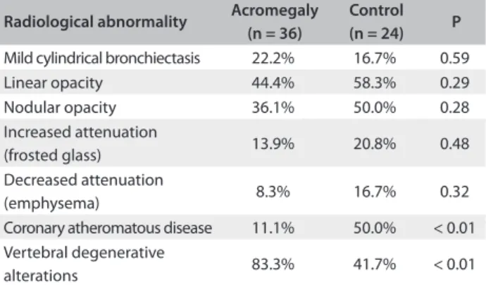
Table 2. Proportion of radiographic abnormalities in the acromegaly and control groups obtained by means of high-resolution computed tomography of the thorax
Radiological abnormality Acromegaly (n = 36)
Control (n = 24) P
Mild cylindrical bronchiectasis 22.2% 16.7% 0.59
Linear opacity 44.4% 58.3% 0.29
Nodular opacity 36.1% 50.0% 0.28
Increased attenuation
(frosted glass) 13.9% 20.8% 0.48
Decreased attenuation
(emphysema) 8.3% 16.7% 0.32
Coronary atheromatous disease 11.1% 50.0% < 0.01 Vertebral degenerative
cross-sectional analytical study with a control group | ORIGINAL ARTICLE
emphysema-type lesions were seen. No nodular opacity had a diameter greater than 1 cm. he entire set of lesions observed was of small extent. If it had been possible to arrange each set of lesions in contiguity for a given patient, the area taken would have been less than one bronchopulmonary segment, except for emphysema, which was difuse and extensive. When compared with the proportion of parenchymal lesions found in the control group, no statistically signiicant diferences were found.
With regard to non-pulmonary alterations seen on thorax tomography, a signiicant diference in the proportion of atheromatous alterations was observed. hese were greater in the control group, and vertebral degenerative alterations were more pronounced in patients with acromegaly (Table 2).
Of the 36 patients initially included in the study, 3 refused to undergo arterial puncture for blood gas analysis. Of the 33 patients who underwent arterial blood gas analysis, 7 (21%) had PaO2 levels below the lower limit of normality, but none had levels of respira-tory failure (PaO2 < 60 mmHg). Among the 7 patients with hypox-emia, 1 had bronchial asthma with symptoms on the examination day, 2 had chronic obstructive pulmonary disease, 2 were obese, 1 had bronchial infection and 1 had no clinically apparent respira-tory disease in the assessment of symptoms or lung structure from the CT scan. In absolute terms, these patients showed PaO2 levels from 0.5 to 4.8 mmHg, i.e. below the lower limit.
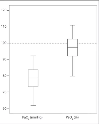
Figure 1 shows the distribution of absolute PaO2 values in mmHg, and they are relatively low. However, this was not con-irmed when the values were corrected for the altitude of Brasília (PaO2 as % of predicted values).
Four patients did not undergo exercise oximetry: 1 refused to do it, 1 was excluded due to knee arthralgia; 1 was excluded due to severe sequelae from pelvic trauma; and 1 was excluded due to the presence of coronary disease. None of the 32 patients who did undergo exercise oximetry showed decreased SPO2 during exercise.
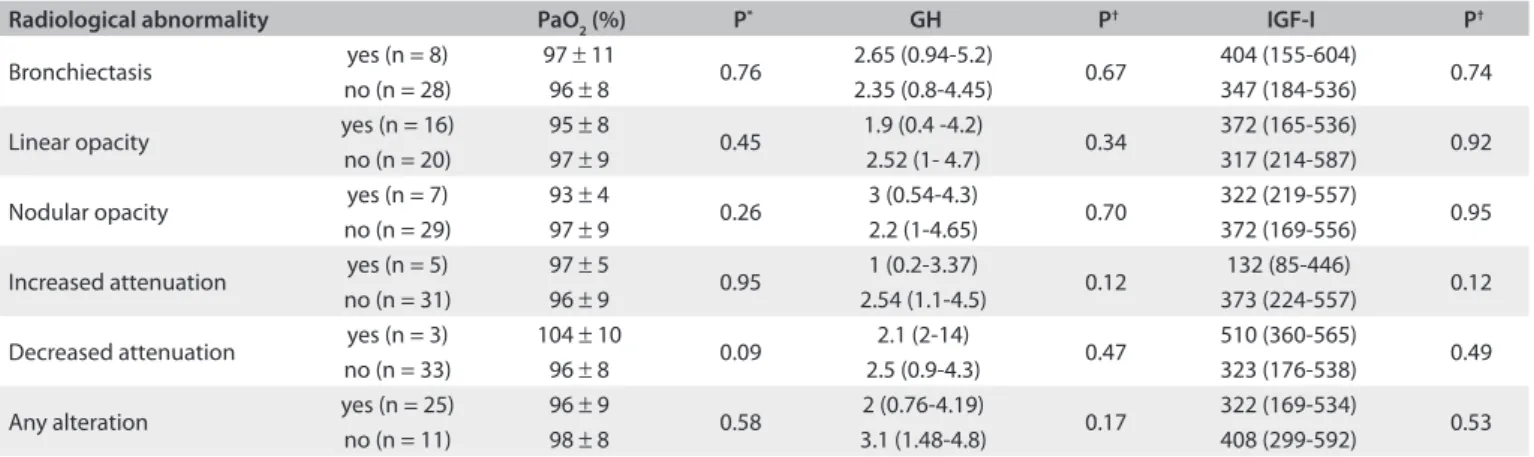
As shown in Table 3, the comparisons between PaO2, GH and IGF-I values as a function of the presence or absence of parenchymal lung abnormalities in each group of tomographic alterations were not statistically signiicant.
DISCUSSION
It has been well established that respiratory alterations in patients with acromegaly may be involved in higher morbidity and mor-tality rates associated with this disease.1,15 Acromegaly patients
develop various respiratory alterations as a result of anatomical changes involving skull and facial bones and sot tissues, carti-lage, respiratory mucosa, lung volume, rib cage geometry and respiratory muscle activity.15
In previous studies on the acromegaly patients who were the subject of the present study,16,17 it was observed that hypoxia
during sleep (deined as more than ive episodes of desaturation per hour) occurred frequently. It afected 41% of the enrolled patients. It was demonstrated that craniofacial abnormalities, obesity and GH and IGF-1 alterations have similar efects on the magnitude of the development of nocturnal hypoxemia in patients with acromegaly.16 Moreover, in evaluating clinical data
as predictors of this condition, it was found that neck circumfer-ence greater than 44 cm was the main factor involved, in compar-ison with other commonly implicated factors such as body mass index (BMI), daytime sleepiness and snoring.17
In this study, the parenchymal lung abnormalities seen on high-resolution thorax tomography showed no statistically sig-niicant diference between the patients with acromegaly and the control group. herefore, no pulmonary indings from this study can be associated with acromegaly. It is likely that factors com-mon to both groups could be responsible, such as smoking. For instance, among other abnormalities related to smoking, emphy-sema is observed in both groups. In addition, it has been shown that the severity of sleep apnea syndrome in acromegaly patients is related to smoking and lung disease.18
Nevertheless, the association of pulmonary lesions with deaths among acromegaly patients is unclear.15 Out of 256 cases
reviewed, chronic bronchitis and emphysema were found in ive
Figure 1. Distribution of absolute and relatively low PaO2 values, in mmHg. This was not conirmed when the values
were corrected for the altitude of Brasília (PaO2 as % of
predicted values). 120
110
100
90
80
70
60
patients and bronchiectasis in three other patients.19 In another
study on 1,362 acromegaly patients, only 11 of them died due to smoking, chronic bronchitis, emphysema and chronic obstruc-tive pulmonary disease.2 hese results are consistent with those
observed in the present study, i.e. no increased frequency of these alterations was observed in either the acromegaly or the control patients. Corroborating this observation of a lack of relationship between acromegaly and lung structural alterations, there were also no signiicant diferences in GH and IGF-I levels (Table 3), based on the presence or absence of radiological abnormalities in any of the lesion groups studied.
Two additional extrapulmonary observations were noted on high-resolution thorax tomography scans. he irst was the much lower frequency of aortic and coronary atheromatous disease in the group with acromegaly, compared with that of the control group. In another study, no diference in the frequency of ath-eromatous plaques was found between acromegaly and control patients, although greater thickening of the intima and media layers of the carotid arteries was observed among those with active or inactive disease.20 It is probable that the results observed
in the present study relect signiicant selection bias in the con-trol group, since the sample mainly included patients who had already been selected for investigation of vascular alterations.
The second observation was the much higher preva-lence of vertebral degenerative alterations in the acromeg-aly patients, in comparison with the control group. Indeed, arthropathy is an important morbidity factor in these patients, affecting the axial skeleton about 52%,21 which is in agreement
with the data observed.
In this study, the PaO2 levels were below the lower reference limit in seven patients (21%), of whom six had clinical conditions other than acromegaly to justify such values. Only one patient had no apparent reason for hypoxemia. It might be that this was a discrepant isolated case with an unusually low value, which is
not uncommon in interpreting pulmonary function tests,22 and
this may also have been associated with acromegaly or another unapparent condition.
he low hypoxemia magnitude found allows us to question its clinical relevance, particularly because of the hypothetical association between hypoxemia and respiratory morbidity and mortality in patients with acromegaly. In this study, no patient showed respiratory failure or decreased SpO2 during exercise and there was no relationship between hypoxemia and any radiologi-cal alterations in the lung structure.
Other authors have studied the behavior of PaO2 in acro-megaly.8,9,12 One study8 showed only the normal average value for
the entire group. In another two studies, all of the individual val-ues were described.9,12 Hypoxemia was present in only one of the
ten acromegaly patients studied,9 when the same normality
cri-teria used in the present study were applied.23 he other study
included eleven acromegaly patients who were asymptomatic and nonsmokers, and who had normal thorax X-rays.12 Perfusion
lung scans showed abnormal results in four out of ive patients. Ten patients for whom PaO2 data were available showed hypox-emia prevalence of 80%. However, the authors did not deine a lower normal limit criterion. Four of the patients were obese; one presented BMI of 39.3 kg/m2. When the normality criteria used
in that study23 were applied to these patients, the hypoxia
preva-lence dropped to 70%.12 If PaO
2 were to be corrected for BMI, 24
the hypoxemia prevalence would be 50%, which is still a very high rate. It is possible that the patients studied by Luboshitzky et al.12
showed some pulmonary vascular involvement whose nature has not yet been correlated with the presence of acromegaly.
Hypoxemia has intra or extrapulmonary causes. he intra-pulmonary causes are difusion and ventilation-perfusion distur-bances. An association of hypoxemia with pulmonary alterations due to acromegaly in a cross-sectional study should be viewed with extreme caution, since ventilation-perfusion mismatches
Table 3. PaO2, growth hormone (GH) and insulin-like growth factor I (IGF-I) values based on the presence or absence of radiological pulmonary abnormalities, obtained by means of high-resolution thorax computed tomography
Radiological abnormality PaO2 (%) P* GH P† IGF-I P†
Bronchiectasis yes (n = 8) 97 ± 11 0.76 2.65 (0.94-5.2) 0.67 404 (155-604) 0.74
no (n = 28) 96 ± 8 2.35 (0.8-4.45) 347 (184-536)
Linear opacity yes (n = 16) 95 ± 8 0.45 1.9 (0.4 -4.2) 0.34 372 (165-536) 0.92
no (n = 20) 97 ± 9 2.52 (1- 4.7) 317 (214-587)
Nodular opacity yes (n = 7) 93 ± 4 0.26 3 (0.54-4.3) 0.70 322 (219-557) 0.95
no (n = 29) 97 ± 9 2.2 (1-4.65) 372 (169-556)
Increased attenuation yes (n = 5) 97 ± 5 0.95 1 (0.2-3.37) 0.12 132 (85-446) 0.12
no (n = 31) 96 ± 9 2.54 (1.1-4.5) 373 (224-557)
Decreased attenuation yes (n = 3) 104 ± 10 0.09 2.1 (2-14) 0.47 510 (360-565) 0.49
no (n = 33) 96 ± 8 2.5 (0.9-4.3) 323 (176-538)
Any alteration yes (n = 25) 96 ± 9 0.58 2 (0.76-4.19) 0.17 322 (169-534) 0.53
no (n = 11) 98 ± 8 3.1 (1.48-4.8) 408 (299-592)
cross-sectional analytical study with a control group | ORIGINAL ARTICLE
are present in a myriad of conditions. herefore, it is necessary to rule out the coexistence of other diseases, particularly non-pulmonary diseases that cause hypoxia. Obesity, for example, can cause hypoxia through a zone of low ventilation-perfusion ratio formed in the lower regions due to changes in compliance of the thoracic cage.25
When functional respiratory alterations are subtle, a cross-sectional clinical investigation based on reference values makes it harder to distinguish whether alterations are present. Creating speciic cutof values to deine functional alterations in acromeg-aly cases would irst require deinition of which abnormalities are clearly associated with this condition.
Longitudinal study designs are particularly interesting in this regard, since these make it possible to assess the impact of treat-ment on disease progression and simplify the issue of control, which may be due to the individual himself. However, it is very diicult to deine a homogeneous cohort with regard to the length of time of disease progression because the insidious nature of the disease leads to highly variable times between disease onset and diagnosis.
Although there is higher morbidity and mortality relating to lung diseases in patients with acromegaly, in comparison with healthy individuals,1 it is not clear which respiratory changes can
contribute to this situation.17,18 he results reported in the
pres-ent paper may contribute towards understanding the pulmonary function abnormalities of patients with acromegaly and may pro-vide data for future research on this topic.
CONCLUSION
In conclusion, no changes in lung structure were observed on thoracic tomography, in comparison with control subjects. he abnormalities in respiratory function found were largely explained by alternative diagnoses or had subclinical manifesta-tions, without showing any plausible relationship with the abnor-mality of the lung structure.
REFERENCES
1. Wright AD, Hill DM, Lowy C, Fraser TR. Mortality in acromegaly. Q J
Med. 1970;39(153):1-16.
2. Orme SM, McNally RJ, Cartwright RA, Belchetz PE. Mortality and
cancer incidence in acromegaly: a retrospective cohort study.
United Kingdom Acromegaly Study Group. J Clin Endocrinol Metab.
1998;83(8):2730-4.
3. Brody JS, Fisher AB, Gocmen A, DuBois AB. Acromegalic
pneumonomegaly: lung growth in the adult. J Clin Invest.
1970;49(6):1051-60.
4. Evans CC, Hipkin LJ, Murray GM. Pulmonary function in acromegaly.
Thorax.1977;32(3):322-7.
5. Harrison BD, Millhouse KA, Harrington M, Nabarro JD. Lung function
in acromegaly. Q J Med. 1978;47(188):517-32.
6. De Troyer A, Desir D, Copinschi G. Regression of lung size in adults
with growth hormone deiciency. Q J Med. 1980;49(195):329-40.
7. Donnelly PM, Grunstein RR, Peat JK, Woolcock AJ, Bye PT. Large lungs
and growth hormone: an increased alveolar number? Eur Respir J.
1995;8(6):938-47.
8. García-Río F, Pino JM, Díez JJ, et al. Reduction of lung distensibility
in acromegaly after suppression of growth hormone hypersecretion.
Am J Respir Crit Care Med. 2001;164(5):852-7.
9. Iandelli I, Gorini M, Duranti R, et al. Respiratory muscle function
and control of breathing in patients with acromegaly. Eur Respir J.
1997;10(5):977-82.
10. Trotman-Dickenson B, Weetman AP, Hughes JM. Upper airlow
obstruction and pulmonary function in acromegaly: relationship to
disease activity. Q J Med. 1991;79(290):527-38.
11. Criner GJ. Efects of long-term oxygen therapy on mortality and
morbidity. Respir Care. 2000;45(1):105-18.
12. Luboshitzky R, Barzilai D. Hypoxemia and pulmonary function in
acromegaly. Am Rev Respir Dis. 1980;121(3):471-5.
13. Vieira Neto L, Abucham J, Araujo LA, et al. Recomendações do
Departamento de Neuroendocrinologia da Sociedade Brasileira de
Endocrinologia e Metabologia para o diagnóstico e tratamento da
acromegalia no Brasil. [Recommendations of Neuroendocrinology
Department from Brazilian Society of Endocrinology and Metabolism
for diagnosis and treatment of acromegaly in Brazil]. Arq Bras
Endocrinol Metabol. 2011;55(2):91-105.
14. Austin JH, Müller NL, Friedman PJ, et al. Glossary of terms for CT of
the lungs: recommendations of the Nomenclature Committee of the
Fleischner Society. Radiology. 1996;200(2):327-31.
15. Colao A, Ferone D, Marzullo P, Lombardi G. Systemic complications of
acromegaly: epidemiology, pathogenesis, and management. Endocr
Rev. 2004;25(1):102-52.
16. Rodrigues MP, Naves LA, Casulari LA, et al. Craniofacial abnormalities,
obesity, and hormonal alterations have similar efects in magnitude
on the development of nocturnal hypoxemia in patients with
acromegaly. J Endocrinol Invest. 2008;31(12):1052-7.
17. Rodrigues MP, Casulari LA, Naves LA, et al. Utilização de achados
clínicos para predizer hipoxemia durante o sono em pacientes
com acromegalia [Using clinical data to predict sleep hypoxemia in
patients with acromegaly]. Arq Neuropsiquiatr. 2007;65(2A):234-9.
18. Vannucci L, Luciani P, Gagliardi E, et al. Assessment of sleep apnea
syndrome in treated acromegalic patients and correlation of its
severity with clinical and laboratory parameters. J Endocrinol Invest.
2013;36(4):237-42.
19. Nabarro JD. Acromegaly. Clin Endocrinol (Oxf ). 1987;26(4):481-512.
20. Colao A, Spiezia S, Cerbone G, et al. Increased arterial intima-media
thickness by B-M mode echodoppler ultrasonography in acromegaly.
Clin Endocrinol (Oxf ). 2001;54(4):515-24.
21. Scarpa R, De Brasi D, Pivonello R, et al. Acromegalic axial arthropathy: a
22. Lung function testing: selection of reference values and
interpretative strategies. American Thoracic Society. Am Rev Respir
Dis. 1991;144(5):1202-18.
23. Crapo RO, Jensen RL, Hegewald M, Tashkin DP. Arterial blood gas
reference values for sea level and an altitude of 1,400 meters. Am J
Respir Crit Care Med. 1999;160(5 Pt 1):1525-31.
24. Cerveri I, Zoia MC, Fanfulla F, et al. Reference values of arterial oxygen
tension in the middle-aged and elderly. Am J Respir Crit Care Med.
1995;152(3):934-41.
25. Kryger MH. Restrictive lung disorders. In: Kryger MH, Roth T, Dement
WC, editors. Principles and practice of sleep medicine. Philadelphia:
Elsevier Saunders; 2005. p. 1136-44.
Acknowledgements: The authors would like to thank Ana Paula
Wanderley, Antônio Wanderley, Denise Vieira, Mírian Morbeck and
Rodrigo Carvalho for secretarial and technical support. They also thank
Laboratório Sabin for performing growth hormone and insulin-like
growth factor I determinations, and José Nogueira de Aguiar Junior, who
greatly encouraged this research
Sources of funding: None
Conlict of interest: None
Date of irst submission: September 5, 2013
Last received: March 23, 2014
Accepted: March 26, 2014
Address for correspondence: Luiz Augusto Casulari
CLINEN - SCN
Quadra 1 — bloco F — sala 1105
Brasília (DF) — Brasil
CEP 70711-905
Tel. (+55 61) 3328-0228