Bladder carcinogenesis in rats subjected to ureterosigmoidostomy
and treated with L-lysine
Carcinogênese de bexiga em ratas submetidas à ureterossigmoidostomia
tratadas com L-lisina.
ConCeição apareCida dornelas1; alessandra Marques dos santos2; antonio luCas oliveira Correia3; CaMila de Carvalho Juanes2; João paulo Ferreira Coelho4; BianCa lopes Cunha4; andré viniCius vieira MaCiel4; FranCisCo vagnaldo FeChine JaMaCaru5.
A B S T R A C T
Objective: to evaluate the effect of L-lysine in the bladder and intestinal epithelia in rats submitted to vesicosigmoidostomy.
Methods: we divided forty Wistar rats into four groups: group I – control group (Sham); group II – submitted to vesicosig-moidostomy and treated with L-lysine 150mg/kg; group III – submitted only to vesicosigvesicosig-moidostomy; and group IV – received L-lysine 150mg/kg. After eight weeks the animals were sacrificed. Results: in the bladders of all operated animals we observed simple, papillary and nodular hyperplasia of transitional cells, transitional cell papillomas and squamous metaplasia. As for the occurrence of aberrant crypt foci in the colons of operated animals, we did not observe statistically significant differences in any of the distal, proximal and medium fragments, or in all fragments together (p=1.0000). Conclusion: Although statistically there was no promotion of carcinogenesis in the epithelia of rats treated with L-lysine in the observed time, it was clear the histogenesis of bladder carcinogenesis in its initial phase in all operated rats, this being probably associated with chronic infec-tion and tiny bladder stones.
Keywords: Lysine. Carcinogenesis. Urinary Bladder Neoplasms. Epithelium. Therapeutics.
INTRODUCTION
H
ammer described the first case of carcinoma in ureterosigmoidostomy in 19291. The risk of cancer in anastomoses areas or bowel of patients undergoing surgery for urinary derivations, bladder expansions or bladder replacements with intestinal segments is known for a long time. Most are adeno-carcinomas, but transitional cell carcinomas have also been described. Although the lag time between uri-nary bypass surgery and the onset of cancer is long, the risk of cancer after ureterosigmoidostomy is esti-mated at 200 to 500 times compared with the gen-eral population. However, the exact pathophysiolo-gy of this carcinogenesis process is not known. The earliest pre-neoplastic lesions in colorectal carcino-genesis are the dysplastic aberrant crypts foci (ACF), mucin depleted foci (MDF) and b-catenin-acumulat-ed crypts (BCAC)2. Dornelas et al.3 found that L-ly-sine has promotes bladder chemical carcinogenesis in rats. The objective of this study is to evaluate the effect of this amino acid in the bladder and intestinal epithelia of animals undergoing urinary diversion by vesicosigmoidostomy, a classic experimental surgical carcinogenesis model4.
METHODS
The research project was conducted at the Department of Pathology and Forensic Medicine of Faculty of Medicine of the Universidade Federal do Ceará (UFC) and developed according to the protocol approved by the Ethics in Animal Research Commit-tee (CEPA) of the Universidade Federal do Ceará.
We divided 40 Wistar rats weighing 150 grams into four groups: Group I (6 animals) was submitted to the opening and closing of the lateral walls of rectum and bladder; Group II (14 mice) was
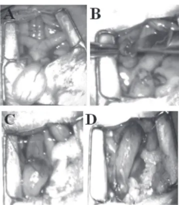
subjected to vesicosigmoidostomy according to the Crissey technique4 (Figure 1), and subsequently treat-ed with L-lysine 150 mg/kg diluted in 0.5ml distilled water by gavage; Group III (14 animals) was submit-ted only to vesicosigmoidostomy; Group IV received only L-lysine 150mg/kg diluted in 0.5ml distilled wa-ter by gavage. Afwa-ter eight weeks we sacrificed the animals and carried out histological analysis (haema-toxylin and eosin) of specimens from the the areas of rectal and bladder anastomosis; The colons was fixed and then stained with 0.1% methylene blue for eval-uation of aberrant crypt foci in stereomicroscopy5,6.
RESULTS
In all operated animals we macroscopically observed polypoid lesions located in the anastomo-ses regions and bladder epithelium. There were nu-merous tiny calculi inside the bladders of individuals submitted to vesicosigmoidostomy. In one animal, there was dilation of the right ureter and faceted cal-culi therein, forming a Stone Street (Figure 2).
On histopathology, the bladder segment of all the operated animals displayed transitional cells simple, nodular and papillary hyperplasia, transitional cell papilloma, (Figure 3) and transitional cell papilloma with squamous metaplasia. In the intestinal segment in operated rats, there were rare aberrant crypts and foci of chronic colitis near the anastomosis area, and some animals presented with atrophy of the epithelium with mucin reduction in areas distant from the anastomosis.
Mortality among operated animals was 45% (18 animals). The main causes were kidney and lung abscesses.
Except by group IV, in all other groups, even in the one in which the animals were not operated, the stereoscopic microscopy identified rare ACF. All ACF contained only one crypt. There were no multi-plicity of crypts or dysplasia signs in the ACF.
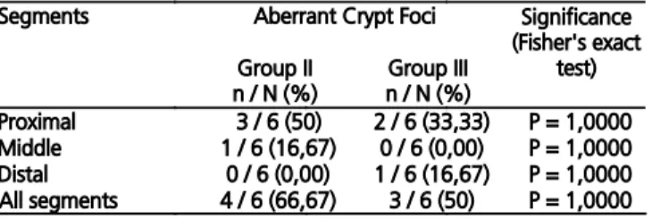
As for the occurrence of ACF in stereoscopic evaluation, when comparing Groups II and III (oper-ated animals) with the Fisher’s exact test, we did not find statistically significant differences considering the proximal (p=1.0000), medium (p=1.0000) and distal (p=1.0000) fragments, or all fragments togeth-er (p=1.0000) (Table 1).
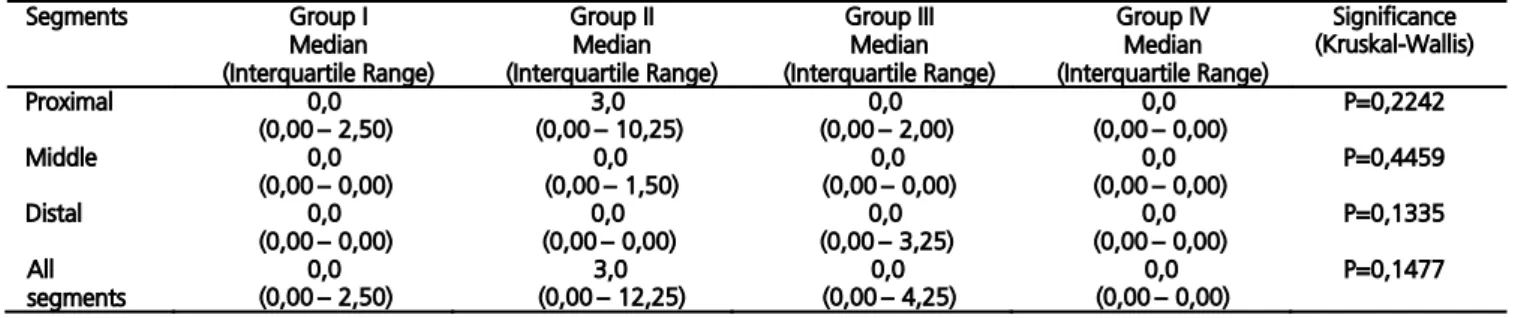
Regarding the presence or absence of aber-rant crypts, the data were expressed as median, in-terquartile range and minimum and maximum values of measurements made in five animals of Groups I and IV and six rats in Groups II and III. We used the Kruskal-Wallis test to compare the four groups, asso-ciated with the multiple comparison test of Dunn, to check for differences between groups in pairs. The
Figure 1. Surgical steps of vesicosigmoidostomy by the Crissey 1980 te-chnique: A) Bladder exposure, bicornuate uterus and cervix; B) bladder dome opening and longitudinal incision of colon wall; C) 7-0 Vycril continuous running suture of bladder dome to the colon; and D) suture of bladder neck and urethral section.
results of the evaluation of the number of aberrant crypts found in the proximal, middle and distal colon segments are described in table 2 and in figure 4.
DISCUSSION
The carcinogenesis of anastomoses in uri-nary derivations with gastrointestinal segments remains unclear. Some mechanisms have been proposed to explain the pathophysiology, such as chronic inflammation, recurrent infections, hydro-genionic potential changes (PH) and production of carcinogens by bacteria, among other causes7. The first urinary diversion was the ureterosigmoidostomy, followed years later by colocystoplasty, ileocystoplas-ty and gatrocystoplasileocystoplas-ty. Then, there was the appear-ance of malignancy in a higher percentage than in the general population in the different derivations and urinary bladder enlargements with gastrointesti-nal segments. Some authors postulated that patients under 25 years of age undergoing ureterosigmoid-ostomy had 7,000 times greater risk for developing cancer than the same age population8.
Many experimental studies were performed in rats using the Crissey vesicosigmoidostomy model4,
searching for histopathologic changes, without using carcinogens, and identifying chronic inflammation, hyperplasia and dysplasia with sulfomucins reduction and increased sialomucins9. Gitlin et al.7 held gastro-cystoplasty and ileogastro-cystoplasty in dogs and observed overgrowth of transitional epithelium in enterovesical and gastrovesical anastomoses. These cells expressed not only uroplakins (a molecular marker for urothelial differentiation), but also mucosubstance. They then suggested that these anastomotic cells possessed alterations and hybrid characteristics, possibly being vulnerable to neoplastic transformation.
In 2012, in a study of 44 different centers between 1970 and 2007 with 17,758 patients un-dergoing urinary derivations and bladder plasty with intestinal loops, German researchers found 32 tu-mors10. The risk of tumors in ureterosigmoidostomy was 22 times higher, and in cystoplasty, 13 times higher, than in other forms of continent urinary di-version, such as neobladder, with statistical signifi-cance (p < 0.0001). The risk of tumors in ileocecal derivations (1.26%) and in colon derivation with neo-bladder was 1.43%, significantly higher (p = 0.0001) than in ileal neobladder (0.5%)10.
Our approach for evaluation of colorectal carcinogenesis was the research of aberrant crypts foci (ACF). The ACF was originally described by Bird5 in rats subjected to chemical carcinogenesis of the colon. However, some years later the same author suggested, and others have concluded that, the focus of aberrant crypts is in fact part of the sequential evolution of colon carcinogenesis, which can become dysplastic and may cause adenomas and later carcinomas, thus serving as a model of early, or pre-neoplastic, lesion in colorec-tal carcinogenesis11-13. The dysplastic ACF may present microsatellite instability, methylation with epigenetic si-lencing14. For this reason, it has been consecrated as a model for trials of new anticancer molecules, using the model in rats subjected to chemical carcinogenesis13,15.
Do urinary derivations follow the course of this aberrant crypt foci model? The answer could be interesting if ACF research could be used in the preven-tive clinical evaluation of patients undergoing urinary derivations. Does ACF occur in vesicosigmoidostomy in rats? There are no reports in the literature. And
fur-Figure 3. A) histologic section showing colovesical anastomosis with transitional cell papilloma; and B) transitional cell papillary hyperplasia with squamous metaplasia. Hematoxylin and eo-sin 10x increase.
n: number of rats with aberrant crypt foci in the group; N: number of animals in the group.
ther, can L-lysine promote carcinogenesis of the colon and/or bladder submitted to urinary diversion in rats? This occurrence is carried out by promoters in primed cells. Promoters are able to take primed cells to prolif-eration and, therefore, develop additional mutations. Promoters are not capable of producing mutation, but the condition of maintaining cell proliferation is re-quired so that they can contribute to carcinogenesis16. In a recent study, Dornelas et al.3 found that L-lysine has promoting action of bladder carcinogenesis in rats subjected to chemical carcinogenesis by BBN.
In our study we observed rare ACF. There were no multiplicities of aberrant crypts. All ACF con-tained only one crypt and there were no dysplasias in the ACF. Furthermore, despite a greater number of ab-errant crypts in rats submitted to vesicosigmoidostomy, there was no statistically significant difference between groups II and III, so there was no promotion of carcino-genesis in rats treated with L-lysine. In addition, we observed ACF in non-operated animals. Although the significance of finding isolated ACF without dysplasias is still unknown, there are reports of ACF involution. There has also been described the spontaneous emer-gence of ACF in 344 Fisher rats without the use of a car-cinogens17,18. The commercial diet can promote ACF in-duction in modified animals19. We should note that the observation time in our experiment was eight weeks. Some authors found that advanced age can influence the spontaneous appearance of ACF in humans20.
When analyzing the graph showing the presence of ACF in the proximal (Figure 4A) seg-ment and in proximal, middle and distal segseg-ments together (Figure 4D), we noted that there are greater numbers of ACF in animals undergoing surgery and treated with L-lysine than in those who were only operated without L-lysine treatment. However, there was no statistically significant difference.
The bladder segment, on the other hand, proved extremely reactive when subjected to deriva-tion. All operated animals showed histological lesions in the bladder epithelium of transitional cell simple, papillary and nodular hyperplasia, transitional cells papilloma and squamous metaplasia, lesions already described as sequential in the bladder histogenesis/ carcinogenesis in rodents21.
Calcium salts precipitate at high pH and urate salts precipitate at low pH. In humans, urolithi-asis is very common, but the association between cal-culi and bladder cancer is rare. However, in rats and mice, crystals and urolithiasis increase the likelihood of bladder carcinogenesis22,23.
When the bladder epithelium is subjected to mechanical irritative processes (calculi, foreign body) and recurrent infection, it can evolve into reactive histologic lesions and also sequentially to carcinogenesis in humans.
The bladder epithelium is similar among spe-cies. However, there are anatomical differences that may pathophysiologically explain the association be-tween calculi and carcinogenesis in animals. During voiding, the urinary system of rodents is in horizontal position, whilst the human is in vertical one. When the bladder contracts, it all “wrinkles” except by the trigone region. When there are stones in the bladder of rats, these loose crystals in the anterior wall pro-mote mucosal damage throughout the bladder with contraction. In humans, foreign objects are located in the trigone region, which does not contract during urination. Mucosal damage is then decreased. For this same anatomical reason, human quickly eliminate the crystals that are within the bladder. When calculi
cause obstruction in humans, they cause pain in most cases, what makes treatment and desobstruction to be arranged. Thus, anatomical factors can make ro-dents more susceptible to bladder carcinogenesis23,24.
Our animals did not have a functioning bladder trigone. We ligated the bladder neck and sectioned it, but the bladder was still kept in the horizontal position and saccular, not eliminating the bladder contents through the rectum. This occurred in all operated animals of our experiment in a similar way (groups II and III). Urine, feces, repeated infec-tions may have originated the miniature calculi and the histological lesions. All animals presented with miniature calculi in the bladder, and once the bladder of the animal is positioned ventrally due to four-paw ambulation, the irritating stimulus keeps constant and may explain the magnitude of the histological changes observed in bladder epithelium.
In conclusion, although statistically there was no promotion of carcinogenesis in the epithelia of rats treated with L-lysine in the observed time, the histogenesis of bladder carcinogenesis is clear in its initial phase in the bladder epithelium in all operated rats, this being probably associated with chronic in-fection and tiny bladder stones.
Table 2. Number of Aberrant crypt foci found in all segments in groups I, II, III and IV.
A B S T R A C T
Objetivo: o objetivo deste trabalho é avaliar o efeito da L-lisina nos epitélios vesical e intestinal de ratas submetidas à vesicossigmoid-ostomia. Métodos: quarenta ratas Wistar, foram divididas em quatro grupos: grupo I- grupo controle (Sham); grupo II- submetido à vesicossigmoidostomia e tratado com L-lisina 150mg/kg; grupo III- submetido apenas à vesicossigmoidostomia; e grupo IV- recebeu L-lisina 150mg/kg. Após oito semanas os animais foram sacrificados. Resultados: na bexiga de todos os animais operados observou-se hiperplasia simples, papilar e nodular de células transicionais, papiloma de células transicionais e metaplasia escamosa. Quanto à ocorrência de focos de criptas aberrantes nos colos dos animais operados, não foi evidenciado diferença estatística significante em nenhum dos fragmentos distal, proximal e médio, e todos juntos (P=1,0000). Conclusão: apesar de, estatisticamente, não ter havido promoção de carcinogênese nos epitélios dos ratos tratados com L-lisina, no tempo observado, é nítida a histogênese da carcinogênese de bexiga em sua fase inicial, no epitélio vesical, em todos os ratos operados, estando esta provavelmente associada à infecção crônica e aos diminutos cálculos vesicais.
REFERENCES
1.
Hammer E. Cancer du colon sigmoide dix ans aprèsimplantation des uretères d’une vessie exstrophiée. J Urol. 1929;28:260.
2.
Femia AP, Paulsen JE, Dolara P, Alexander J, CaderniG. Correspondence between flat aberrant crypt foci and mucin-depleted foci in rodent colon carcinogene-sis. Anticancer Res. 2008;28(6A):3771-5.
3.
Dornelas CA, Fechine-Jamacaru FV,Albuquer-que IL, Magalhães HIF, Souza AJS, Alves LA, et al. Chemoprevention with green propolis extracted in L-lysine versus carcinogenesis promotion with L-lysine in N-butyl-N-[4-hydroxybutyl] nitrosamine (BBN) induced rat bladder cancer. Acta Cir Bras. 2012;27:(2);185-92.
4.
Crissey MM, Steele GD, Gittes RF. Rat model forcarcinogenesis in ureterosigmoidostomy. Science. 1980;207(4435):1079-80.
5.
Bird RP. Observations and quantification ofaber-rant crypts in the murine colon treated with a co-lon carcinogen: preliminary findings. Cancer Lett. 1987;37(2):147-51.
6.
Burlamaqui IMB, Dornelas CA, Escalante RD, MotaDMC, Mesquita FJC, Carvalho ER, et al. Optimiza-tion of visibility and quantificaOptimiza-tion of aberrant crypt foci in colonic mucosa in Wistar rats. Acta Cir Bras. 2010;25(2):148-52.
7.
Gitlin JS, Wu XR, Sun TT, Ritchey ML, Shapiro E. Newconcepts of histological changes in experimental aug-mentation cystoplasty: insights into the development of neoplastic transformation at the enterovesical and gastrovesical anastomosis. J Urol. 1999;162(3 Pt 2):1096-100.
8.
Eraklis AJ, Folkman MJ. Adenocarcinoma at the site ofureterosigmoidostomies for exstrophy of the bladder. J Pediatr Surg. 1978;13(6D):730-4.
9.
Castro MA, Ferreira U, Martins MH, StoppigliaRM, Rodrigues Netto Jr N. Histological and histo-chemical changes of the intestinal mucosa at the urothelial-enteric anastomotic site. Int braz j urol. 2006;32(2):222-7.
10.
Kälble T, Hofmann I, Thüroff JW, Stein R, HautmannR, Riedmiller H, et al. Secondary malignancies in uri-nary diversions. Urologe A. 2012;51(4):500, 502-6.
11.
McLellan EA, Medline A, Bird RP. Sequentialanaly-ses of the growth and morphological characteristics of aberrant crypt foci: putative preneoplastic lesions. Cancer Res. 1991;51(19):5270-4.
12.
Hurlstone DP, Cross SS. Role of aberrant crypt focidetected using high-magnification-chromoscopic colonoscopy in human colorectal carcinogenesis. J Gastroenterol Hepatol. 2005;20(2):173-81.
13.
Alrawi SJ, Schiff M, Carroll RE, Dayton M, Gibbs JF,Kulavlat M, et al. Aberrant crypt foci. Anticancer Res. 2006;26(1A):107-19.
14.
Orlando FA, Tan D, Baltodano JD, Khoury T, GibbsJF, Hassid VJ, et al. Aberrant crypt foci as precur-sors in colorectal cancer progression. J Surg Oncol. 2008;98(3):207-13.
15.
Burlamaqui IMB, Dornelas CA, Valença Júnior JT,Mota DMC, Mesquita FJC, Veras LB, et al. Effect of a hyperlipidic diet rich in omegas 3, 6 and 9 on aber-rant crypt formation in rat colonic mucosa. Acta Cir Bras. 2012;27(1):30-6.
16.
Pitot HC. The molecular biology of carcinogenesis.Cancer. 1993;72(3 Suppl):962-70.
17.
Furukawa F, Nishikawa A, Kitahori Y, TanakamaruZ, Hirose M. Spontaneous development of aber-rant crypt foci in F344 rats. J Exp Clin Cancer Res. 2002;21(2):197-201.
18.
Tanakamaru Z, Mori I, Nishikawa A, FurukawaF, Takahashi M, Mori H. Essential similarities be-tween spontaneous and MeIQx-promoted aber-rant crypt foci in the F344 rat colon. Cancer Lett. 2001;172(2):143-9.
19.
Svendsen C, Alexander J, Paulsen JE, Knutsen HK,Hjertholm H, Brantsæter AL, et al. The impact of commercial rodent diets on the induction of tu-mours and flat aberrant crypt foci in the intestine of multiple intestinal neoplasia mice. Lab Anim. 2012;46(3):207-14.
20.
Rudolph RE, Dominitz JA, Lampe JW, Levy L, QuP, Li SS, et al. Risk factors for colorectal cancer in relation to number and size of aberrant crypt foci in humans. Cancer Epidemiol Biomarkers Prev. 2005;14(3):605-8.
21.
Oyasu R. Epithelial tumours of the lower urinary22.
Cohen SM. Role of urinary physiology andchemis-try in bladder carcinogenesis. Food Chem Toxicol. 1995;33(9):715-30.
23.
Urinary bladder carcinogenesis: implications for riskassessment. Rodent Bladder Carcinogenesis Working Group. Food Chem Toxicol.1995;33(9):797-802.
24.
DeSesso JM. Anatomical relationships of urinarybla-dders compared: their potential role in the develop-ment of bladder tumours in humans and rats. Food Chem Toxicol. 1995;33(9):705-14.
Received: 08/10/2015
Accepted for publication: 16/03/2016 Conflict of interest: none.
Source of funding: none.
Mailing address: