Volume 2013, Article ID 128594,8pages http://dx.doi.org/10.1155/2013/128594
Research Article
Assessment of Cytotoxicity, Fetotoxicity, and Teratogenicity of
Plathymenia reticulata
Benth Barks Aqueous Extract
Lia de Barros Leite Albuquerque,
1Cháriston André Dal Belo,
2Marcio Galdino dos Santos,
3Patricia Santos Lopes,
4Marli Gerenutti,
1and Yoko Oshima-Franco
11Post-Graduate Program in Pharmaceutical Sciences, University of Sorocaba, UNISO, Rodovia Raposo Tavares, km 92.5, 18023-000 Sorocaba, SP, Brazil
2Federal University of Pampa, CIPBiotec, UNIPAMPA, Avenida Antonio Trilha 1847, 97300-000 S˜ao Gabriel, RS, Brazil 3Post-Graduate Course in Environmental Sciences, PGCiamb, Federal University of Tocantins, UFT, Avenida NS 15 ALC NO 14,
109 Norte, 77001-090 Tocantins, Brazil
4Federal University of S˜ao Paulo, UNIFESP, R. Prof. Artur Riedel 275, 09972-270 Diadema, SP, Brazil Correspondence should be addressed to Ch´ariston Andr´e Dal Belo; charistonbelo@unipampa.edu.br
Received 29 August 2013; Revised 12 November 2013; Accepted 13 November 2013
Academic Editor: Marina Sokovi´c
Copyright © 2013 Lia de Barros Leite Albuquerque et al. his is an open access article distributed under the Creative Commons Attribution License, which permits unrestricted use, distribution, and reproduction in any medium, provided the original work is properly cited.
Scientiic assessment of harmful interactions of chemicals over the entire reproductive cycle are divided into three segments based on the period: from premating and mating to implantation (I), from implantation to major organogenesis (II), and late pregnancy and postnatal development (III). We combined the segments I and II to assessPlathymenia reticulataaqueous extract safety. In order to investigate reproductive toxicity (segment I), pregnant rats received orally 0.5 or 1.0 g/kg of extract, daily, during 18 days. hese concentrations were determined by a preliminaryin vitroLD50 test in CHO-k1 cells. A control group received deionized water. he ofspring was removed at the 19th day, by caesarean, and a teratology study (segment II) was carried out. he corpora lutea, implants, resorptions, live, and dead fetuses were then counted. Placenta and fetuses were weighted. External and visceral morphology were provided by the ixation of fetuses in Bouin, whereas skeletal analysis was carried out on the diaphanizated ones. he increase in the weights of placenta and fetuses was the only abnormality observed. Since there was no sign of alteration on reproduction parameters at our experimental conditions, we conclude thatP. reticulataaqueous extract is safe at 0.5 to 1.0 g/kg and is not considered teratogenic.
1. Introduction
Plathymenia reticulataBenth (Leguminosae) is a plant pop-ularly known as “vinh´atico” (wine-like), found in “Cerrado” region in Brazil, and represents a good source of high quality wood. he plant coevolution with other species has led to the development of secondary metabolites for its self-defenses against pathogens (viruses, bacteria, and fungi) and predators like insects and mammals.
Among several secondary metabolites identiied inP. re-ticulatatrunk heartwood, plathyterpol [1], vinhaticyl acetate,
and methyl vinhaticoate [2], 16,18-diacetoxycass-13(15)-ene and 16-hidroxy-18-acetoxycass-13(15)-ene [3] are the most common. he medicinal potential of P. reticulata as anti-inlammatory [4], antimicrobial [5, 6], and depurative of blood [7] is also highlighted by the inherent presence of tan-nins, lavonoids [5], and cassane diterpenes in its constitution [3].
bladder, blood, skeletal muscle, and central and peripheral nervous systems including the neuromuscular junctions [8].
In vitrostudies showed thatP. reticulatabarks hydroalco-holic extract inhibits the irreversible neuromuscular blockade induced byBothrops jararacussu(79.3 ± 7.5%) andCrotalus durissus terriicus(73.2±6.7%) venoms, on mouse neuromus-cular junctions. his antivenom activity was mainly related to protein precipitation caused by the high content of tannins (4%) present in the extract [9].
Using mouse skeletal muscles, further investigations upon the anti-snake venom proile of P. reticulata barks secondary metabolites showed that the dichloromethane extract (0.4 mg/mL) inhibited the throphic muscle efects induced byBothrops jararacussuvenom [10].
In an attempt of investigating the risk assessment of
P. reticulata, its mutagenic potential was evaluated by the
Salmonellamutagenicity assay (Ames test) and the micronu-cleus test in CHO-K1 cells. Although the hydroalcoholic extract ofP. reticulatabarks showed mutagenicity, the Ames test also unveiled its anticarcinogenic potential [11].
In Brazil, local markets frequently sell herbal medicinal plants, in which tannin-rich trees, like P. reticulata, are commonly found. Plants rich in tannins are also described for treating diarrhea, hypertension, wounds, burns, kidney and stomach diseases, and inlammation [12]. However, in spite of their obvious clinical beneits, the oral administration of these remedies, associated with the lack of scientiic proof of safety, put in risk the population health. Besides the potential adverse efects caused by the herb itself, teratogenicity is another important concern.
During pregnancy, one of the results of acute or chronic exposure to naturally occurring chemical agents can be an abnormal ofspring development. Manifestations of the developmental toxicity include structural malformations, growth retardation, functional impairment, and/or death of the organism [13].
In this work we showed the safety evaluation ofP. retic-ulataaqueous extract using developmental and reproductive toxicology protocols (segments I and II).
2. Materials and Methods
2.1. Vegetal Material and Extraction Procedure. Samples of
P. reticulataBenth bark were collected from Miracema city herbarium (Miracema, Tocantins, Brazil) in December 2007. he specimens were deposited (protocols NRHTO 3327) at the herbarium of Federal University of Tocantins. he bark was dried at 40∘C in an incubator with forced air circulation apparatus for 48 hours. he material was ground in a mill (MA 340, Marconi, Brazil), macerated for seven days (1276.32 g) in70% ethanol (14.5 liters), and the suspension was protected from light and percolated at 20 drops/minute, resulting in a20% (w/v) hydroalcoholic extract. his pro-cedure was previously described by Farrapo et al. [10] and Della Torre et al. [11]. Briely, the obtained extract contained
3.75% polyphenols and0.16% lavonoids and showed positive reactions to tannins. he resulting material was concentrated in a rotary evaporator (TE-210, Tecnal, Brazil) and lyophilized
(Multitasking Freeze Drying S, SNL216V-115, hermo Fisher Scientiic, USA).
2.2. Cell Line and Culture Conditions. As described by Della Torre et al. [11], Chinese hamster ovary cells (CHO-K1 lineage, American Type Cell Culture, ATCC number CCL-61) were maintained at 37∘C in5% CO2 and97% humidity in RPMI 1640 culture medium (Gibco, USA), supplemented with10% (v/v) fetal bovine serum (FBS, Gibco),1% (v/v) L-glutamine (L-Glu, Gibco), 1% (v/v) penicillin streptomycin (PS, Gibco) and 0.1% (v/v) amphotericin B (Gibco). For subculturing and experiments, the cells were harvested using
0.05% (w/v) trypsin and 0.02% (w/v) ethylene diamine tetracetic acid (EDTA) in a saline phosphate-bufered solu-tion, pH 7.4. Each trypsinization was recorded as one passage. he test was performed at the third passage.
2.3. Cytotoxicity Evaluation (IC10 and IC50). he cytotoxicity evaluation was carried out by using the CellTiter 96 AQueous Non-radioactive Cell Proliferation Assay (Promega, Madison, WI, USA), in which 3-(4,5-dimethylthiazol- 2-yl)-5-(3-carboxymethoxyphenyl)-2-(4-sulphophenyl)-2H-tetrazolium, inner salt (MTS) is bioreduced to formazan by dehydrogenase enzymes in metabolically active cells. he amount of formazan produced by the cells was determined by measuring sample absorbance at 490 nm with a spectrophotometer SpectraMax 190 (Molecular Devices, S˜ao Paulo, SP, Brazil). Statistical analysis of data was performed by using one-way analysis of variance (ANOVA) between two diferent sample curves and solvent control curve. he binomial proportion conidence interval was adopted.
2.4. In Vitro LD50 of P. reticulata Barks Lyophilized Extract.
he value of the LD50 (Lethal Dose to kill50% of animals), an essential test for the controlled use of animals of assaysin vivo, was determined inP. reticulatabarks lyophilized extract. In this assay, the inhibitory concentration that kills 50% of the cells (IC 50), was (0.331 mg mL−1), as describe by Della Torre et al. [15]. Applying the formula: log [LD50 (mg mL−1)]
= 0.372 ×log IC50 (�g mL−1)+2.024[16,17], the value of the LD50 was calculated in order to orientate the in vivo
experimental assays.
2.5. In Vivo Experiment
2.5.1. P. reticulata Aqueous Extract Preparation. heP. retic-ulataaqueous extract, to be administered via gavage in rats, was prepared daily using the previous lyophilized extract (see plant material and extraction) dissolved in deionized water.
(protocol number A011/CEP/2008) was approved by the institutional Committee for Ethics in Research of Vale do Paraiba University (UNIVAP), and the experiments were carried out according to the guidelines of the Brazilian College for Animal Experimentation.
2.5.3. he Reproduction and Fertility Study (Segment I). he method for reproductive evaluation was previously described elsewhere [18–20]. Briely, 15 sexually naive rat females were mated with males (ive females with two males per cage). Pregnancy was conirmed through the presence of sperma-tozoids in vaginal-washing rubbing observed by microscopy analysis [21]. he presence of spermatozoids was considered as the irst day of pregnancy. Each pregnant female was kept in separate cage. hree experimental groups were analyzed, two treated and one control. he animals had free access to water and food during all the experiment and the consumption was monitored daily. For reproductive evaluation, each group of 5 females received 0.5 g/kg/day (group 1) or 1.0 g/kg/day (group 2) ofP. reticulataextract or deionized water (group 3, control), from the irst to the 18∘day of pregnancy. he weight gain of pregnant females was monitored during the pregnancy.
2.5.4. he Teratology Study (Segment II). For the teratogenic study each group of 5 females received by gavage 0.5 g/kg/day (group 1), 1.0 g/kg/day (group 2) ofP. reticulataextract, or deionized water (group 3) from days 1 to 18 of pregnancy. Pregnant rats were anesthetized with halothane (Halotano, Cristalia, Brazil), killed, and submitted to a rapidly exci-sion of their uterus. he following macroscopic parameters were evaluated in order to observe the reproductive perfor-mance of rats [22]: (1) placenta weight (grams); (2) fetus weight (grams); (3) preimplantation loss (%)=corpora lutea number−implantation number/corpora lutea number; (4) postimplantation loss (%) = implantation number − alive fetus/implantation number; and (5) ofspring vitality (%).
he ofspring was anesthetized with halothane, killed, and ixed in Bouin’s solution for 24–48 h, replaced by70% hydroalcoholic solution. he following parameters were mea-sured (cm): A: craniocaudal; B: tail; C: anteroposterior of cranio; D: laterolateral of cranio; E: anteroposterior of thorax; and F: laterolateral of thorax. he other ofspring group was anesthetized with halothane, killed, eviscerated, and diaphanizated for posterior skeletal examination. he fetuses selected were ixed in ethanol, then “cleared” and stained by a KOH alizarin red-S technique [23]. Examination included enumeration of the vertebra, ribs, and other bone structures, degree of ossiication, and any fusions or abnormalities in bone shape or position [24].
2.6. Statistical Analysis. Data from the diferent assays were irst analyzed regarding distribution and variance homogene-ity. Normally distributed data were submitted to comparison between both groups by using Student’s �-test. Nonnor-mally distributed data were irst transformed (log). One-way ANOVA or Fisher’s exact tests were used for evaluation of physical development parameters. Signiicance level was set as5%.
IC50:0.331
140.00
120.00
100.00
80.00
60.00
40.00
20.00
0.00
0.005 0.0158 0.05 0.158 0.4993 1.577 4.985 15.754
Cell viability (%) Xextract concentration (mg/mL)
Figure 1: Efect of P. reticulata hydroalcoholic extract against Chinese hamster ovary cell viability (%). he graph shows the cell viability versus diferent concentrations ofP. reticulataextract (mg/mL). he IC10 value was estimated in 0.113 mg/mL and the IC50 in 0.331 mg/mL, calculated via Phototox sotware program [14].
3. Results and Discussion
his study was designed to evaluate the safety of oral administration of P. reticulataaqueous extract in pregnant rats.
A prerequisite for understanding the abnormal ment in mammals is the evaluation of the normal develop-ment that, in turn, is characterized by changes such as size, biochemistry and physiology, and in shape and functionality. In this view, gametogenesis is the process of forming the haploid germ cells, the egg, and sperm. hese gametes fuse in the process of fertilization to form the diploid zygote,the embryo. It is well known that, in developmental toxicity studies, the major efects of prenatal exposure of a chemical compound are observed at the time of birth as embryolethal-ity, malformations, and growth retardation. A disturbance on a single cell may induce an abnormal development at the zygote (one-cell) stage, the blastocyst stage (when only a few cells in the inner cell mass are embryo progenitors), or during organogenesis, when organ rudiments may consist of only few cells. Nevertheless, the relationship between these efects is too complex to evaluate and varies with the type of agent, the time of exposure, and the dose of the toxic compound [13]. Figure1shows the cell viability (%) compared to diferent plant extract concentrations. he value of IC10 was found to be 0.113 mg/mL, accounting for the concentration at which approximately 90% of cells survived (noncytotoxic concentration). he IC50 of 0.331 mg/mL corresponds to the concentration at which approximately 50% of the cells survived.
De Toledo et al. [6] evaluated Plathymenia reticulata
cytotoxicity using VERO cells and found a cytotoxic concen-tration (CC50) of 156.67�g/mL. A possible explanation for these diferent values may relay in the diferent cell line used and also the diferent colorimetric assays.
W eig h t ga in o f t h e me an (g) Pregnancy (days) Control group
0.5g/kg P. reticulata aqueous extract
1.0g/kg P. reticulata aqueous extract
30 27 24 21 18 15 12 9 6 3 0
1–5 6–10 11–15 16–19
Figure 2: Efect ofP. reticulataaqueous extract on weight gain of pregnant rats. On the graph each bar is the mean±S.E.M. of ive experiments. Note that there were no signiicant changes (� > 0.05,
�-test, and one-way ANOVA test were applied in this assay) between theP. reticulata-treated and control groups.
A B C D E F
M easur es (cm) 25 20 15 10 5 0 ∗ Control group
0.5g/kg P. reticulata aqueous extract
1.0g/kg P. reticulata aqueous extract
∗ ∗ ∗ ∗ ∗ ∗∗ ∗ ∗ ∗ ∗
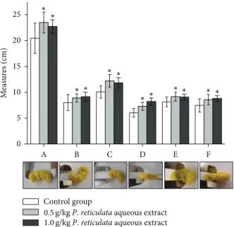
Figure 3: Efect of P. reticulata aqueous extract (0.5 g/kg and 1.0 g/kg) against the external morphological parameters of ofspring. he graph is the mean±S.E.M. of ive experiments. (A: craniocau-dal; B: tail; C: anteroposterior of cranio; D: laterolateral of cranio; E: anteroposterior of thorax; and F: laterolateral of thorax.)∗� < 0.05 compared to control group.
of the initial dose for in vivo studies, mainly when LD50 tests for acute oral toxicity, is required. Nowadays, most of the toxicological studies involving new drugs combine in vivo and in vitroassays in order to increase safety (i.e., in the case of a further approval for clinical use). For example, the evaluation of the development and safety of medicinal products require the estimation of IC50 values [17]. In our experimental conditions, together with the IC50 values in CHO-K1 cells, it was calculated the LD50 as 915 mg/kg.
Table 1: Reproductive performance of pregnant rats exposed to Plathymenia reticulataaqueous extract.
Teratogenicity parameters Control Experimental 0.5 g/kg Experimental 1.0 g/kg Preimplantation loss
(%) 0 0 0
Postimplantation loss
(%) 0 1.69 5.55
Placenta weight (grams)
0.494±0.07
(� = 59)
0.542±0.09∗
(� = 58)
0.530±0.07∗
(� = 51)
Fetus weight (grams) 1.336±0.25
(� = 59)
1.433±0.20∗
(� = 58)
1.456±0.15∗
(� = 51)
∗� < 0.05.
he above calculations applied to thein vivoexperimental assays permitted the determination of two concentrations (0.5 and 1 g/kg) ofP. reticulataextract, that mimics the human consumption.
Figure 2 shows the graph of mean (±S.E.M.) weight gain during the gestation, considering water ingestion and food consumptionad libitum. At the 95% conidence level the two means (control compared toP. reticulata extracts) are not signiicantly diferent. he weight losses of control pregnant rats and treated groups and also of 6–10th day of pregnancy compared to 1–5th day can be explained by the habituation phenomenon [25], since rats are very sensitive to manipulation.
he relationship between maternal and developmental toxicity is not only a result of an insult to the conceptus at the cellular level. Insults may occur through diferent routes, including a direct efect on the embryo/fetus, indirect toxicity of the agent to the mother through the placenta, or a combination of direct and indirect efects. Maternal conditions could potentially harm the developing organism by altering the nutritional status, among several diferent factors [26,27].
It is well known that intergenerational malnutrition is responsible for reducing the gain of weight during pregnancy in rats [28]. herefore, the distinction between direct and indirect developmental toxicity is important to understand safety assessment tests in pregnant animals. In our experi-mental conditions, all animals had access to food and water
ad libitum, in order to exclude this variable (Figure3). Here, the concentrations assayed did not induce maternal toxicity. According to Rogers and Kavlock [13], a decrease in food or water intake would induce weight loss and other clinical signs. As an example, they have shown a signiicant maternal weight reduction at the end of pregnancy in the sibutramine nonoverweight drug-treated group, compared to the control (nonoverweight, no drug). his data can be linked to a signiicant increase in post-implantation loss and placental index, suggesting that sibutramine alone or the condition of excess weight in the absence of drugs has altered the reproductive performance [15].
CR
CL
VS M
PE
(a)
CR CL
VS PE
(b)
S M
(c)
Figure 4: Representative pictures of 19th days gestation fetuses for teratogenicity test. Pregnant rats were treated daily withP. reticulata (1.0 g/kg) and the ofspring removed surgically prior to birth. Diaphanizated fetuses were analyzed by lateral (a), posterior (b), and frontal (c) views. he parameters of sternum ossiication (S), clavicule (CL), cranio (CR), pelvis (PE), mandible (M), and vertebral spine (VS) were examined. Notice that no abnormality was observed.
fetus weight (grams), preimplantation loss (%), postimplan-tation loss, and ofspring vitality (%). Regarding this later information, the data ater administration of P. reticulata
aqueous extract (0.5 g/kg or 1 g/kg) did not difer from control group (Table 1), except for placenta and fetuses weights. Under these parameters,P. reticulatatreatment increased the gain (in grams) of placenta and fetuses. According to Langley and Jackson [29], low-protein intake induces intrauterine protein restriction during diet that could explain the gain (in grams) of placenta and fetuses inP. reticulatatreatment. However, in our experimental conditions animals had access to food and waterad libitum. According to des Robert et al. [30], high protein intake via the enteral route could explain the enhanced weight gain inP. reticulatatreatment.
he external morphological parameters of ofspring were measured and compared to control group. All parameters evaluated were statistically diferent to the control, but not between the experimental treated-groups, via mother, that received 0.5 g/kg or 1.0 g/kg ofP. reticulataaqueous extract (Figure3).
No abnormality was seen in fetuses, except with the of-spring sizes, demonstrating the safety ofP. reticulataaqueous extract, in our experimental conditions. When cyclophos-phamide (40 mg/kg), a well-known teratogenic agent, was used as a positive control, a strong teratogenic activity was observed (a dose 12.5 and 25 times lower than 0.5 and 1.0 g/kg
P. reticulata, resp.). At this concentration cyclophosphamide was able to induce high resorption rate (approximately,80%) and severe fetal malformations with retarded growth. hese later phenomena were traduced by craniofacial alterations such as severe microcephaly, agnathia, open eyes, limb reduc-tion, and trunk anomalies such as phocomelia or amelia, as well as, eventration of the abdominal wall and absence of the tail. Even at 15 mg/kg of cyclophosphamide, the teratogenic efect was observed in 70% of fetuses, which exhibited external tail and digit anomalies, such as short or crooked tail, syndactyly and ectrodactyly [31].
Nasal region
Oral region
Cerebral hemispheres
Eyes region 1
2
3
4
5
6
7
8
9
10
11
12
13
14
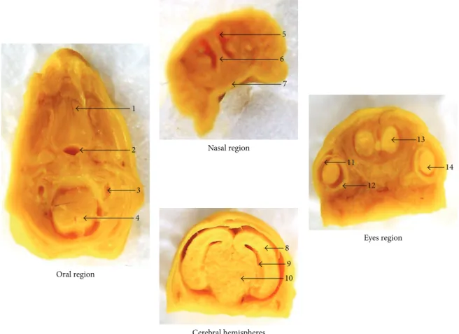
Figure 5: Representative sections from head and neck regions of fetuses exposed toP. reticulataaqueous extract (1.0 g/kg). Oral region transversally sectioned: 1: palate; 2: trachea; 3: inner ear; 4: marrow. Nasal region frontally sectioned: 5: nasal cavity; 6: nasal septum; 7: palate. Cerebral hemispheres region frontally sectioned: 8: cerebral hemisphere; 9: ventricles; 10: diencephalon. Eyes region frontally sectioned: 11: cornea; 12: retina; 13: olfactory bulb; 14: crystalline. Ater a careful analysis of the anatomical parameters, no abnormality was observed among the groups.
not obstructed in all fetuses examined, treated groups, and control group (Figure5).
his study also included the daily observation of pregnant rats to a previous oral administration ofP. reticulataaqueous extract (0.5 g/kg or 1.0 g/kg). In this protocol there were no signs of increased hair loss, excessive salivation, alteration in respiration, and abnormal gaits, tremors, or seizures. Although the gain of weight (grams) of pregnant rats was not statistically diferent (treated groups compared to the control group), there were signiicative changes in the weights of placenta and fetuses (� = S.E.M.,∗� < 0.05). he fetal evaluation between treated and control groups showed no malformations, deined as “those structural anomalies that alter general body conformity, disrupt or interfere with body function, or are generally thought to be incompatible with life” [32].
4. Conclusions
Overall the results suggest that oral administration of P. reticulataaqueous extract at 0.5 and 1.0 g/kg is safe, related to reproduction and fertility parameters or even in terms
of inducing teratogenicity. his paper also shows that the combination ofin vitroassays to select the dosage, within vivoexperiments, which involved the segment I (the period of premating and mating to implantation) and segment II (the period from implantation to major organogenesis), can be useful for assessing safety parameters of new medicinal compounds.
Conflict of Interests
he authors declare that there is no conlict of interests regarding the publication of this paper.
Acknowledgments
Course in Pharmaceutical Sciences (Master Level) from UNISO and has a scholarship from CAPES/PROSUP.
References
[1] T. J. King and S. Rodrigo, “Plathyterpol, a diterpene from Plathy-menia reticulata,”Chemical Communications, no. 12, article 575, 1967.
[2] F. J. A. Matos, A. A. Craveiro, and M. A. M. A. De Maurera, “Furan diterpenes of thePlathymeniagenus,”Journal of Natural Products, vol. 47, no. 4, pp. 581–584, 1984.
[3] R. D. S. Leal, M. A. S. Lima, and E. R. Silveira, “Cassane diterpenes fromPlathymenia reticulata,”Journal of the Brazilian Chemical Society, vol. 14, no. 1, pp. 120–125, 2003.
[4] A. Pott, V. J. Pott, and A. A. Bueno Sobrinho, “Plantas ´uteis `a sobrevivˆencia no Pantanal,” Proceedings of the Simp´osio sobre Recursos Naturais e S´ocio-econˆomicos do Pantanal, Corumb´a, Brazil, 2004.
[5] T. T. Fernandes, A. T. Santos, and F. C. Pimenta, “Atividade antimicrobiana das plantasPlathymenia reticulata, Hymenaea courbaril e Guazuma ulmifolia,”Revista de Patologia Tropical, vol. 34, pp. 113–122, 2005.
[6] C. E. M. De Toledo, E. A. Britta, L. F. Ceole et al., “Antimicrobial and cytotoxic activities of medicinal plants of the Brazilian cerrado, using Brazilian cachaa as extractor liquid,”Journal of Ethnopharmacology, vol. 133, no. 2, pp. 420–425, 2011.
[7] M. G. Santos, S. F. Lolis, and C. A. Dal Belo, “Levantamentos Etnobotˆanicos realizados em duas comunidades de remanes-centes de negros da regi˜ao do Jalap˜ao, Estado do Tocantins,” in Sociabilidade Negras. Comunidades Remanescentes, Escravid˜ao e Cultura, A. L. Pires, S. Cardoso, and R. Oliveira, Eds., pp. 29– 49, Daliana, Belo Horizonte, Brazil, 2006.
[8] S. Norton, “Toxic efects of plants,” in Casarett and Doull’s Toxicology: the basic Science of poisons, pp. 965–976, McGraw-Hill, New York, NY, USA, 2001.
[9] R. F. Melo, N. M. Farrapo, D. S. Rocha Jr. et al., “Antiophidian mechanisms of medicinal plants,” inFlavonoids: Biosynthesis, Biological Efects and Dietary Sources, R. B. Keller, Ed., pp. 249– 262, Nova Science Publishers, New York, NY, USA, 2009. [10] N. M. Farrapo, G. A. A. Silva, K. N. Costa et al., “Inhibition of
Bothrops jararacussuvenom activities byPlathymenia reticulata Benth extracts,”Journal of Venom Research, vol. 2, pp. 52–58, 2011.
[11] A. Della Torre, L. B. L. Albuquerque, N. M. Farrapo et al., “Mutagenicity induced by the hydroalcoholic extract of the medicinal plantPlathymenia reticulatabenth,”Journal of Ven-omous Animals and Toxins Including Tropical Diseases, vol. 17, no. 2, pp. 190–198, 2011.
[12] A. R. Carvalho, T. J. Lacerda, F. E. Oliveira, and S. E. Santos, “Extratos de plantas medicinais como estrat´egia para o controle de doenc¸as f´ungicas do inhame (Dioscoreasp.) no nordeste,” in II Simp´osio Nacional sobre as Culturas do Inhame e do Taro—Empresa Estadual de Pesquisa Agropecu´aria da Para´ıba, EMEPA, Jo˜ao Pessoa, Brazil, 2002.
[13] J. M. Rogers and R. J. Kavlock, “Developmental toxicology,” in Casarett and Doull’s Toxicology: the basic Science of poisons, pp. 351–386, McGraw-Hill, New York, NY, USA, 2001.
[14] OECD, “Phototox Version 2. 0,” in Chemical Testing— Guidelines, section 4: sotware, Organisation for Economic Co-operation and Development, Paris, France, 2010,
http://www.oecd.org/document/62/0,3343,en 2649 34377
44706494 1 1 1 1,00.html.
[15] L. A. D. Francia-Farje, D. S. Silva, G. T. Volpato et al., “Sibu-tramine efects on the reproductive performance of pregnant overweight and non-overweight rats,”Journal of Toxicology and Environmental Health A, vol. 73, no. 13-14, pp. 985–990, 2010. [16] ICCVAM, “Peer review panel report: the use of in vitro basal
cytotoxicity test methods for estimating starting doses for acute oral systemic toxicity testing,” NIH publication no. 07-4519, Research Triangle Park, National Toxicology Program, 2006,
http://iccvam.niehs.nih.gov/docs/acutetox docs/ATpanelrpt06/ ATpanelrpt.pdf#search=peer%20review%20panel%20report:%
20the%20use%20or%20in%20vitro%20vasal%20cytotoxicity.
[17] N. M. Esteves-Pedro, A. C. D. Rodas, C. A. Dal Belo, Y. Oshima-Franco, M. G. Dos Santos, and P. S. Lopes, “Implementation of the three Rs in the human hazard assessment of Brazilian medicinal plants: an evaluation of the cytotoxic and genotoxic potentials ofDipteryx alatavogel,”ATLA Alternatives to Labo-ratory Animals, vol. 39, no. 2, pp. 189–196, 2011.
[18] M. Gerenutti, F. S. Del Fiol, and F. C. Groppo, “Reproduc-tive performance of pregnant rats and embryotoxic efects of ciproloxacin,”Pharmazie, vol. 61, no. 1, pp. 79–80, 2006. [19] M. Gerenutti, A. F. Rollo Oliveira Prestes, M. Glauzer Silva et al.,
“he efect of Cecropia glazioui Snethlage on the physical and neurobehavioral development of rats,”Pharmazie, vol. 63, no. 5, pp. 398–404, 2008.
[20] N. M. Esteves-Pedro, T. Borim, V. S. Nazato et al., “In vitroand in vivosafety evaluation ofDipteryx alataVogel extract,”BMC Complementary and Alternative Medicine, vol. 12, article 9, 2012. [21] U.S. EPA, “Guidelines for the Health Assessment of Suspected Developmental Toxicants,” Federal Register 51 FR 34040, 1986, Proposed Amendments Federal Register 54 FR 9386, 1989. [22] P. Randazzo-Moura, M. G. Silva, Y. Oshima-Franco, F. C.
Groppo, and M. Gerenutti, “he efect of aqueous extract of Cecropia glazioui Snethlage (Embauba) in the rat fetal development,”Chinese Medicine, vol. 2, pp. 115–119, 2011. [23] D. C. Damasceno and W. G. Kempinas,Anomalias Congˆenitas—
Estudos Experimentais, Coopmed, Belo Horizonte, Brazil, 2008. [24] K. A. Keller, “Developmental and reproductive toxicology,” in Toxicology Testing Handbook: Principles, Applications and Data Interpretation, D. Jacobson-Kram and K. A. Keller, Eds., pp. 195– 252, Marcel Dekker, New York, NY, USA, 2006.
[25] A. M. McNamara, P. D. Magidson, C. Linster, D. A. Wilson, and T. A. Cleland, “Distinct neural mechanisms mediate olfactory memory formation at diferent timescales,”Learning and Mem-ory, vol. 15, no. 3, pp. 117–125, 2008.
[26] G. P. Daston, “Relationships between maternal and develop-mental toxicity,” inDevelopmental Toxicology, C. A. Kimmel and J. Buelke-Sam, Eds., pp. 189–212, Raven Press, New York, NY, USA, 1994.
[27] N. Chernof, J. M. Rogers, A. J. Alles et al., “Cell cycle alterations and cell death in cyclophosphamide teratogenesis,” Teratogenesis Carcinogenesis and Mutagenesis, vol. 9, no. 4, pp. 199–209, 1989.
[28] J. R. Galler and G. Zartarian, “Reproductive performance in rats with diferent histories of malnutrition,”British Journal of Nutrition, vol. 45, no. 2, pp. 251–255, 1981.
[31] A. Torchinsky, L. Lishanski, O. Wolstein et al., “NF-�B DNA-binding activity in embryos responding to a teratogen, cyclophosphamide,”BMC Developmental Biology, vol. 2, article 1, pp. 1–11, 2002.
Submit your manuscripts at
http://www.hindawi.com
Pain
Research and Treatment
Hindawi Publishing Corporation
http://www.hindawi.com Volume 2014
World Journal
Hindawi Publishing Corporation
http://www.hindawi.com Volume 2014
Hindawi Publishing Corporation http://www.hindawi.com
Volume 2014
Toxins
Journal of
Vaccines
Journal ofHindawi Publishing Corporation
http://www.hindawi.com Volume 2014
Hindawi Publishing Corporation
http://www.hindawi.com Volume 2014
Antibiotics
Toxicology
Journal ofHindawi Publishing Corporation
http://www.hindawi.com Volume 2014
Stroke
Research and Treatment
Hindawi Publishing Corporation
http://www.hindawi.com Volume 2014
Drug Delivery
Journal ofHindawi Publishing Corporation
http://www.hindawi.com Volume 2014
Hindawi Publishing Corporation
http://www.hindawi.com Volume 2014 Advances in Pharmacological Sciences
Tropical Medicine
Hindawi Publishing Corporation
http://www.hindawi.com Volume 2014
Medicinal ChemistryInternational Journal of
Hindawi Publishing Corporation
http://www.hindawi.com Volume 2014
Addiction
Journal ofHindawi Publishing Corporation
http://www.hindawi.com Volume 2014
Hindawi Publishing Corporation
http://www.hindawi.com Volume 2014
BioMed
Research International Emergency Medicine International
Hindawi Publishing Corporation
http://www.hindawi.com Volume 2014
Hindawi Publishing Corporation
http://www.hindawi.com Volume 2014
Diseases
Hindawi Publishing Corporation
http://www.hindawi.com Volume 2014
Anesthesiology Research and Practice
Scientifica
Hindawi Publishing Corporation
http://www.hindawi.com Volume 2014
Journal of
Hindawi Publishing Corporation
http://www.hindawi.com Volume 2014
Pharmaceutics
Hindawi Publishing Corporation
http://www.hindawi.com Volume 2014