tion is characterized by an area of abnormal, nonfunctional pulmonary tissue, which receives its blood supply via a systemic artery and has no connection with the tracheobronchial tree. In intralobar pulmonary sequestration, this tissue is located within the visceral pleura of a pulmo-nary lobe, and its venous drainage is into the pulmonary veins. Radiographic images play a fundamental role in establishing the diagnosis and provide the medical team with a vascular map for surgical planning.(3) We report a case of intralobar pulmonary sequestration in which diagnosis was delayed and hemoptysis was the clinical manifestation.
Introduction
The development of the tracheobronchial tree begins on the 24th day of embryogenesis. The ventral wall of the pharynx of the fetus migrates caudally, forming the larynx and the trachea, as well as the right and left lung buds. The buds elongate, developing all of the lung segmenta-tion by the 36th day. In the same period, the vascular supply, originating from branches of the splenic vascular plexus, accompanies the bronchial generations.(1) Congenital pulmonary anomalies are rare, the most common being lobar emphysema, cysts in the lungs, cystic adenomatoid malformation and pulmonary sequestration.(2) Bronchopulmonary
sequestra-Usefulness of chest CT in the
diagnosis of pulmonary sequestration*
A utilidade da TC de tórax no diagnóstico do sequestro pulmonar
José Gustavo Pugliese, Thiago Prudente Bártholo, Heron Teixeira Andrade dos Santos, Eduardo Haruo Saito, Cláudia Henrique da Costa, Rogério Rufino
Abstract
Pulmonary sequestration is a rare congenital anomaly, characterized by nonfunctional embryonic pulmonary tissue. Pulmonary sequestration accounts for 0.15-6.40% of all congenital pulmonary malformations. This anomaly, which is classified as intralobar or extralobar, involves the lung parenchyma and its vascularization. We report the case of a 56-year-old male presenting with hemoptysis. A chest X-ray showed an area of opacity behind the cardiac silhouette in the base of the left hemithorax. Chest CT scans with intravenous contrast revealed pulmonary sequestration. The patient underwent surgery, in which the anomalous tissue was successful resected. The postoperative evolution was favorable, and the patient was discharged to outpatient treatment.
Keywords: Hemoptysis; Bronchopulmonary sequestration; Tomography, spiral computed.
Resumo
O sequestro pulmonar é uma rara anomalia congênita, caracterizada por tecido pulmonar embrionário não funcionante, perfazendo 0,15-6,40% de todas as malformações pulmonares congênitas. Essa anomalia envolve o parênquima e a vascularização pulmonar, sendo classificado como intralobar ou extralobar. Neste relato, descre-vemos o caso de um paciente de 56 anos com hemoptise e imagem hipotransparente retrocardíaca em base de hemitórax esquerdo na radiografia de tórax. Após a realização de TC com contraste endovenoso, foi eviden-ciada a presença de sequestro pulmonar. O paciente foi submetido à cirurgia para a retirada do tecido anômalo, que foi realizada com sucesso. Apresentou boa evolução pós-operatória e recebeu alta com acompanhamento ambulatorial.
Descritores: Hemoptise; Sequestro broncopulmonar; Tomografia computadorizada espiral.
* Study carried out in the Department of Thoracic Surgery and in the Department of Pulmonology and Phthisiology, Universidade do Estado do Rio de Janeiro – UERJ, Rio de Janeiro State University – Pedro Ernesto University Hospital, Rio de Janeiro, Brazil. Correspondence to: Rogério Rufino. Rua Mário Pederneiras, 10/121, Humaitá, CEP 22261-020, Rio de Janeiro, RJ, Brasil. Tel 55 21 2286-9333. E-mail: [email protected]
Financial support: None.
was 32 mm/h. Serology for HIV was negative. A chest X-ray at admission revealed opacity in the posterior segment of the left lower lobe (Figure 1). A CT scan of the chest with two-dimensional reconstruction revealed an aortic branch directed toward the pulmonary opacity (Figure 2), which is consistent with a diagnosis of pulmonary sequestration. The patient was referred for surgery, and a left lower lobectomy was performed to resect the anomalous pulmo-nary tissue (Figure 3). After the macroscopic analysis of the surgical sample, the patient was diagnosed with intralobar sequestration. The postoperative evolution was favorable, and the patient was discharged to outpatient treatment.
Discussion
When faced with a 56-year-old patient, admitted for investigation of hemoptysis, with no clinical evidence of respiratory infection, with no history of smoking and having an area of opacity in the posterior segment of the left lower lobe on chest X-ray, one of the hypotheses to be considered is that it is a case of pulmonary sequestration. Bronchopulmonary sequestra-tion was first described in 1946 by Pryce as “an abnormal artery arising from the aorta and supplying a bronchopulmonary mass or cyst that is dissociated from the normal bronchial tree”.(4) Since then, a number of cases have been reported in the literature,(5,6) and, today, pulmo-nary sequestration, as described by Pryce, is known to be an anomalous lung formation that usually does not have bronchial
communica-Case report
A 56-year-old nonsmoking hypertensive male receiving captopril (75 mg/day) was admitted with hemoptysis. The patient reported that the episodes of hemoptysis had begun one month earlier, at which point he sought medical care and was treated with amoxicillin and clavulanic acid. While on the antibiotics, the patient had a new episode of lower airway bleeding and was referred for hospitalization. Immediately after admission, he had another episode of bleeding, totaling three episodes of hemoptysis. In those episodes, the production ranged from 100 to 200 mL and there were no hemodynamic changes.
The patient reported no dyspnea, fever, weight loss or chest pain. He reported two episodes of pneumonia, one at 16 and one at 30 years of age, both treated with antibiotics.
Physical examination revealed good general health, no fever, an HR of 88 bpm and an arte-rial pressure of 90-145 mmHg. The cardiac auscultation was normal, and examination of the lung revealed reduced expansion and dimin-ished breath sounds in the lower third of the left hemithorax, as well as dullness to percus-sion in this same region. The rest of the physical examination revealed no abnormalities. The results of the laboratory tests performed at admission, such as blood workup, lipid profile, coagulation profile, determination of electro-lyte concentrations, renal function test and hepatic function test, were normal. The ESR
a b
disorder, which would explain the persistence of the anomalous blood supply.(8)
There are two forms of pulmonary seques-tration: intralobar and extralobar.(7)
The intralobar form accounts for 75% of cases(6) and is characterized by sharing the visceral pleural membrane of the normal lung.(8) This form is rarely accompanied by other anomalies. It is most commonly located in the posterior segment of the lower lobe. In 85% of cases, intralobar sequestration is supplied by an artery arising from the infradiaphragmatic aorta.(7) Most patients are asymptomatic, living with the anomaly for years, and the diagnosis is considered during a chest X-ray. Half of the patients are diagnosed after they are 20 years of age. Respiratory symptoms are nonspecific and uncommon, and the patient can report chest pain and bronchospasm during infectious processes. In rarer cases, intralobar sequestra-tion can present as frank hemoptysis.(9) Many times, the diagnosis is made when the anoma-lous region is infected, and most such patients have pneumonia.(10) Given the risk of hemoptysis and recurrent pulmonary infection, the seques-tration is often removed by segmentectomy or lobectomy.(7)
The extralobar form is enclosed within its own membrane, usually in close proximity with the normal lung.(6) This form predominates in male neonates and is more frequently found in the left hemithorax,(7) usually just above the diaphragm.(6) This form is often associated with other congenital anomalies,(7) such as hydropsy, tion and is typically associated with an aberrant
blood supply.(7) Its etiology remains a contro-versial issue. The two most often mentioned hypotheses are the following: formation and caudal migration of a supernumerary lung bud that accompanies the esophagus, which would explain the preferential localization of pulmo-nary sequestration in the lower third of the left hemithorax; and primary pulmonary vascular
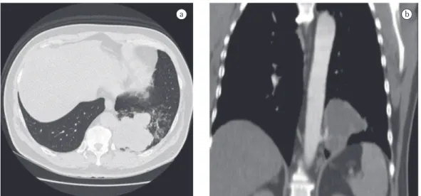
Figure 2 - Chest CT scan. In the lung window (in a), there is an area of lobulated opacity in the posterior segment of the left lower lobe. In the two-dimensional reconstruction (in b), note the thoracic aorta branch directed toward the opacity.
a b
case presented here is rare. The postoperative evolution was favorable, and the patient was discharged to outpatient treatment.
References
1. Desir A, Ghaye B. Congenital abnormalities of intrathoracic airways. Radiol Clin North Am. 2009;47(2):203-25. 2. Hashemzadeh S, Aslanabadi S, Jafari Rouhi AH, Azhough
R, Kaleibar NA. Congenital malformations of the lung. Indian J Pediatr. 2007;74(2):192-4.
3. Abbey P, Das CJ, Pangtey GS, Seith A, Dutta R, Kumar A. Imaging in bronchopulmonary sequestration. J Med Imaging Radiat Oncol. 2009;53(1):22-31.
4. Peyce DM. Lower accessory pulmonary artery with intralobar sequestration of lung. A report of seven cases. J Pathol Bacteriol. 1946;58(3):457-67.
5. Torres LF, Jacob GV, de Noronha L, Seade M, Artigas JL. Congenital pulmonary extralobar intra-abdominal sequestration [Article in Portuguese]. J Pediatr (Rio J). 1997;73(1): 51-3.
6. Pego-Fernandes PM, Freire CH, Jatene FB, Beyruti R, Suso FV, Oliveira SA. Pulmonary sequestration: a series of nine cases operated on [Article in Portuguese]. J Pneumol. 2002; 28(4):175-9.
7. De Nicola AL, Frigerio MV, Zanforlin Filho SM, Gollop TR. Prenatal diagnosis of pulmonary sequestration by ultrasound: a case report [Article in Portuguese]. Rev. Bras. Ginecol. Obstet. 2003;25(3):207-10.
8. Vieira J, Rego A, Oliveira A, Sá Ferreira D, Furtado A, Couceiro A, et al. Bronchopulmonary sequestration--a 12-year experience. Rev Port Pneumol. 2006;12(5):489-501.
9. Cooke CR. Bronchopulmonary Sequestration. Respir Care. 2006; 51(6):661-4.
10. Zhang XQ, Zhou JY. Bronchopulmonary sequestration: review of 27 cases [Article in Chinese]. Zhonghua Jie He He Hu Xi Za Zhi. 2008;31(6):421-4.
11. Nacif MS, Lima Filho HS, Mello RA, Jauregui GF, Miranda BJ, Caramel JM, et al. Seqüestro broncopulmonar intralobar: relato de caso. Radiol Bras. 2005;38(1):65-7.
12. Wu N, Sun Y, Zheng QF, Lü C, Yan S, Zhang LJ, et al. Diagnosis and treatment of intralobar pulmonary sequestration [Article in Chinese]. Zhonghua Yi Xue Za Zhi. 2007;87(37):2627-31.
13. Fumino S, Iwai N, Kimura O, Ono S, Higuchi K. Preoperative evaluation of the aberrant artery in intralobar pulmonary sequestration using multidetector computed tomography angiography. J Pediatr Surg. 2007;42(10):1776-9.
14. Kang M, Khandelwal N, Ojili V, Rao KL, Rana SS. Multidetector CT angiography in pulmonary sequestration. J Comput Assist Tomogr. 2006;30(6):926-32.
15. Schussler JM, Dockery WD, Gilbey JG, Lal VR. An alternate route: 64-slice CT diagnosis of pulmonary pseudosequestration. Am J Med. 2007;120(1):23-5. 16. Truong MT, Sabloff BS, Ko JP. Multidetector CT of
solitary pulmonary nodules. Radiol Clin North Am. 2010;48(1):141-55.
17. Bolca N, Topal U, Bayram S. Bronchopulmonary sequestration: radiologic findings. Eur J Radiol. 2004;52(2):185-91.
diaphragmatic hernia, pectus excavatum, cystic adenomatoid malformation of the lung, tracheoesophageal fistulas and gastric dupli-cations or cramps, and its arterial supply, in the vast majority of cases, originates from the abdominal aorta.(9-11) Extralobar sequestration is usually asymptomatic and, since there is no bronchial communication, the risk of infection is low.(7)
In the investigation of a suspected case of pulmonary sequestration, imaging studies have two principal objectives: to rule out other pathologies; and to confirm the presence of an anomalous arterial supply.(12-15) The most common radiological presentation is a homogeneous opacity in the posterior basal segment of the left lower lobe. Arteriography allows the char-acterization of anomalous arteries, providing valuable information for preoperative plan-ning.(13) Similarly to nuclear magnetic resonance imaging, spiral CT allows the visualization of the source and course of the vessels in most cases. In such cases, angiography is unnecessary.(10,15) The diagnosis of pulmonary sequestration is confirmed by multichannel CT scans of the chest with intravenous contrast and reconstruction, there being no need for aortography or magnetic resonance imaging of the chest and abdomen, since CT scans of the chest and upper abdomen allows the visualization of the arterial vessel communicating with the sequestration and of the changes in the lung parenchyma.(14,15)
Currently, multichannel CT scanners, with faster software for reconstruction, have made aortography practically disappear in the context of diagnosis and surgical planning, since these CT scanners allow the reconstruction of vessels that are just a few millimeters in diameter.(16,17)
About the authors
José Gustavo Pugliese
Resident in Pulmonology and Phthisiology. Universidade do Estado do Rio de Janeiro – UERJ, Rio de Janeiro State University – Pedro Ernesto University Hospital, Rio de Janeiro, Brazil.
Thiago Prudente Bártholo
Resident in Pulmonology and Phthisiology. Universidade do Estado do Rio de Janeiro – UERJ, Rio de Janeiro State University – Pedro Ernesto University Hospital, Rio de Janeiro, Brazil.
Heron Teixeira Andrade dos Santos
Resident in Thoracic Surgery. Universidade do Estado do Rio de Janeiro – UERJ, Rio de Janeiro State University – Pedro Ernesto University Hospital, Rio de Janeiro, Brazil.
Eduardo Haruo Saito
Adjunct Professor of Thoracic Surgery. Faculdade de Ciências Médicas da Universidade do Estado do Rio de Janeiro – FCM/UERJ, Rio de Janeiro State University School of Medical Sciences – Rio de Janeiro, Brazil.
Cláudia Henrique da Costa
Adjunct Professor of Pulmonology and Phthisiology. Faculdade de Ciências Médicas da Universidade do Estado do Rio de Janeiro – FCM/UERJ, Rio de Janeiro State University School of Medical Sciences – Rio de Janeiro, Brazil.
Rogério Rufino