Occurrence of hepatopulmonary syndrome in patients with
cirrhosis who are candidates for liver transplantation*
Ocorrência de síndrome hepatopulmonar em pacientes cirróticos candidatos a transplante de fígado
Liana Gonçalves Macêdo, Edmundo Pessoa de Almeida Lopes, Maria de Fátima Pessoa Militão de Albuquerque, Brivaldo Markman-Filho, Flávio Henrique Amaral Pires Véras, Ana Carolina Chiappetta Correia de Araújo,
Álvaro Antônio Bandeira Ferraz
Abstract
Objective: To determine the occurrence of hepatopulmonary syndrome (HPS) in patients with cirrhosis who are candidates for liver transplantation; to compare demographic, clinical, laboratory, and spirometric characteristics, as well as echocardiography results, arterial blood gas analysis, and severity of liver disease between the groups of patients with and without HPS; and to describe the occurrence of HPS in the subgroup of patients with cirrhosis and schistosomiasis mansoni (mixed liver disease). Methods: Between January and November of 2007, we evaluated 44 patients under treatment at the Liver Transplant Outpatient Clinic of the Federal University of Pernambuco Hospital das Clínicas, in the city of Recife, Brazil. The diagnostic criteria for HPS were intrapulmonary vascular dilatation, identified by transthoracic echocardiography, and an alveolar-arterial oxygen tension difference
≥ 15 mmHg or a PaO2 < 80 mmHg. Results: The mean age of the patients was 52 years, and 31 patients (70%) were males. The most common cause of cirrhosis was alcohol use. Schistosomiasis was present in 28 patients (64%). Of the 44 patients, 20 (45.5%) were diagnosed with HPS. No significant differences were found between those patients and the patients without HPS in terms of any of the characteristics studied. Of the 28 patients with cirrhosis and schistosomiasis, 10 (35.7%) were diagnosed with HPS. Conclusions: In the population studied, HPS was highly prevalent and did not correlate with any of the variables analyzed.
Keywords: Hepatopulmonary syndrome; Liver transplantation; Liver cirrhosis; Hypertension, portal; Schistosomiasis mansoni; Echocardiography.
Resumo
Objetivo: Verificar a ocorrência da síndrome hepatopulmonar (SHP) em pacientes cirróticos candidatos a transplante de fígado; comparar as características demográficas, clínicas, laboratoriais e espirométricas, resultados de ecocardiografia, análise de gases arteriais e da gravidade da doença hepática nos pacientes com e sem SHP; e descrever a ocorrência de SHP no subgrupo de pacientes com cirrose associada à esquistossomose mansônica (doença hepática mista). Métodos: Entre janeiro e novembro de 2007, foram avaliados 44 pacientes inscritos no Ambulatório de Transplante Hepático do Hospital das Clínicas da Universidade Federal de Pernambuco, em Recife (PE). Os critérios diagnósticos para SHP foram a presença de dilatações vasculares intrapulmonares, identificadas por ecocardiografia transtorácica, assim como diferença alveoloarterial de oxigênio ≥ 15 mmHg ou PaO2 < 80 mmHg. Resultados: A idade média foi 52 anos, e 31 pacientes (70%) eram do sexo masculino. A
causa mais frequente de cirrose foi uso de etanol. A esquistossomose esteve presente em 28 pacientes (64%). Dos 44 pacientes, 20 (45,5%) foram diagnosticados com SHP. Não foram observadas diferenças significativas em relação às características estudadas. No subgrupo de pacientes com cirrose associada à esquistossomose, 10/28 (35,7%) receberam o diagnóstico de SHP. Conclusões: A SHP apresentou elevada prevalência nesta população estudada, não sendo observadas associações entre a sua ocorrência e as variáveis analisadas.
Descritores: Síndrome hepatopulmonar; Transplante de fígado; Cirrose hepática; Hipertensão portal; Esquistossomose mansoni; Ecocardiografia.
* Study carried out under the auspices of the Postgraduate Program in Health Sciences. Health Sciences Center, Universidade Federal de Pernambuco – UFPE, Federal University of Pernambuco – at the Liver Transplant Outpatient Clinic and at the Department of Cardiology, Universidade Federal de Pernambuco – UFPE, Federal University of Pernambuco – Hospital das Clínicas, Recife, Brazil. Correspondence to: Liana Gonçalves de Macedo. Hospital Otávio de Freitas, Comissão de Residência Médica (COREME), Avenida Aprígio Guimarães, s/n, Tejipió, CEP 50920-640, Recife, PE, Brasil.
Tel 55 81 3182-8529. E-mail: [email protected] Financial support: None.
the Hospital das Clínicas da Universidade Federal de Pernambuco (HC-UFPE, Federal University of Pernambuco Hospital das Clínicas) and who were on the waiting list for liver transplantation at the beginning of the study. Between January and November of 2007, data were collected in accordance with the following exclusion criteria: having received a liver transplant before the conclusion of the evaluation; having failed to return for follow-up before the final phase of data collection; and having refused to participate in any of the stages of the study.
Patients were evaluated consecutively in accordance with the demand for treatment at the outpatient clinic. After having given written informed consent, patients were interviewed, all by the same physician, in order to gather information regarding age, gender, date of inclusion on the waiting list for liver transplantation (waiting time), and etiology of liver cirrhosis, as well as history of HSS, lung disease, smoking, platypnea, and dyspnea. Dyspnea was measured by the modified Medical Research Council dyspnea scale.(14) In addition,
physical examination was performed in order to screen for ascites, encephalopathy, jaundice, telangiectasias (spider veins), and digital clubbing.
The medical charts were analyzed in order to collect data regarding HSS-related liver impairment, which was determined by an abdominal ultrasound showing Symmers’ fibrosis accompanied by positive epidemiology for schistosomiasis, which was defined as “having had contact with river water in endemic areas”.(10,11,15)
A total of 10 mL of blood was drawn from the peripheral vein in order to determine levels of albumin, aminotransferases, total bilirubin, and creatinine, as well as the international normalized ratio (INR) of prothrombin time, in order to determine the MELD score and the Child-Pugh classification. In addition, 2 mL of blood were drawn from the radial artery, using the puncture and sample handling techniques recommended by the Brazilian Thoracic Association, for the analysis of pH, PaCO2, PaO2, and A-aDO2 with a GEM 3000 blood gas analyzer (Instrumentation Laboratory, Bedford, MA, USA).(16) In addition, A-aDO
2 was calculated
by applying a standardized equation,(1) with the
patient in the sitting position breathing room
Introduction
Hepatopulmonary syndrome (HPS) is defined as a triad, represented by intrapulmonary vascular dilatation (IPVD) associated with an alveolar-arterial oxygen tension difference (A-aDO2) ≥ 15 mmHg or a PaO2 < 80 mmHg and liver disease.(1) Although HPS is a common
complication of liver cirrhosis,(1-4) it has also been
reported in patients with non-cirrhotic portal hypertension (PH).(5-7)
In the northeastern region of Brazil, hepatosplenic schistosomiasis (HSS)—the hepatosplenic form of schistosomiasis mansoni—is considered to be a leading cause of PH.(8) It is known that HSS causes presinusoidal
PH through increased blood flow from the splenic vein and increased resistance due to liver fibrosis, without significant destruction of hepatocytes, thus preserving the architecture and function of the liver.(9) Liver fibrosis in HSS, also known as
Symmers’ fibrosis, is not diffuse and is located in the periportal region.(10,11) A study conducted
in Brazil reported that 5 (10.2%) of 49 patients with HSS had HPS without cirrhosis.(5)
Due to PH and the formation of collateral circulation, patients with HSS commonly present with digestive bleeding, requiring blood transfusions. Blood transfusions performed some decades ago resulted in infection with the hepatitis B or C virus; currently, many patients present with mixed liver disease (MLD), defined as HSS accompanied by viral hepatitis.(12,13)
There have been no studies investigating the occurrence of HPS in this group of patients.
The objective of the present study was to determine the occurrence of HPS in candidates for liver transplantation; to compare demographic, clinical, and laboratory characteristics, as well as echocardiography results, spirometry results, arterial blood gas analysis, and severity of liver disease (Child-Pugh class and Model for End-Stage Liver Disease [MELD] score) between the groups of patients with and without HPS; and to describe the occurrence of HPS in the subgroup of patients with MLD.
Methods
in accordance with the recommendations of the American Society of Echocardiography.(18)
An upper-limb peripheral vein was punctured, and a three-way stopcock was placed. Two 10-mL syringes with 9.5 mL of the solution were connected to the stopcock. Microbubbles were produced manually by agitating the solution between the two syringes 10 times, and the solution was subsequently injected. This procedure was performed three times in each patient, with an interval between injections in order to ensure that the cardiac chambers were completely free of contrast. The images were acquired simultaneously with the injection of the contrast, with the transducer in the apical four-chamber position and the patient in the left lateral decubitus position. Two specialists, who were only aware that the patients had cirrhosis air at sea level. All tests were performed at
the Central Laboratory of the HC-UFPE using routine methods.
Concurrently with arterial blood gas analysis, pulse oximetry was performed using a pulse oximeter (Onyx II 9550; Nonin, Plymouth, MN, USA) to determine SpO2. Spirometry was subsequently performed with a Microlab 3300 spirometer (Micro Medical Ltd., Kent, England) in order to measure FEV1, FVC, and FEV1/FVC,
the predicted values for these maneuvers being adjusted to the Brazilian population.(17)
Chest X-rays were taken in order to rule out other lung diseases.
Contrast transthoracic echocardiography (TTE; contrast agent, 0.9% saline solution) was performed with an HDI 1500 echocardiograph (Philips Medical Systems, Bothell, WA, USA),
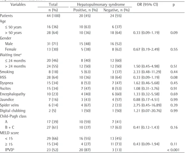
Table 1 - Univariate analysis of the demographic characteristics, clinical characteristics, liver disease classification and presence of intrapulmonary vascular dilatation in 44 patients with cirrhosis who were candidates for liver transplantation, according to the presence of hepatopulmonary syndrome. Federal University of Pernambuco
Hospital das Clínicas, 2007.
Variables Total Hepatopulmonary syndrome OR (95% CI) p
n (%) Positive, n (%) Negative, n (%)
Patients 44 (100) 20 (45) 24 (55)
Age
≤ 50 years 16 (36) 10 (63) 6 (37)
> 50 years 28 (64) 10 (36) 18 (64) 0.33 (0.09-1.19) 0.09
Gender
Male 31 (71) 15 (48) 16 (52)
Female 13 (30) 5 (38) 8 (62) 0.67 (0.19-2.49) 0.55
Waiting timea
≤ 24 months 20 (46) 8 (40) 12 (60)
> 24 months 24 (55) 12 (50) 12 (50) 1.50 (0.45-4.98) 0.51
Smoking 8 (18) 5 (63) 3 (37) 2.33 (0.48-11.29) 0.44
HSS 28 (64) 10 (36) 18 (64) 0.33 (0.09-1.19) 0.08
Dyspnea 15 (34) 8 (53) 7 (47) 1.62 (0.46-5.68) 0.45
Ascites 15 (34) 7 (47) 8 (53) 1.08 (0.31-3.76) 0.91
Encephalopathy 10 (23) 4 (40) 6 (60) 1.33 (0.32-5.58) 0.69
Jaundice 7 (16) 3 (43) 4 (57) 0.88 (0.17-4.51) 0.99
Spider veins 6 (14) 4 (67) 2 (33) 2.75 (0.45-16.89) 0.39
Digital clubbing 2 (5) 1 (50) 1 (50) 1.21 (0.07-20.76) 0.99
Child-Pugh class
A 17 (39) 10 (59) 7 (41)
B + C 27 (61) 10 (37) 17 (63) 0.41 (0.12-1.43) 0.16
MELD score
< 15 29 (66) 16 (55) 13 (45)
≥ 15 15 (34) 4 (27) 11 (73) 0.43 (0.09-1.94) 0.11
IPVDb 23 (52) 20 (87) 3 (13) < 0.001
and without HPS. For the qualitative variables, we used Pearson’s chi-square test or Fisher’s exact test, ORs and 95% CIs being calculated.
The study design was approved by the Research Ethics Committee of the Federal University of Pernambuco Health Sciences Center.
Results
Of the 61 patients with cirrhosis who were on the HC-UFPE waiting list for liver transplantation, and were liver transplant candidates, analyzed
the images simultaneously. The images were recorded for reviewed in cases of uncertainty. The test was considered to be indicative of IPVD when contrast was observed in the left atrium after four to six cardiac cycles, after contrast had been observed in the right atrium, during any of the injections, in the absence of intracardiac communication. Intracardiac communication was considered to be present when the contrast was observed in the left atrium for three cardiac cycles following the opacification of the right atrium.(19)
In order to diagnose HPS, we used a criterion that is currently recommended by guidelines published in 2004; the criterion consists of identifying IPVD through TTE, together with changes in arterial blood gases, which were defined as an A-aDO2≥ 15 mmHg or a PaO2 <
80 mmHg (both adjusted for age).(1) The severity
of HPS was classified according to the degree of hypoxemia: mild (PaO2 ≥ 80 mmHg); moderate
(60 mmHg ≤ PaO2 < 80 mmHg); severe (50
mmHg ≤ PaO2 < 60 mmHg); or extremely severe
HPS (PaO2 < 50 mmHg). (1)
All of the data were submitted to statistical analysis with the program Statistical Package for the Social Sciences, version 12.0 (SPSS Inc., Chicago, IL, USA). For the quantitative variables, mean, standard deviation, and range were used in order to indicate the variability of the data. The Student’s t-test was used in order to compare the means between the groups of patients with
Table 2 - Mean age, waiting time for liver transplantation, spirometric parameter values, pulse oximetry values, arterial blood gas values and liver disease classification for 44 liver transplant candidates, according to the presence of hepatopulmonary syndrome. Federal University of Pernambuco Hospital das Clínicas, 2007.
Variables Total Variation Hepatopulmonary syndrome p
Positive Negative
mean ± SD mean ± SD mean ± SD
Age, years 51.77 ± 9.03 29-67 51.15 ± 9.80 52.29 ± 8.51 0.69
Time,a months 26.64 ± 11.97 10-63 28.75 ± 13.77 24.87 ± 10.19 0.29
FVC, L 2.95 ± 0.81 1.03-4.74 3.07 ± 0.74 2.85 ± 0.86 0.38
FEV1, L 2.39 ± 0.59 0.97-3.57 2.53 ± 0.54 2.30 ± 0.63 0.18
SpO2, % 98.07 ± 1.40 94-100 97.60 ± 1.60 98.45 ± 1.10 0.04b
PaO2, mmHg 87.66 ± 9.28 60-100 83.55 ± 9.48 91.08 ± 7.72 0.006b
PaCO2, mmHg 32.15 ± 4.77 21-43 30.60 ± 4.64 33.45 ± 4.56 0.05b
A-aDO2, mmHg 22.07 ± 11.98 0-63 27.87 ± 11.39 17.25 ± 10.37 0.002b
Child-Pugh, n 7.55 ± 2.20 5-14 7.20 ± 2.04 7.83 ± 2.33 0.35
MELD, n 14.10 ± 3.56 8-25 13.34 ± 2.95 14.73 ± 3.95 0.20
A-aDO2: alveolar-arterial oxygen tension difference; and MELD: Model for End-Stage Liver Disease.
aWaiting time for liver
transplantation. bDiagnostic criterion for hepatopulmonary syndrome.
Table 3 - Distribution of the etiology of liver disease in 44 patients with cirrhosis who were candidates for liver transplantation. Federal University of Pernambuco
Hospital das Clínicas, 2007.
Etiology All
patients
Patients with HSS
n (%) n
(% of the total)
Alcohol 18 (40.9) 10 (22.7)
Alcohol + HCV 3 (6.8) 2 (4.5)
HCV 7 (15.9) 5 (11.4)
HBV 4 (9.1) 2 (4.5)
HCV + HBV 2 (4.5) 2 (4.5)
NASH 2 (4.5) 2 (4.5)
Secondary biliary cirrhosis
2 (4.5) 0 (0.0)
Cryptogenic 5 (11.4) 5 (11.4)
Autoimmune 1 (2.2) 0 (0.0)
Total 44 (100) 28 (64.0)
17 were excluded: 6 because they died before undergoing echocardiography; 5 because they declined to participate in the study; 3 because they failed to appear at the outpatient clinic before the final phase of data collection; and 3 because they were transferred to other facilities. Of the remaining 44 patients, 31 (70%) were male (Table 1). The mean age was 52 years (range, 29-67 years), and the mean waiting time for liver transplantation was 27 months (range, 10-63 months; Table 2).
The etiology of cirrhosis in the sample as a whole and in the subgroup of patients also presenting with HSS are shown in Table 3.
Of the 44 patients under study, 20 (45.5%) met the criteria for the diagnosis of HPS (positive TTE results for IPVD and an A-aDO2 ≥ 15 mmHg or a PaO2 < 80 mmHg). All 7 of
the patients with a PaO2 < 80 mmHg and IPVD
presented an A-aDO2 ≥ 15 mmHg (Table 4).
The classification according to the degree of hypoxemia demonstrated that 13 (65%) of the patients had mild HPS (PaO2≥ 80 mmHg) and 7 (35%) had moderate HPS (60 mmHg ≤ PaO2 < 80 mmHg). None of the patients had severe or extremely severe HPS.
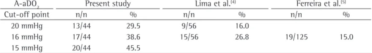
In order to facilitate comparisons with other studies, other A-aDO2 cut-off points were used in order to diagnose the syndrome. Those cut-off points can be seen in Table 5.
No significant differences were found between the groups of patients with and without HPS in terms of clinical history or physical examination (Table 1). In addition, no significant differences were found between the groups of patients with and without HPS in terms of the mean age, waiting time for transplantation, or classification of liver disease (Table 2).
Of the 28 patients with MLD, 10 (35.7%) had HPS (A-aDO2 ≥ 15 mmHg and positive TTE results for IPVD; Table 4). Of those, 7 had mild HPS (PaO2 ≥ 80 mmHg), and 3 had moderate
HPS (60 mmHg ≤ PaO2 < 80 mmHg). When an
A-aDO2 cut-off point of 20 mmHg was used,
the frequency of HPS was 25%, compared with 28.6% when the cut-off point was 15 mmHg.
Discussion
Until recently, the diagnostic criteria for HPS were not standardized, which resulted in a wide variation in the reported prevalence of the syndrome, as well as in difficulties in comparing
Table 4 - Demonstration of the alveolar-arterial oxygen tension difference and PaO2 in 44 patients with cirrhosis who were candidates for liver transplantation and in the subgroup of patients with mixed liver disease. Federal University of Pernambuco Hospital das Clínicas, 2007.
Patient Cirrhosis Mixed liver disease
A-aDO2, mmHg
PaO2, mmHg
A-aDO2, mmHg
PaO2, mmHg
1 30 80
2a 20 90
3 16 95
4a 34 72
5a 28 83
6 24 87
7 4 98
8 2 100
9a 33 78
10 24 79
11 12 98
12 27 84
13a 15 90
14 15 86
15 11 100
16 22 83
17 14 88
18 22 81
19a 37 74
20a 20 95
21a 36 76
22 14 93
23 14 99
24a 38 72
25a 26 77
26a 15 92
27 24 91
28 21 94
29a 20 88
30 45 75
31a 36 87
32a 17 93
33a 22 84
34 5 100
35 15 97
36 4 97
37a 27 91
38a 25 95
39 0 99
40 26 94
41a 30 82
42 23 88
43a 15 92
44a 63 60
A-aDO2: alveolar-arterial oxygen tension difference.
aPresence of hepatopulmonary syndrome, defined as an
A-aDO2≥ 15 mmHg and positive transthoracic
in liver volume and distortion of the liver architecture.(15) The diagnostic, epidemiologic,
and ultrasound criteria adopted in the present study allowed us to suggest but not confirm the presence of HSS. Therefore, the presence of HSS can only be confirmed after transplantation, by means of anatomopathological analysis of the liver.
In this context, when the subgroup of patients with MLD was analyzed separately, the occurrence of HPS was 35.7% (10/28), 28.6% (8/28), and 25% (7/28), respectively, when A-aDO2 cut-off points of 15 mmHg, 16 mmHg, and 20 mmHg were used. To date, there have been no studies investigating HPS in patients presenting with combinations of various PH mechanisms. However, the analysis of studies that evaluated PH patients without cirrhosis reveals that the occurrence of HPS is lower than is that described in patients with cirrhosis. In fact, a study investigating patients with noncirrhotic portal fibrosis and using an A-aDO2 cutoff of
20 mmHg showed the frequency of HPS to be 8% (2/25).(7) In another study, in which patients
with HSS were evaluated and an A-aDO2 cut-off
point of 15 mmHg was used, the occurrence of HPS was 10.2% (5/49).(5)
Taking into consideration that the pathophysiology of PH in HSS differs from that observed in cirrhosis (increased blood flow being a relevant factor in the former and increased resistance being a relevant factor in the latter), the combination of the two conditions might increase the pressure in the portal vein and result in a higher occurrence of HPS. In the present study, the PH triggered by HSS might have been an additional factor for the development of the syndrome, although we were unable to demonstrate differences between the groups of patients with and without HPS in terms of the occurrence of this helminthiasis (Table 1). Underscoring the role of schistosomiasis-related PH (without associated cirrhosis) in the etiology studies.(20) According to the 2004 arterial blood
gas standards for the diagnosis of HPS,(1) the
frequency of the syndrome in the present study was 45.5%.
A study involving liver transplant candidates and using an A-aDO2 of 20 mmHg as a cut-off point reported that the occurrence of HPS was 16% (9/56); however, when the A-aDO2 cut-off point was lowered to 15 mmHg, the frequency of HPS was 27% (15/56).(4) In another study
involving liver transplant candidates and using an A-aDO2 cut-off point of 15 mmHg, the prevalence of HPS was reported to be 15% (19/125).(21)
In the present study, the occurrence of HPS was 45.5% (20/44), 38.6% (17/44), and 29.5% (13/44) when A-aDO2 cut-off points of 15 mmHg, 16 mmHg, and 20 mmHg, respectively, were used. For all of the different A-aDO2 parameters analyzed, the occurrence of HPS was greater than that reported in other studies (Table 5).(4,21) The occurrence of MLD in 64%
of the patients (Table 1) is one of the factors in which the present study differs from other studies investigating the prevalence of HPS in liver transplant candidates. However, the number of patients was too small to demonstrate that there was an association between this variable and the occurrence of HPS.
In the present study, the diagnosis of the HSS-cirrhosis combination was based on an abdominal ultrasound showing Symmers’ fibrosis, together with positive epidemiology for schistosomiasis.(15) The use of liver ultrasound
in the diagnosis of HSS has recently increased, since it is a noninvasive method that evaluates practically the entire organ.(10,11) This is superior
to needle liver biopsy, which can result in sampling error because it allows the analysis of only a small liver fragment. In the present study, however, we were unable to quantify Symmers’ fibrosis in this subgroup of patients due to the concomitant cirrhosis, which leads to a reduction
Table 5 - Comparison between the present study and others in terms of the frequency of hepatopulmonary syndrome, according to the cut-off point for the alveolar-arterial oxygen tension difference.
A-aDO2 Present study Lima et al.(4) Ferreira et al.(5)
Cut-off point n/n % n/n % n/n %
20 mmHg 13/44 29.5 9/56 16.0
16 mmHg 17/44 38.6 15/56 26.8 19/125 15.0
15 mmHg 20/44 45.5
have resulted in a reduction in PH, leading to a lower occurrence of cirrhosis complications, such as ascites and digestive bleeding, which are generally associated with death. A recent survey revealed an association between HPS and severe liver disease (MELD) without increased short-term mortality, indirectly suggesting that the syndrome does not lead to death.(21)
Regarding liver function impairment, whether defined by the Child-Pugh class or the MELD score), we found no differences between the groups of patients with and without HPS, which is in accordance with the findings of another study.(4) In fact, HPS has been shown to
be more common in patients with milder liver dysfunction (Child-Pugh class A).(30) In contrast,
HPS has also been shown to be more common in patients with hepatic changes that were more severe (Child-Pugh classes B and C).(3,21)
In summary, the frequency of HPS in the liver transplant candidates analyzed in the present study was high, even when different A-aDO2 cut-off points were used. In addition, we found no association between the occurrence of HPS and the variables analyzed. Studies involving larger numbers of patients with cirrhosis, HSS (without cirrhosis), and MLD could confirm these findings.
Acknowledgments
The authors would like to thank Dr. Roberto Rodríguez-Roisin for his suggestions regarding the manuscript; fellow HC-UFPE physicians Dr. Izolda Moura and Dr. Luiz Loureiro Júnior (Central Laboratory) for their aid in performing the laboratory tests; Nurse Helena Lins (Liver Transplant Outpatient Clinic) for her aid in treating the patients; and the statisticians Carlos Luna (Oswaldo Cruz Foundation, Recife, Brazil) and Ulisses Ramos Montarroyos (Department of Tropical Medicine of the Federal University of Pernambuco) for their aid in performing the statistical analysis.
References
1. Rodríguez-Roisin R, Krowka MJ, Hervé P, Fallon MB. Pulmonary-Hepatic vascular Disorders (PHD). Eur Respir J. 2004 Nov;24(5):861-80.
2. Parolin MB, Coelho JCU, Puccinelli V, Schulz GJ, de Souza AM, de Barros JA. Prevalência da síndrome hepatopulmonar em candidatos a transplante hepático. Arq Gastroenterol. 2002 Jan-Mar;39(1):11-6.
of HPS, this syndrome has been diagnosed only in patients with the hepatosplenic form, not in those with the hepatointestinal form.(5)
In the present study, no differences were observed between the groups of patients with and without HPS in terms of the demographic and clinical characteristics (Table 1), which is in accordance with the findings reported by other authors.(2,4,22) However, in studies investigating
larger samples, digital clubbing, spider veins, and ascites were reported to be associated with HPS.(23,24) It is possible that the number of
patients involved in the present study was not sufficient to demonstrate differences that were more significant.
Studies using TTE with saline solution as the contrast agent, together with second harmonic imaging, obtained conflicting results regarding the occurrence of IPVD. This can be explained by differences in the underlying diseases investigated and by variations in echogenicity or in the interpretation of test results.(19) In the
present study, the TTE results were suggestive of IPVD in 52% of the patients, which is in accordance with the results of other studies, in which rates ranging from 30 to 56% were reported.(4,25-27)
It is of note that, in the present study, the waiting time for transplantation was long (mean, 27 months). This is similar to that reported for other liver transplant centers before the MELD score came into use as a means of determining the position on the waiting list.(28) It should
be highlighted that before the introduction of the MELD scale in Brazil in mid-2006, the position on the waiting list was determined on a chronological (first-come, first-served) basis. A longer waiting time translated to a potential advantage for receiving a transplant, since patients with early-stage liver disease were included in the list.(29)
18. Lang RM, Bierig M, Devereux RB, Flachskampf FA, Foster E, Pellikka PA, et al. Recommendations for chamber quantification: a report from the American Society of Echocardiographys Guidelines and Standards Committee and the Chamber Quantification Writing Group, developed in conjunction with the European Association of Echocardiography, a branch of the European Society of Cardiology. J Am Soc Echocardiogr. 2005 Dec;18(12):1440-63.
19. Vedrinne JM, Duperret S, Bizollon T, Magnin C, Motin J, Trepo C, et al. Comparison of transesophageal and transthoracic contrast echocardiography for detection of an intrapulmonary shunt in liver disease. Chest. 1997 May;111(5):1236-40.
20. Collisson EA, Nourmand H, Fraiman MH, Cooper CB, Bellamy PE, Farmer DG, et al. Retrospective analysis of the results of liver transplantation for adults with severe hepatopulmonary syndrome. Liver Transpl. 2002 Oct;8(10):925-31.
21. Ferreira PP, Camara EJN, Paula RLPD, Zollinger CC, Cavalcanti AR, Bittencourt PL. Prevalence of hepatopulmonary syndrome in patients with decompensated chronic liver disease and its impact on short-term survival. Arq Gastroenterol. 2008 Mar;45(1):34-7.
22. Przybyłowski T, Krenke R, Fangrat A, Nasilowski J, Grabczak EM, Styczynski G, et al. Gas exchange abnormalities in patients listed for liver transplantation. J Physiol Pharmacol. 2006 Sep;57(Suppl 4):313-23. 23. Arguedas MR, Drake BB, Kapoor A, Fallon MB.
Carboxyhemoglobin levels in cirrhotic patients with and without hepatopulmonary syndrome. Gastroenterology. 2005 Feb;128(2):328-33.
24. Martínez GP, Barberà JA, Visa J, Rimola A, Paré JC, Roca J, et al. Hepatopulmonary syndrome in candidates for liver transplantation. J Hepatol. 2001 May;34(5):651-7. 25. Ferreira MAP, Barreto SSM, Knorst MM, da Silva
MRA, Pinotti AF. Semiquantitative echocardiographic evaluation of intrapulmonary vascular dilatations: correlation with evaluation of shunt levels and pulmonary function parameters. J Bras Pneumol. 2009 Feb;35(2):106-13.
26. Santa-Cruz RA, Pearson MD, Cohen MG, Shrestha R, Willis PW4, Hinderliter A, et al. Clinical predictors and characteristics of patients with chronic liver disease and intrapulmonary shunts. Clin Cardiol. 2005 Sep;28(9):437-41.
27. Pavarino PR, Corbucci HA, Marchi CH, Mata PF, Godoy MF. A ecocardiografia com contraste no diagnóstico de dilatações vasculares intrapulmonares em candidatos ao transplante hepático. Arq Bras Cardiol. 2004 Apr;82(4):327-36.
28. Martin AP, Bartels M, Hauss J, Fangmann J. Overview of the MELD score and the UNOS adult liver allocation system. Transplant Proc. 2007 Dec;39(10):3169-74. 29. Everson GT. MELD: the answer or just more questions?
Gastroenterology. 2003 Jan;124(1):251-4.
30. Arguedas MR, Abrams GA, Krowka MJ, Fallon MB. Prospective evaluation of outcomes and predictors of mortality in patients with hepatopulmonary syndrome undergoing liver transplantation. Hepatology. 2003 Jan;37(1):192-7.
3. Schenk P, Fuhrmann V, Madl C, Funk G, Lehr S, Kandel O, et al. Hepatopulmonary syndrome: prevalence and predictive value of various cut offs for arterial oxygenation and their clinical consequences. Gut. 2002 Dec;51(6):853-9.
4. Lima BLG, França AVC, Pazin-Filho A, Araújo WM, Martinez JAB, Maciel BC, et al. Frequency, clinical characteristics, and respiratory parameters of hepatopulmonary syndrome. Mayo Clin Proc. 2004 Jan;79(1):42-8.
5. Ferreira RDCDS, Domingues ALC, Markman Filho B, Veras FHAP, Batista LJDB, Albuquerque Filho ES. Hepatopulmonary syndrome in patients with Schistosoma mansoni periportal fibrosis. Acta Trop. 2009 Aug;111(2):119-24.
6. Kaymakoglu S, Kahraman T, Kudat H, Demir K, Cakaloglu Y, Adalet I, et al. Hepatopulmonary syndrome in noncirrhotic portal hypertensive patients. Dig Dis Sci. 2003 Mar;48(3):556-60.
7. De BK, Sen S, Sanyal R. Hepatopulmonary syndrome in noncirrhotic portal hypertension. Ann Intern Med. 2000 Jun;132(11):924.
8. Favre TC, Ximenes RAA, Galvão AF, Pereira AP, Wanderlei TN, Barbosa CS, et al. Reliability of current estimates of schistosomiasis prevalence in the Rainforest Zone of the state of Pernambuco , Northeastern Brazil. Mem. Inst. Oswaldo Cruz. 2006 ;101(Suppl 1):73-8.
9. Strauss E. Hepatosplenic schistosomiasis: a model for the study of portal hypertension. Ann Hepatol. 2002 Jan-Mar;1(1):6-11.
10. Abdel-Wahab MF, Esmat G, Milad M, Abdel-Razek S, Strickland GT. Characteristic sonographic pattern of schistosomal hepatic fibrosis. Am J Trop Med Hyg. 1989 Jan;40(1):72-6.
11. Homeida M, Abdel-Gadir AF, Cheever AW, Bennett JL, Arbab BM, Ibrahium SZ, et al. Diagnosis of pathologically confirmed Symmers periportal fibrosis by ultrasonography: a prospective blinded study. Am J Trop Med Hyg. 1988 Jan;38(1):86-91.
12. Aquino RT, Chieffi PP, Catunda SM, Araújo MF, Ribeiro MC, Taddeo EF, et al. Hepatitis B and C virus markers among patients with hepatosplenic mansonic schistosomiasis. Rev Inst Med Trop Sao Paulo. 2000 Nov-Dec;42(6):313-20.
13. Conceição MJ, Argento CA, Chagas VL, Takiya CM, Moura DC, Silva SC. Prognosis of schistosomiasis mansoni patients infected with hepatitis B virus. Mem Inst Oswaldo Cruz. 1998 ;93(Suppl 1):255-8.
14. Jardim JR, Oliveira JA, Nascimento O, (editors). II Consenso brasileiro sobre doença pulmonar obstrutiva crônica – DPOC – 2004. J Bras de Pneumol. 2004 Nov;30(Suppl 5).
15. Pereira LM, Domingues AL, Spinelli V, McFarlane IG. Ultrasonography of the liver and spleen in Brazilian patients with hepatosplenic schistosomiasis and cirrhosis. Trans R Soc Trop Med Hyg. 1998 Nov-Dec;92(6):639-42.
16. Viegas CAA. Gasometria arterial. J Bras de Pneumol. 2002 Oct;28(Suppl 3):S233-8.
About the authors
Liana Gonçalves Macêdo
Doctoral Student. Postgraduate Program in Tropical Medicine, Universidade Federal de Pernambuco – UFPE, Federal University of Pernambuco – and Pulmonologist. Otávio de Freitas Hospital, Pernambuco State Health Department, Recife, Brazil.
Edmundo Pessoa de Almeida Lopes
Adjunct Professor. Health Sciences Center, Universidade Federal de Pernambuco – UFPE, Federal University of Pernambuco – Recife, Brazil.
Maria de Fátima Pessoa Militão de Albuquerque
Full Researcher. Aggeu Magalhães Research Center, Oswaldo Cruz Foundation, and Associate Professor I. Universidade Federal de Pernambuco – UFPE, Federal University of Pernambuco – Recife, Brazil.
Brivaldo Markman-Filho
Adjunct Professor. Health Sciences Center, Universidade Federal de Pernambuco – UFPE, Federal University of Pernambuco – Recife, Brazil.
Flávio Henrique Amaral Pires Véras
Cardiologist. University Hospital, Rio Grande do Norte University, Natal, Brazil.
Ana Carolina Chiappetta Correia de Araújo
Radiologist. Esperança Hospital, Recife, Brazil.
Álvaro Antônio Bandeira Ferraz