Nasolacrimal Duct Mucocele: Case Report and
Literature Review
Fernanda Carneiro Corujeira de Britto
1Vitor Veloso Rosier
1Tovar Vicente Luz
2Raquel Crisóstomo Lima Verde
2Clara Mônica Figueiredo de Lima
3Marcus Miranda Lessa
41Department of Otolaryngology, Hospital Universitário Professor
Edgard Santos–Universidade Federal da Bahia (HUPES-UFBA),
Salvador, BA, Brazil
2Department of Otolaryngology, Endoscopic Sinus Surgery,
HUPES-UFBA, Salvador, BA, Brazil
3Department of Otolaryngology, Health Sciences, HUPES-UFBA,
Salvador, BA, Brazil
4Department of Otolaryngology, Universidade de São Paulo,
São Paulo, SP, Brazil; HUPES-UFBA, Salvador, BA, Brazil Int Arch Otorhinolaryngol 2015;19:96–98.
Address for correspondence Fernanda Carneiro Corujeira de Britto, MD, Hospital Universitário Professor Edgard Santos–Universidade Federal da
Bahia, Ambulatório Magalhães Neto Rua Padre Feijó, 240, 2° andar (Amb. de Otorrino), Canela, Salvador, BA CEP: 40.110-170 (e-mail: [email protected]).
Introduction
Nasolacrimal duct obstruction (NLDO) is a common congeni-tal abnormality that occurs in 30% of neonates (range 6
to 84%).1Only 2 to 4% of these children become symptomatic, and most cases of NLDO resolve spontaneously in thefirst year of life.1–4
Dacryocystocele or congenital mucocele of nasolacrimal duct is a relatively rare variant of NLDO, representing0.1% of
children with congenital obstruction, and results from the coexistence of a distal and a proximal obstruction.1,5
Nasolacrimal duct mucocele typically presents as a bulging in the lower medial canthus of the eye, associated with
epiphora. The cyst may extend inferiorly, herniating into the nasal cavity.
In this article, we report a case of congenital nasolacrimal duct mucocele, which recurred after surgery and was treated with endoscopic approach, followed by a literature review.
Case Report
The patient was a 30-year-old man with a history of bilateral congenital cyst of nasolacrimal duct diagnosed by the pres-ence of a bulge in both lacrimal sac topographies since birth, without associated symptoms. He underwent surgery on the left side at 9 years of age and on the right side at 21 years of
Keywords
►
mucocele
►
nasolacrimal duct
►
dacryocysto-rhinostomy
Abstract
Introduction
Mucoceles are benign expansive cystic formations, composed of a
mucus-secreting epithelium (respiratory or pseudostrati
fi
ed epithelium). Nasolacrimal
mucocele occurs in a small proportion of children with nasolacrimal duct obstruction
and is characterized by a cystic mass in the medial canthus with dilation of the
nasolacrimal duct; although dacryocystoceles are rare in adults, they have been
reported in patients with trachoma.
Objective
Discuss clinical aspects, diagnosis, and therapeutic management of
muco-cele of nasolacrimal duct based on literature review.
Resumed Report
The authors report a case of bilateral congenital nasolacrimal duct cysts
in a 30-year-old man, identi
fi
ed as a tumor in the topography of both lacrimal sacs since
birth without associated symptoms. The patient underwent successive surgical treatments,
leading to recurrence of the tumor at the right side and recurrent local infections.
Conclusion
Endoscopic dacryocystorhinostomy has been increasingly used with good
results and success rates similar to the external access.
received June 25, 2013 accepted
November 18, 2013 published online February 13, 2014
DOI http://dx.doi.org/ 10.1055/s-0034-1366978. ISSN 1809-9777.
Copyright © 2015 by Thieme Publicações Ltda, Rio de Janeiro, Brazil
Case Report
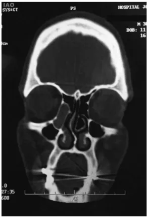
age, but the tumor recurred on the right side, without tearing, pain, discharge, or other symptoms. In subsequent evaluation with an ophthalmologist, a lack of upper and lower right lacrimal ducts was identified and indicated reconstruction surgery lacrimal spot, which was done in December 2007. However, he developed ipsilateral epiphora later, requiring another two procedures—dacryocystectomy in July 2011 and August 2011—but without success, leading to recurrent local infections. He denied loss of visual acuity or nasal symptoms during the whole period. After the last procedure, computed tomography (CT) showed a cystic expansion in the right lacrimal sac topography and dilatation of the bony canal of the nasolacrimal duct (►Figs. 1and2).
The patient was then referred for evaluation by the rhinology group of Hospital Universitário Professor Edgard Santos (Salvador, Brazil); nasal endoscopy showed no alter-ations. He had endoscopic dacryocystorhinostomy in Decem-ber 2011, with confection of mucosalflap and osteotomy of nasolacrimal bone, noting that the lacrimal sac was already opened with drainage of thick purulent secretion. The patient remains asymptomatic and without clinical signs 1 year and 8 months after surgery.
Discussion
The nasolacrimal duct is formed by canalization of the caudal extremity of an epithelial cord derived from the ectoderm in the naso-opticfissure, which is often not completed at birth. NLDO at birth is common and usually asymptomatic or presents with epiphora in neonates and infants, which
resolves spontaneously in most cases.2Generally, it results in blockage in the distal end of the nasolacrimal system, at the Hasner valve level, although the blockage can also occur at the lacrimal spot.2,3,5
Nasolacrimal mucocele, on the other hand, occurs in a small proportion of children with NLDO, when there is a distal obstruction (distal membrane perforation failure) associated with a proximal obstruction, which can be functional or mechanical.2,3,5It is characterized by a cystic mass at the medial canthus with dilation of the nasolacrimal duct that can, rarely, extend into nasal cavity.2,3,6Patients with da-cryocystoceles may present with local infection or difficult breathing or breast-feeding in the breast ipsilateral to the mucocele.5
Although dacryocystoceles are rare in adults, recurrent chronic keratitis of bacterial etiology (Chlamydia trachoma-tis) has been reported in patients with trachoma, which, due to repeated infections, can lead scarring of the conjunctiva and even to lacrimal obstruction.7
It is believed that a mixture of mesodermal cells, mucus, amniotic fluid, tears, and colonizing bacteria compose the contents of the lacrimal sac, causing distention of the lacrimal system seen in dacryocystocele.7
Encephalocele, hemangioma, dermoid cysts, and nasal gliomas may present similarly and must be considered in the differential diagnosis.5
Dacryoceles appear as rounded, well-circumscribed lesions centered in the region of lacrimal sac in CT and magnetic resonance imaging (MRI). In CT, the density of Fig. 1 Computed tomography of the paranasal sinuses, axial section,
in bone window, showing hypodense cystic lesion in the right naso-lacrimal duct topography.
Fig. 2 Computed tomography of the paranasal sinuses, coronal section, in bone window, showing hypodense cystic lesion in the right nasolacrimal duct topography.
the lesion is not homogeneous when infected. In MRI, the dacryocele appears hypointense on T1 images and hyperin-tense on T2 images. If infected, it may show a peripheral contrast uptake.8
Conservative treatment of dacryocystocele is based on a short course of topical antibiotics, warm compresses, and local massage three times a day, with a reported resolution rate of 76%. Dacryocystitis may occur within a few days or weeks and requires intravenous antibiotics to prevent sepsis. Most ophthalmologists recommend early surgical interven-tion in cases of respiratory compromise, dacryocystitis, cel-lulitis, large dacryocystoceles inducing astigmatism, or recurrent dacryocystoceles and in cases of failed conservative strategies.1,9,10In cases of infection or respiratory compro-mise, drainage is required 24 to 48 hours after the start of antibioticoterapia.6
Dacryocystorhinostomy is a surgery commonly per-formed, in which afistula is created between the lacrimal sac and the nasal cavity to relieve the epiphora caused by NLDO. External access is still noted as the most effective procedure by many ophthalmologists. The success rates vary from 75 to 95% in external access and 60 to 90% in endoscopic approach. The most common cause of failure in endoscopic surgery is obstruction of the new ostium by granulation or scar tissue.11–13
However, recent studies have shown better results with endoscopic dacryocystorhinostomy with confection muco-sal flap in front of the middle turbinate and subsequent lateral wall osteotomy, with similar rates compared with the external access. The endoscopic technique has advan-tages such as no scars, less surgical trauma, less bleeding, a quicker return to work, and preservation of the medial canthus structure, providing sustainability of lacrimal pump mechanism.11,14
The success of surgery depends on creating a large bony ostium and preventing closure of this stoma. Many techni-ques have been described to avoid or prevent obstruction. Use of mucosalflaps after wide resection of bone surrounding the sac is one technique used to prevent granulation tissue and narrowing of the canal, with good results according to the literature.11,14,15
The main advantage of external dacryocystorhinostomy (DCR) is visualization of the anatomy that facilitates the precise removal of bone in the lacrimal fossa and enables the exact anastomosis of the nasal mucosa and lacrimal sac. Neverthe-less, an intranasal component that is not recognized prior to the external access increases the chance of treatment failure if the catheter does not pass over the wall of the cyst.5,11
Conclusion
Nasolacrimal duct mucocele is a rare occurrence; however, it carries a risk of major complications (local infection, cellulitis, respiratory distress, etc.). Surgery (dacryocystorhinostomy) is considered as the definitive treatment, and external access is still the most widespread and commonly used approach by ophthalmologists. However, the interest in an endoscopic nasal approach has increasingly grown as recent studies have shown good results with this technique, with similar success rates between the two types of procedures.
References
1 Leonard DS, O’Keefe M, Rowley H, Hughes JP. Neonatal respiratory distress secondary to bilateral intranasal dacryocystocoeles. Int J Pediatr Otorhinolaryngol 2008;72(12):1873–1877
2 Gujar SK, Gandhi D. Congenital malformations of the orbit. Neu-roimaging Clin N Am 2011;21(3):585–602, viii
3 Hulka GF, Kulwin DR, Weeks SM, Cotton RT. Congenital lacrimal sac mucoceles with intranasal extension. Otolaryngol Head Neck Surg 1995;113(5):651–655
4 Yee SW, Seibert RW, Bower CM, Glasier CM. Congenital nasolacri-mal duct mucocele: a cause of respiratory distress. Int J Pediatr Otorhinolaryngol 1994;29(2):151–158
5 Wong RK, VanderVeen DK. Presentation and management of congenital dacryocystocele. Pediatrics 2008;122(5):e1108–e1112 6 Koch BL. Case 73: Nasolacrimal duct mucocele. Radiology 2004;
232(2):370–372
7 Bhaya M, Meehan R, Har-El G. Dacryocystocele in an adult: endoscopic management. Am J Otolaryngol 1997;18(2):131–134 8 Castillo BV Jr, Kaufman L. Pediatric tumors of the eye and orbit.
Pediatr Clin North Am 2003;50(1):149–172
9 Edmond JC, Keech RV. Congenital nasolacrimal sac mucocele associated with respiratory distress. J Pediatr Ophthalmol Strabis-mus 1991;28(5):287–289
10 Paysse EA, Coats DK, Bernstein JM, Go C, de Jong AL. Management and complications of congenital dacryocele with concurrent in-tranasal mucocele. J AAPOS 2000;4(1):46–53
11 Kansu L, Aydin E, Avci S, Kal A, Gedik S. Comparison of surgical outcomes of endonasal dacryocystorhinostomy with or without mucosalflaps. Auris Nasus Larynx 2009;36(5):555–559 12 Lueder GT. Endoscopic treatment of intranasal abnormalities
associated with nasolacrimal duct obstruction. J AAPOS 2004; 8(2):128–132
13 Fayet B, Racy E, Assouline M. Complications of standardized endonasal dacryocystorhinostomy with unciformectomy. Ophthalmology 2004; 111(4):837–845
14 Tsirbas A, Wormald PJ. Endonasal dacryocystorhinostomy with mucosalflaps. Am J Ophthalmol 2003;135(1):76–83
15 Çukurova I, Caner Mercan G, Çetinkaya E, et al. Endoscopic dacryocystorhinostomy: outcomes using mucosalflap preserving technique. Eur Arch Otorhinolaryngol 2013;270(5):1661–1666
International Archives of Otorhinolaryngology Vol. 19 No. 1/2015 Nasolacrimal Duct Mucocele Britto et al.