BrazJOtorhinolaryngol.2016;82(1):112---115
www.bjorl.org
Brazilian
Journal
of
OTORHINOLARYNGOLOGY
CASE
REPORT
Giant
sialolith
of
submandibular
gland
duct
treated
by
excision
and
ductal
repair:
a
case
report
夽
,
夽夽
Sialolito
gigante
de
ducto
da
glândula
submandibular
tratado
por
excisão
e
reparo
ductal:
relato
de
caso
Thiago
de
Paula
Oliveira
a,
Isaac
Nilton
Fernandes
Oliveira
a,
Eduardo
Carvalho
Paes
Pinheiro
a,
Renata
Caroline
Ferreira
Gomes
a,
Pietro
Mainenti
b,c,∗aFaculdadedeMedicinadeJuizdeFora,UniversidadePresidenteAntônioCarlos,JuizdeFora,MG,Brazil
bDepartmentofPathology,FaculdadedeMedicinadeJuizdeFora,UniversidadePresidenteAntônioCarlos,JuizdeFora,MG,
Brazil
cDepartmentofOralandMaxillofacialSurgery,CentroMédicoRioBranco,JuizdeFora,MG,Brazil
Received11March2015;accepted27March2015 Availableonline7September2015
Introduction
Sialolithiasis is one of the most common diseases of the salivary glands.1,2 It is a condition characterized by an
obstructivephenomenoninasalivaryglandorinits
excre-tory duct due to a calculus.1 The clinical presentation is
usuallycharacterized by local swelling,pain,infection of
theaffectedarea,anddilationofthesalivaryduct.1
Sialo-lithiasisusuallyaffectsadultsbetweenthethirdandfourth
decades of life, with a frequency of 12:1000.3 The
num-berofcasesinmalepatientsisabouttwicethatoffemale
patients.3Itisestimatedthat80---90%ofcasesoccurinthe
夽
Pleasecitethisarticleas:OliveiraTP,OliveiraINF,PinheiroECP, GomesRCF,MainentiP.Giantsialolithofsubmandibularglandduct treatedbyexcisionandductalrepair:casereport.BrazJ Otorhino-laryngol.2016;82:112---5.
夽夽Institution:FaculdadedeMedicinadeJuizdeFora, Universi-dadePresidenteAntônioCarlos,JuizdeFora,MG,Brazil.
∗Correspondingauthor.
E-mail:[email protected](P.Mainenti).
submandibular gland, while 10---20% occur in the parotid
gland.3Thesizeofthecalculi variesfrom<1mmtoafew
centimeters.Althoughthefrequencyofsialolithiasisis
rel-ativelyhigh,theoccurrenceofgiantsialoliths, largerthan
1.5cminanydiameter,israre.Forthisreasonfewstudies
arefoundinthepertinentmedicalliterature.1,4
This report describes a case of giant sialolith in a
42-years-oldmale,addressingtheclinicalfeatures,the
diag-nosis, and theductal repair surgeryperformed to restore
salivaryflow.
Case
report
The patient,a 42-year-old black man,attended a dental
appointment in Marchof 2014. Afterroutine radiographic
examination,hewasreferredforaconsultationwithanoral
andmaxillofacialsurgeon,inAprilof2014.During
anamne-sisthe patientdenied anyprevious diseases.He reported
only an uneventfulsurgery on the right leg. The physical
examinationshowedanankyloglossiaand,duringpalpation,
ahardnessintherightsubmandibularsalivarygland.To
fur-ther investigate the case, imaging exams wererequested
(Fig.1A).Aprovisionaldiagnosisofsialolithiasisintheright
submandibularglandductwassuggested.
http://dx.doi.org/10.1016/j.bjorl.2015.03.013
Giantsialolithofsubmandibulargland:reportofexcisionandductalrepair 113
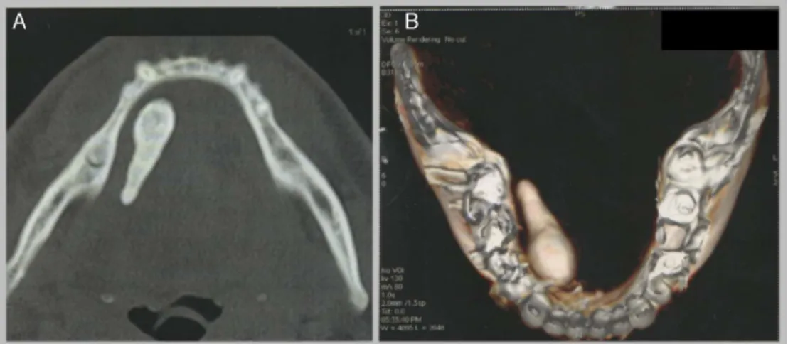
Figure1 (A)Computedtomographyscan(axialaspect)revealingamineralizedtissuewithheterogeneousdensityanddimensions of3.0×1.0cm,approximately.(B)Three-dimensionalimageofthesialolithandthemandible.
Sincethesialolithhadexuberantdimensions,anexcision
followedbythereconstructionofthesubmandibulargland
ductwasproposed.Bloodtestsandsurgicalriskexamswere
requestedforthepatient.
OnMay21,2014,thesurgicalprocedurewasconducted
by an intra-oral approach. The sialolith was removed by
curettage afterdirect incisionof theduct.A partial
min-eralizationfavoredthefragmentationofthedistalportion
ofthecalculus. Atruesalivaryglandcystwasremovedin
associationwiththecalculus(Fig.2).
Forthetreatmentofankyloglossia,atonguefrenectomy
wasperformed. To restore the salivaryflow, a No.8
ure-thral catheter wasplaced in the residual duct path. The
mucosawassuturedaroundthe catheterusingaVicryl
3-0suture inordertorepair theductof thesubmandibular
gland.
Theothertissuesweresuturedinanatomicalplanesand
therewerenocomplicationsduringthesurgicalprocedure.
Twodaysafterthesurgery,anultrasoundshowedthatthe
catheterwasinsidethesubmandibularglandduct(Fig.3).
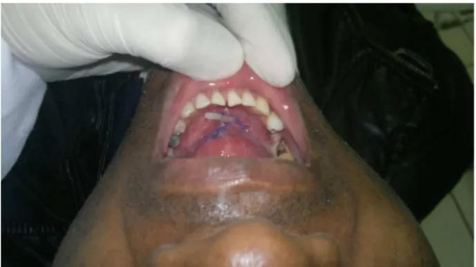
Aftermilkingof thegland,thepresenceof crystalline
liq-uidflowingfromwithinthe tubewasnoted(Fig.4).Eight
daysafterthesurgery,thepatientreportedanincreasein
salivaryvolumeandtheoccurrence ofcontractionsin the
submandibularglandregion.
Figure3 Ultrasoundshowingthecatheterinsidethesalivary glandduct.
Thesutures andthedrainwereremovedfourteen days
afterthesurgery.Aglandmilkingmaneuvershowedcopious
salivation, indicating that the performed surgical
tech-niquesucceededinreconstructingtheductalstructure.The
114 OliveiraTPetal.
Table1 Comparativetableofconsultedcases.
Author Sialolithsize Symptoms Removalmethod Age Gender
Guptaetal. (Case1)
2.8cm×1.1cm Intermittent,dullachingpain,
andswellinginleft submandibularareaduring meals
Surgicallyremovedvia intraoralapproachunder localanesthesiaand transpositionofductal opening
48 Male
Guptaetal. (Case2)
1.9cm×5.0cm Swellinginmouthassociated
withpainoverleftsideofface duringintakeoffood
Surgicallyremovedvia intraoralapproachunder localanesthesiaand transpositionofductal opening
45 Female
Iqbaletal. 3.5cm×3.0cm Asymptomatic Surgeryunderlocal
anesthesia,intra-oral approachwith marsupialization
55 Male
Dalaletal. 1.8cm×6.0cm Pusdischargeandcontinuous
painofprickingandsharp nature,radiatingtothetongue withrestrictedtongue
movement
Sialolithotomyvia intraoralapproachunder localanesthesia
40 Female
Fowell& MacBean
4.1cm Painintherightfloorofmouth andsubmandibularregion, exacerbatedbyswallowing
Excisionoftheright submandibularglandand stoneviaastandard extra-oralapproach
58 Male
Krishnanetal. (Case1)
3.4cm Recurrentpainandswelling overeightyearsthatincreased duringmeals.Inthelasttwo yearspresentedasymptomatic
Sialolithotomyvia intraoralapproachunder localanesthesia.The woundwaslefttoheal bysecondaryintention
41 Male
Krishnanetal. (Case2)
2.5cm Multipleepisodesofpainand swellingintheleftlowerpart ofthemandible,duringthe pastfourtofiveyears, especiallyatmealtimes
Surgicallyremoved throughatransoral approach,withsharp dissectionunderlocal anesthesia
32 Female
follow-upappointments withintwo monthsof thesurgery showednocomplicationsorcomplaints.
Discussion
Sialolithiasis is a disease that can affect any age group, withahigherprevalenceinmaleadults.2,5Itmainlyaffects
Figure 4 The catheter and the sutures are in the correct surgicalplacement.
the submandibular gland.6 Despite being a common
dis-ease, the presence of giant calculus is extremely rare
and most sialoliths do not exceed 1.5cm.3,5 The
calcu-lus in the present case had dimensions of approximately
3.0cm×1.0cm,thusconsideredagiantsialolith.1
The reported symptoms usually are pain and swelling
in the gland, which worsen during the meal time
(Table1).2---4,6,7Inthecurrentreport,thepatientremained
asymptomatic despite the exuberant dimensions of the
stone.
AccordingtoJensen8andCawsonetal.,7salivarystones
canbeassociatedwiththepresenceof truesalivarygland
cysts.Suchlesionsoccurduetotheobstructionofthe
sali-varyflow,followedbyaproliferationoftheductepithelium
thatsurroundsthestone.Thepresentspecimenpresented
asquamousandoncocyticdifferentiationinaccordwiththe
literature.8
Thepathophysiologyofthestoneformationisstillpoorly
understood.3 However, it is believed that the sialolith is
formed after the deposition of calcium salts around a
‘‘niche’’oforganicmaterial.7
In 80% of cases the submandibular gland is affected7
Giantsialolithofsubmandibulargland:reportofexcisionandductalrepair 115
compositionofthe salivaproducedby thegland,whichis
morealkalineandwithamajorconcentrationofcalcium6;
(b) the salivaryflow occursagainst gravity2,9; and(c) the
longandtortuousanatomyoftheductofthesubmandibular
gland.6,9Allthesefactorsworktogetherintheformationof
thecalculusinthesubmandibulargland.2,6,9Intheauthors’
opinion, theoccurrence of the sialoliths presented in the
consultedliteratureisinlinewiththeirunderstanding.
Regarding the treatment, a less invasive procedure is
of utmost importance in order to preserve the gland’s
function.2,4,7,9 The pertinent literature indicated some
surgical procedures such as trans-oral sialolithotomy,
sialoendoscopy, extracorporeal shockwave lithotripsy, and
resectionofthegland.2,3Forsmallsialoliths, conservative
treatmentsusingsialogoguesandmassageoftheglandare
alsopossible.7Thecurrentcaseshowedthetreatmentofan
exuberant calculus throughan intra-oral approach
associ-atedwithaductalrepair.AlthoughFowelletal.2concluded
that sialoplasty is one of the main treatments for giant
sialoliths,thistechniquehasnotbeendescribedorusedby
theauthorsconsulted.Theyperformedtheremovalofthe
sialolithwithclosurebysecondaryintention.
Among the possible surgical complications, one is
injury of the mandibular nerve,2 another is Wharton’s
duct stenosis.2 There was no evidence of any of these
complicationsinthepresentcase.Theductalrepair
main-tainedsalivaryflowbetweentheglandandtheoralcavity.
Thesurgicalremovalofsialolithvariesbetweensurgeons.
Thepreferredapproachismostlyperformedthrough
intra-oralintervention(Table1).
Conclusion
The present case reportdescribed the removalof agiant
sialolith.To thebestoftheauthors’ knowledge,thiscase
isuniquewithregardtothesurgicalductalrepairafterthe
excisionofasalivarystone.
Conflicts
of
interest
Theauthorsdeclarenoconflictsofinterest.
References
1.GuptaA,RattanD,Gupta R.Giantsialolithsofsubmandibular glandduct: reportoftwo caseswithunusualshape.Contemp ClinDent.2013;4:78---80.
2.FowellC,MacbeanA.Giantsalivarycalculiofthesubmandibular gland.JSurgCaseRep.2012;9:1---4.
3.IqbalA,GuptaAK,NatuSS,GuptaAK.Unusuallylargesialolith ofWharton’sduct.AnnMaxillofacSurg.2012;2:70---3.
4.DalalS,JainS,AgarwalS,VyasN.Surgicalmanagementofan unusuallylargesialolithofWharton’sduct:acasereport.King SaudUnivJDentSci.2013;4:33---5.
5.Filho MAO, Almeida LE, Pereira JA. Sialolito gigante asso-ciado à fístula cutânea. Rev Cir Traumatol Buco-Maxilo-Fac. 2008;8:35---8.
6.Branco BLC, Cardoso AB, Caubi AF, Pena GN. Sialolitíase: relatodeumcaso.RevCirTraumatolBuco-Maxilo-Fac.2003;3: 9---14.
7.CawsonRA,OdellEW,PorterSR.Neoplasticandnon-neoplastic diseasesofsalivaryglands.In:Cawson’sessentialsoforal pathol-ogyandoralmedicine.7thed.Edinburgh:ChurchillLivingstone; 2002.p.291---3.
8.JensenJL.Idiopathicdiseases.In:EllisGL,AuclairPL,GneppDR, editors.Surgicalpathologyofthesalivaryglands.Philadelphia: W.B.Saunders;1991.p.60---82.