Nephron-sparing surgery for treatment of reninoma: a rare
renin secreting tumor causing secondary hypertension
_______________________________________________
Fabio Cesar Miranda Torricelli1, Giovanni Scala Marchini1, José Roberto Colombo Junior1, Rafael
Ferreira Coelho1, Willian Carlos Nahas1, Miguel Srougi1
1Division of Urology, University of Sao Paulo Medical School, Sao Paulo, Brazil
ABSTRACT ARTICLE INFO
______________________________________________________________ ______________________
Main findings: A 25-year-old hypertensive female patient was referred to our institu-tion. Initial workup exams demonstrated a 2.8 cm cortical lower pole tumor in the right kidney. She underwent laparoscopic partial nephrectomy without complications. Histo-pathologic examination revealed a rare juxtaglomerular cell tumor known as reninoma. After surgery, she recovered uneventfully and all medications were withdrawn.
Case hypothesis: Secondary arterial hypertension is a matter of great interest to uro-logists and nephrouro-logists. Renovascular hypertension, primary hyperadosteronism and pheocromocytoma are potential diagnosis that must not be forgotten and should be excluded. Although rare, chronic pyelonephritis and renal tumors as rennin-producing tumors, nephroblastoma, hypernephroma, and renal cell carcinoma might also induce hypertension and should be in the diagnostic list of clinicians.
Promising future implications: Approximately 5% of patients with high blood pressure have specific causes and medical investigation may usually identify such patients. Fur-thermore, these patients can be successfully treated and cured, most times by minimally invasive techniques. This interesting case might expand knowledge of physicians and aid better diagnostic care in future medical practice.
Key words:
Kidney; Kidney Neoplasms; Laparoscopy; Nephrectomy
Int Braz J Urol. 2015; 41: 172-6
_____________________
Submitted for publication: June 13, 2014
_____________________
Accepted after revision: July 16, 2014
INTRODUCTION
Approximately 5% of patients with hyper-tension have specific secondary causes, which may be identified after meticulous medical history, physical examination, laboratory tests, and image workup. Secondary hypertension might be caused by several conditions affecting the kidneys, heart, arteries, or the endocrine system. Proper treatment addresses the underlying condition and tends to normalize blood pressure, reducing the risk of se-vere heart, kidney and brain complications.
investigation discovered a renal tumor as a possi-ble cause of hypertension.
Cases hypothesis and rational
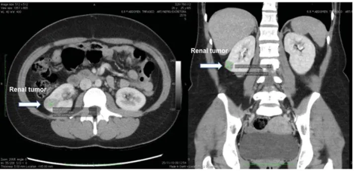
Laboratory screening included serum so-dium, calcium, potassium, aldosterone, renin, cor-tisol, catecholamines, and urinary metanephrines. Initial work-up revealed mild hypokalemia (3.4 mEq/L) and increased renin levels (7.7 ng/mL/h) with normal aldosterone (22 ng/dL). Other hor-mone levels were within normality. Doppler ultra-sound did not reveal any abnormality in renal ves-sels flow. However, a 2.5 cm hypoecogenic mass was found in the right kidney. Contrast-enhanced computed tomography of the abdomen demons-trated a 2.8 cm, well circumscribed, solid, hypo-enhancing cortical lower pole lesion in the right kidney with 100 Hounsfield Units (Figure-1). The patient was counseled on options regarding radi-cal nephrectomy and nephron-sparing surgery, as well as alternative for open or laparoscopic in-tervention. She decided for laparoscopic partial nephrectomy and the procedure was accompli-shed without intra-operative complications. Total surgical time was 120 minutes and renal hilum clamping time was 14 minutes. Bleeding was ne-gligible. The patient had an uneventful recovery and was discharge home in postoperative day one. Histopathologic examination revealed a rare juxtaglomerular cell tumor known as reninoma
(Figure-2) after minucious immunohistochemi-cal analysis, which was negative for Cytokeratin 35BH11, EMA, CD 34, CD 56, S-100, Chromogra-nin A, HMB-45, WT-1, and CD 31; and positive for AML (areas), vimentin (focally), ACTIN HHF35 (rare cels), and CD 117 (focally). Patient’s blood pressure normalized within 2 months of surgery (Systemic Blood Pressure < 120x90 mmHg), allo-wing withdrawn of all medications.
DISCUSSION AND FUTURE PERSPECTIVES
Secondary hypertension is a topic of par-ticular interest for urologists worldwide, mainly because it has potential causes that may be re-cognized and definitively treated by their special-ty. Patients with severe or refractory hyperten-sion, sudden onset of hypertenhyperten-sion, high blood pressure before 20 year-old or after 50 year-old, spontaneous hypokalemia, and unexplained re-nal dysfunction are situations in which complete workup for secondary hypertension and its rela-ted pathologies should be performed. Renovas-cular hypertension, primary hyperadosteronism, and pheocromocytoma are among those causes and should always be ruled out (1-4). Although
rare, chronic pyelonephritis and renal tumors as renin-producing tumors, nephroblastoma, hyper-nephroma, and renal cell carcinoma might also induce hypertension by different mechanisms and should be a part of the clinician diagnos-tic list. The purpose of this ardiagnos-ticle was to report and illustrate an interesting case where radiolo-gical investigation discovered a renal tumor as a possible cause of hypertension. Partial nephrec-tomy and pathological examination confirmed a juxtaglomerular cell tumor known as reninoma. The rationale is to provide the better care for patients with reversible causes of hypertension,
In our case, a young female patient presen-ted with severe hypertension. The above-mentio-ned diagnoses must be remembered so that appro-priate investigation can be started. Renovascular hypertension is a condition in which patients have unilateral or bilateral renal artery stenosis and be-come normotensive when the vessel constriction is treated by angioplasty or surgery (1). The two main etiologies are fibromuscular dysplasia and atherosclerotic plaque. Doppler ultrasound is the most cost-effective study to screen renal artery stenosis, but it is dependent on the operator. The following option is nuclear magnetic angiography
and has worse outcomes than fibromuscular dys-plasia when treated by angioplasty (5).
This case was very illustrative to remind us that approximately 5% of patients with hyper-tension have reversible secondary causes (6). Here, we presented a patient with reninoma, an unusual cause of hypertension that mainly affects young individuals (7-9). The patient was successfully tre-ated by minimally invasive laparoscopic nephron--sparing surgery, reducing the risk of loss of renal function and shorting hospital stay. Gottardo el al (8) reported the case of a 16-year-old hyperten-sive boy who presented with severe hypokalemia and markedly increased plasma renin activity. Ab-dominal ultrasonography and contrast-enhanced computed tomography revealed a 2 cm well-cir-cumscribed, solid, hypoenhancing cortical lesion in the lower pole of the left kidney. The patient underwent open nephron-sparing surgery. Histo-pathologic examination revealed a juxtaglomeru-lar cell tumor. In our case, immunohistochemical (IHC) study comprised vascular markers (CD31 and Vimentin), neuroendocrine tumor marker (Cromo-granin), Mesothelioma marker (WT1), and hema-topietic markers (CD117 and AML). All were ne-gative or focally positive (Vimentin, CD117) and were used to exclude others rare renal tumors. Cases like reninoma do not have a specific pattern and IHC findings are inconsistent in the literature. Our diagnosis was based on microscopic characte-ristics such as tumor cells with mild nuclear aty-pia, inconspicuous nucleoli and pale ill-defined cytoplasm (8, 10).
Mete et al (7) reported a similar case in whi-ch a 14-year-old boy with hypertension and pre-operative diagnosis of reninoma underwent open nephron-sparing surgery for a 2 cm mass in right kidney. The patient became normotensive postope-ratively and follow-up intravenous urography sho-wed bilateral normally functioning kidneys. Althou-gh image exams may find renal masses that could be associated to secondary arterial hypertension, there are other methods to prove such association. Wong et al (9) demonstrated the utility of both ap-propriate imaging studies and selective venous ca-theterization following provocative administration of an ACE-I for diagnosis of reninoma. We do not perform selective venous sampling routinely since it
is a more invasive technique. Nevertheless, it may help in cases where all other diagnostic modalities have been performed and doubt regarding the spe-cific cause of secondary hypertension persists.
General physicians, urologists and nephro-logists are probably the most prone to seeing these patients in an office basis. We hope that our case may help these and other physicians in their futu-re practice. Laboratory testing and image workup may identify the cause of secondary hypertension and guide therapeutic options as in our case. Un-treated secondary hypertension culminates in com-plications such as heart failure, kidney failure and stroke. The detection of a secondary cause provides an opportunity to convert an incurable disease into a potentially curable one. Moreover, even when cure cannot be achieved, early recognition and manage-ment may prevent target organ damage, reduce so-cioeconomic burden and improve quality of life (11).
CONFLICT OF INTEREST
None declared.
REFERENCES
1. Baglivo HP, Sánchez RA. Secondary arterial hypertension: improvements in diagnosis and management in the last 10 years. Am J Ther. 2011;18:403-15.
2. Giacchetti G, Ronconi V, Lucarelli G, Boscaro M, Mantero F. Analysis of screening and confirmatory tests in the diagnosis of primary aldosteronism: need for a standardized protocol. J Hypertens. 2006 ;24:737-45.
3. Espiner EA, Ross DG, Yandle TG, Richards AM, Hunt PJ. Predicting surgically remedial primary aldosteronism: role of adrenal scanning, posture testing, and adrenal vein sampling. J Clin Endocrinol Metab. 2003;88:3637-44. 4. Khorram-Manesh A, Ahlman H, Nilsson O, Friberg P, Odén A,
Stenström G, et al. Long-term outcome of a large series of patients surgically treated for pheochromocytoma. J Intern Med. 2005;258:55-66.
5. Zeller T, Frank U, Müller C, Bürgelin K, Sinn L, Bestehorn HP, et al. Predictors of improved renal function after percutaneous stent-supported angioplasty of severe atherosclerotic ostial renal artery stenosis. Circulation. 2003 4;108:2244-9. 6. Viera AJ, Neutze DM. Diagnosis of secondary hypertension:
7. Mete UK, Niranjan J, Kusum J, Rajesh LS, Goswami AK, Sharma SK. Reninoma treated with nephron-sparing surgery. Urology. 2003;61:1259.
8. Gottardo F, Cesari M, Morra A, Gardiman M, Fassina A, Dal Bianco M. A Kidney tumor in an adolescent with severe hypertension and hypokalemia: an uncommon case--case report and review of the literature on reninoma. Urol Int.2010;85:121-4.
9. Wong L, Hsu TH, Perlroth MG, Hofmann LV, Haynes CM, Katznelson L. Reninoma: case report and literature review. J Hypertens. 2008 ;26:368-73.
10. Mao J, Wang Z, Wu X, Dai W, Tong A. Recurrent hypertensive cerebral hemorrhages in a boy caused by a reninoma: rare manifestations and distinctive electron microscopy findings. J Clin Hypertens (Greenwich). 2012;14:802-5.
11. Sukor N. Secondary hypertension: a condition not to be missed. Postgrad Med J. 2011;87:706-13.
_______________________ Correspondence address:
