C A SE R EPORT
267 J Vasc Bras. 2015 July-Sept.; 14(3):267-270 http://dx.doi.org/10.1590/1677-5449.0080
Surgical treatment of a primary external iliac vein aneurysm
Tratamento cirúrgico de um aneurisma primário de veia ilíaca externa
Márcio Luís Lucas1
*
, Tiago Blaya Martins1, Newton Aerts1,2
Abstract
Primary external iliac vein aneurysms are rare and can be complicated by thrombosis, pulmonary embolism, or rupture. To date, there are only 14 cases reported in the literature. In this paper we report a case of a 25-year-old man who presented with left lower limb edema and cyanosis. Vascular ultrasonography revealed a cystic tumor in the left iliac fossa. Computed tomography angiography conirmed that the inding was an external iliac vein aneurysm, measuring 3.8 cm at its largest diameter. he patient underwent surgical treatment with resection followed by longitudinal venorrhaphy, with no complications. After the procedure left limb symptoms improved. he patient has exhibited no late complications over 44 months of follow-up.
Keywords: aneurysm; iliac vein; edema.
Resumo
Aneurismas primários da veia ilíaca são extremamente raros e podem complicar com trombose, embolia pulmonar ou ruptura. Acredita-se que existam apenas 14 casos descritos na literatura. Neste artigo, descrevemos um caso de um jovem de 25 anos, que apresentava edema e cianose do membro inferior esquerdo. A ecograia vascular revelou uma massa cística em fossa ilíaca esquerda. A angiotomograia conirmou o diagnóstico de um aneurisma da veia ilíaca externa esquerda de 3,8 cm, no maior diâmetro. O paciente foi submetido ao tratamento cirúrgico através da ressecção do aneurisma seguida de venorraia longitudinal. Teve boa evolução pós-operatória, com um seguimento clínico de 44 meses. Houve uma melhora do edema no membro inferior esquerdo, sem complicações tardias.
Palavras-chave: aneurisma; veia ilíaca; edema.
1Santa Casa de Misericórdia de Porto Alegre, Porto Alegre, RS, Brazil.
2Universidade Federal de Ciências da Saúde de Porto Alegre - UFCSPA, Porto Alegre, RS, Brazil.
Financial support: None.
Conlicts of interest: No conlicts of interest declared concerning the publication of this article. Submitted: March 30, 2014. Accepted: May 05, 2015.
268 J Vasc Bras. 2015 July-Sept.; 14(3):267-270 External iliac vein aneurysm
INTRODUCTION
Venous aneurysms are uncommon and primary aneurysms involving the iliac vein are extremely rare, but those that do occur may be asymptomatic or complicated by venous thrombosis, pulmonary embolism or rupture.1-3 Aneurysms of the iliac vein
can be primary (with no apparent deinitive cause)
or secondary to a proximal obstruction (for example,
May-Thurner Syndrome), arteriovenous istula, trauma
or cardiovascular anomalies.2 To date, there are just
14 reported cases of primary aneurysm of the iliac vein.1-14 Diagnosis is dificult and treatment has not
yet been standardized. In this article we describe the case of a 25-year-old man with a primary venous aneurysm involving the left external iliac vein who underwent surgical treatment with satisfactory results.
CASE REPORT
A 25-year-old, otherwise healthy male patient was admitted via emergency, presenting with spontaneous and insidious pain, swelling and a feeling of heaviness in the left lower limb. He had no history of traumas or of prior surgery. On clinical examination he exhibited edema and cyanosis of the entire left lower limb and the ipsilateral gluteal region. There were no palpable masses, murmurs or thrills anywhere in the abdominal area. Pulses in the lower limbs were symmetrical and normal. Laboratory tests found no abnormalities, but Doppler venous ultrasonography revealed a fusiform cystic mass with a maximum diameter of approximately 4.0 cm involving the left external iliac vein and with no sign of extrinsic
compression. Venous angiotomography conirmed
a diagnosis of a saccular venous aneurysm of the left external iliac vein with a maximum diameter of 3.8 cm and a small quantity of thrombi within the aneurysm sac (Figure 1).
The patient was given anticoagulant treatment with intravenous heparin until the time of surgery. Surgical access was obtained via a midline incision and the proximal and distal portions of the aneurysm
were carefully identiied and dissected, to avert the
risk of embolization (Figure 2a). After systemic administration of intravenous heparin, the proximal and distal portions of the external iliac vein were clamped and longitudinal venotomy was conducted, followed by venous thrombectomy, resection of the excess vein wall and primary and continuous
suture (longitudinal venorrhaphy) with Prolene 5.0 thread. After removal of the clamps, venous low
was reestablished (Figures 2c and 2c). There were no complications during surgery and no associated vascular intra-abdominal anomalies were observed. Histopathological examination of the resected venous
segment did not detect any speciic abnormalities.
The patient recovered well with no intercurrent
Figure 2. Sequence of intraoperative photographs. a) Aneurysm of left external iliac vein with repair to proximal and distal portions.
b) Venotomy and resection of excess venous wall. c) Appearance after longitudinal venorrhaphy.
Figure 1. Reconstruction of venous angiotomography showing
269 J Vasc Bras. 2015 July-Sept.; 14(3):267-270
Márcio Luís Lucas, Tiago Blaya Martins et al.
events during his stay in hospital and was discharged 5 days after surgery. At an outpatients follow-up visit,
30 days after hospital discharge, signiicant reduction
of the edema was observed and the patients’ initial complaints had completely resolved over 44 months of follow-up.
DISCUSSION
Aneurysms of the iliac vein are extremely rare. Ysa et al.2 conducted a systematic review, identifying
just 21 cases published in the literature, among which secondary aneurysms were most common, with ten cases related to arteriovenous fistulas caused by traumas, two patients with an associated cardiovascular anomaly, one patient with Iliac Vein Compression Syndrome, and another who developed the aneurysm after a heat injury to the iliac vein. At the time that that review was written, there were just seven published cases of primary aneurysms.2
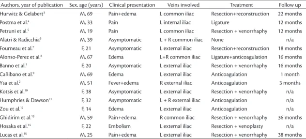
We identiied 14 cases of primary venous aneurysms
involving the iliac vein described in the literature, in eight of which an external iliac vein was involved1-14
(Table 1). Our patient, a young man, presented with pain and edema of the left lower limb as the most important complaints. A review of cases described
in the literature shows that the majority (57%) were
in male patients, aged from 19 to 69 years. The other
published cases (43%) were in women aged 14 to 38.
The majority of patients exhibited some type of
symptom related to the venous aneurysm and ive patients (38%) were asymptomatic. As with our
patient, the most common clinical manifestations
were pain and edema (57%) in the affected limb.
These symptoms could be related to increased venous pressure, as has been reported by other authors.3 In the
great majority of cases (63%) only the left iliac vein was affected, while two patients (14%) only had right
iliac vein involvement and another three patients
(21.5%) had bilateral involvement. Although there
was a small quantity of mural thrombi, our patient’s venous aneurysm was not completely thrombosed, in common with some of the cases described.1,4,6,7,10,11
However, in other situations, the aneurysm may be thrombosed at the time of diagnosis.2,3,5,8,9 Additionally,
some authors have described patients with pulmonary thromboembolism secondary to an aneurysm of the iliac vein.4,12,14
Since there are so few cases described, treatment of this type of aneurysm has not yet been standardized. There are a range of possibilities, depending on the patient’s clinical presentation, the size of the aneurysm,
extent of venous thrombosis (when present) and
presence of collateral circulation. Our patient was
young and exhibited signiicant symptoms without
complete thrombosis of the venous aneurysm. We therefore chose surgical treatment, with resection of the aneurysm by partial resection of the aneurysm wall followed by longitudinal venorrhaphy. The same technique was used by other authors with satisfactory results.1,5,10,13 In some situations other authors have
chosen complete resection of the aneurysm and reconstruction of the vein with synthetic3 or venous7
grafts. Fully thrombosed aneurysms causing minor clinical manifestations and/or with abundant collateral venous networks can be treated clinically using anticoagulation.2,9,12 In cases in which extensive
Table 1. Published cases of patients with primary aneurysms of the iliac vein.
Authors, year of publication Sex, age (years) Clinical presentation Veins involved Treatment Follow up
Hurwitz & Gelabert3 M, 69 Pain+edema L common iliac Resection+reconstruction 22 months
Postma et al.4 M, 33 Pain L internal iliac Ligature 12 months
Petruni et al.5 M, 19 Pain L common iliac Resection + venorrhaphy 12 months
Alatri & Radicchia6 M, 39 Asymptomatic L + R common iliac None n/a
Fourneau et al.7 F, 21 Asymptomatic L external iliac Resection+reconstruction 18 months
Alonso-Perez et al.8 M, 67 Edema L+R common iliac Ligature+anticoagulation 16 months
Banno et al.1 F, 20 Asymptomatic L external iliac Resection + venorrhaphy 16 months
Cañibano et al.9 M, 69 Edema L external iliac Anticoagulation 1 month
Ysa et al.2 M, 51 Fever+edema R external iliac Anticoagulation 3 months
Kotsis et al.10 F, 38 Asymptomatic L external iliac Resection + venorrhaphy n/a
Humphries & Dawson11 F, 32 Asymptomatic L + R external iliac Anticoagulation n/a
Zou et al.12 F, 14 Edema L external iliac Anticoagulation n/a
Ghidirim et al.13 M, 59 Pain+edema R common iliac Resection + venorrhaphy 36 months
Hosaka et al.14 F, 22 Embolism L external iliac Resection + venoplasty n/a
Lucas et al.15 M, 25 Pain+edema L external iliac Resection + venorrhaphy 38 months
270 J Vasc Bras. 2015 July-Sept.; 14(3):267-270 External iliac vein aneurysm
thrombosis involves the iliofemoral axis or cases in which the thrombosed segment involves the internal iliac vein, it is possible to proceed with ligature of the aneurysm only.4,8
Clinical follow-up of these patients is of fundamental importance, because venous reconstructions can develop thrombosis and patients may suffer relapse of symptoms.3 Our patient has been in follow-up for
44 months with total remission of the initial complaints of edema and pain in the left lower limb, and the most recent color Doppler venous ultrasonography did not detect thrombosis. Clinical follow up periods described in the literature vary from 3 to 36 months
and the majority of these patients exhibited signiicant
improvement of symptoms; but a certain proportion of residual edema may remain, primarily in cases in which thrombosis of the venous aneurysm had occurred.2
From the perspective of pathology, the wall of the
venous aneurysm may exhibit intimal ibrosis and
thickening, and a reduction in the number of smooth muscle cells and thickening of the tunica media.1,14
Normal histopathological results have also been reported by other authors.3
In conclusion, this is a description of a rare case of primary aneurysm of the external iliac vein in which
the patient exhibited symptoms of venous insuficiency
in the left lower limb and was successfully treated with resection of the aneurysmal venous wall followed by longitudinal venorrhaphy, with no later complications.
REFERENCES
1. Banno H, Yamanouchi D, Fujita H, et al. External iliac venous aneurysm in a pregnant woman: a case report. J Vasc Surg. 2004;40(1):174-8. http://dx.doi.org/10.1016/j.jvs.2004.02.043. PMid:15218481.
2. Ysa A, Bustabad MR, Arruabarrena A, Pérez E, Gainza E, Alonso JAG. Thrombosed iliac venous aneurysm: a rare form of presentation of a congenital anomaly of the inferior vena cava. J Vasc Surg. 2008;48(1):218-22. http://dx.doi.org/10.1016/j.jvs.2008.02.008. PMid:18589237.
3. Hurwitz RL, Gelabert H. Thrombosed iliac venous aneurysm: a rare cause of left lower extremity venous obstruction. J Vasc Surg. 1989;9(6):822-4. http://dx.doi.org/10.1016/0741-5214(89)90092-X. PMid:2724468.
4. Postma MP, McLellan GL, Northup HM, Smith R. Aneurysm of the internal iliac vein as a rare source of pulmonary thromboembolism. South Med J. 1989;82(3):390-2. http://dx.doi.org/10.1097/00007611-198903000-00029. PMid:2922632.
5. Petrunić M, Kruzić Z, Tonković I, Augustin V, Fiolić Z, Protrka N. Large iliac venous aneurysm simulating a retroperitoneal soft tissue tumour. Eur J Vasc Endovasc Surg. 1997;13(2):221-2. http:// dx.doi.org/10.1016/S1078-5884(97)80024-X. PMid:9091160. 6. Alatri A, Radicchia S. [Bilateral aneurysm of the common iliac vein:
a case report]. Ann Ital Med Int. 1997;12(2):92-3. PMid:9333318.
7. Fourneau I, Reynders-Frederix V, Lacroix H, Nevelsteen A, Suy R. Aneurysm of the iliofemoral vein. Ann Vasc Surg. 1998;12(6):605-8. http://dx.doi.org/10.1007/s100169900208. PMid:9841694. 8. Alonso-Pérez M, Segura RJ, Vidal ED. Thrombosed aneurysm of
the infrarenal vena cava: diagnosis and treatment. J Cardiovasc Surg. 2002;43(4):507-10. PMid:12124563.
9. Cañibano C, Acín F, Martinez E, Medina FJ, Bueno A, Lopez A. Primary iliac venous aneurysm: a case report and review of the literature. Angiologia. 2007;59:277-82. http://dx.doi.org/10.1016/ S0003-3170(07)75054-X.
10. Kotsis T, Mylonas S, Katsenis K, Arapoglou V, Dimakakos P. External iliac venous aneurysm treated with tangential aneurysmatectomy and lateral venorrhaphy: a case report and review of the literature. Vasc Endovascular Surg. 2008;42(6):615-9. http://dx.doi. org/10.1177/1538574408320171. PMid:18662910.
11. Humphries MD, Dawson DL. Asymptomatic bilateral external iliac vein aneurysms in a young athlete: case report and literature review. Vasc Endovascular Surg. 2010;44(7):594-6. http://dx.doi. org/10.1177/1538574410366488. PMid:20519280.
12. Zou J, Yang H, Ma H, Wang S, Zhang X. Pulmonary embolism caused by a thrombosed external iliac venous aneurysm. Ann Vasc Surg. 2011;25(7):982.e15-8. http://dx.doi.org/10.1016/j. avsg.2011.03.013. PMid:21680142.
13. Ghidirim G, Mişin I, Gagauz I, Condraţchi E. [Iliac venous aneurysm: a case report and review of literature]. Chirurgia. 2011;106(2):269-72. PMid:21698869.
14. Hosaka A, Miyata T, Hoshina K, Okamoto H, Shigematsu K. Surgical management of a primary external iliac venous aneurysm causing pulmonary thromboembolism: report of a case. Surg Today. 2014;44(9):1771-3. http://dx.doi.org/10.1007/s00595-013-0776-1. PMid:24201597.
15. Lucas ML, Martins TB, Aerts N. Tratamento cirúrgico de um aneurisma primário de veia ilíaca externa. J. Vasc. Bras. No prelo.
*
Correspondence
Márcio Luís Lucas Rua Passo da Pátria, 515/1001, Bela Vista CEP 90460-060 - Porto Alegre (RS), Brazil E-mail: mlucasvascular@hotmail.com
Author information
MLL - Vascular surgeon and preceptor of the Service of Vascular Surgery, Santa Casa de Misericórdia de Porto Alegre. TBM - Former resident in Vascular Surgery, Universidade Federal de Ciências da Saúde de Porto Alegre (UFCSPA), Service of Vascular Surgery, Santa Casa de Misericórdia de Porto Alegre. NA - Adjunct professor of Vascular Surgery at Universidade Federal de Ciências da Saúde de Porto Alegre (UFCSPA); Chief of the Service of Vascular Surgery, Santa Casa de Misericórdia de Porto Alegre.
Author contributions
Conception and design: MLL Analysis and interpretation: MLL, TBM, NA Data collection: MLL, TBM Writing the article: MLL Critical revision of the article: MLL, TBM, NA Final approval of the article*: MLL, TBM, NA Statistical analysis: N/A Overall responsibility: MLL

