Arq Neuropsiquiatr 2010;68(2):312-314
312
Letter
Vertebrobasilar artery junction
aneurysm associated with fenestration
Javier Espinoza Bentura1, Eberval G. Figueiredo2,Bernardo A. de Monaco3, Manoel J. Teixeira4
Correspondence
Eberval G. Figueiredo
Rua Enéas de Carvalho Aguiar 255 05403-000 São Paulo SP - Brasil E-mail: [email protected]
Received 1 June 2009
Received in final form 17 July 2009 Accepted 31 July 2009
ANEURISMA DA JUNÇÃO DA ARTÉRIA VERTEBROBASILAR ASSOCIADO A FENESTRAÇÃO
1MD, Resident, Honorio Delgado Hospital Universidad Nacional de San Augustin, Orequipa, Peru; 2MD, PHD, Supervisor, Head
of Cerebrovacular Surgery, Division of Neurological Surgery University of São Paulo School of Medicine, São Paulo SP, Brazil;
3MD, Resident, Division of Neurological Surgery University of São Paulo School of Medicine, São Paulo SP, Brazil; 4MD, PHD,
Chairman and Director, Division of Neurological Surgery University of São Paulo School of Medicine, São Paulo SP, Brazil. Fenestration of an intracranial artery is
a rare occurrence. After the vertebral ar-tery, the basilar artery is the second most frequent site of fenestration of intracra-nial arteries1,2. Alike arterial bifurcations,
fenestrations resulting from developmen-tal anomalies have a tendency to develop aneurysms3.
Fenestration of the proximal basilar artery aneurysms located at the verte-brobasilar junction may be associated with
aneurysms1,4,5. Aneurysms arising from the
posterior circulation are estimated to be less than 15% of all intracranial aneurysms. he morbidity and mortality of open sur-gery for posterior circulation aneurysms are higher than those of the anterior
circu-lation6. Endovascular treatment of
verte-brobasilar junction aneurysms with Gug-lielmi Detachable Coils (GDC) can provide an alternative method of treatment1,2.
We present a fenestration of vertebral artery associated with intracranial aneu-rysm and discuss its pathogenesis and management.
CASE
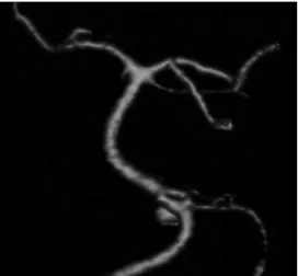
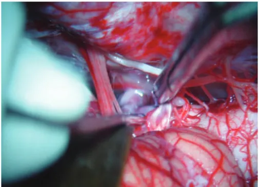
A 45-years-old female complained of intense and sudden headache. At the emer-gency room she was awake and neurolog-ical examination revealed neck stifness. CT scan displayed subarachnoid hemor-rhage. Vertebral angiogram revealed a ver-tebral aneurysm arising from a fenestra-tion at the vertebral juncfenestra-tion (Fig 1). Pa-tient underwent a far lateral approach. he vertebral artery was dissected, and the IX, x and XI were exposed at the jugular fora-men. Aneurism was exposed (Fig 2) and
clipped (Fig 3). Postoperative period was uneventful. Patient has given consent to publish this case.
DISCUSSION
he basilar artery is formed as a result of the regression of the trigeminal arter-ies. When the embryo is about 4mm long, the bilateral longitudinal neural arteries are connected laterally at multiple loca-tions with the primitive hindbrain plexus. At this stage, two fusion phenomena take place, one at the pontomesencephalic sul-cus that is related to the caudal divisions of the internal carotid arteries (ICAs), while the other fusion process involves the lon-gitudinal neural artery system and verte-brobasilar maturation. Caudal basilar fu-sion occurs with late trigeminal involution and cranial basilar fusion with early invo-lution, because the trigeminal artery is re-sponsible for the low changes in the
Arq Neuropsiquiatr 2010;68(2)
313
Vertebrobasilar artery junction aneurysm Bentura et al.
bral system during arterial development. he fusion pro-cess is typically completed when the embryo is 9 mm long (i.e. around the 5th fetal week). Failure of fusion of
the neural arteries with regression of the bridging vessels between longitudinal arteries is an explanation for the phenomenon of basilar artery fenestration.
A fenestration is deined as a single artery with two lu-minal channels. his is typically encountered in the ver-tebral artery or the ICAs in the neck. A lack of fusion of embryologically paired vessels, leads to segmentally
un-fused arteries. his condition can only exist where two embryological arteries fuse during development. here-fore, the basilar artery or the anterior spinal artery can harbor unfused segments.
Duplication can occur where the ‘‘double lumen’’ is due to two embryologically diferent vessels that fuse dur-ing development; an additional vessel persists, whereas in fenestrations the two lumina correspond to a single artery. Non-fusion of the basilar artery usually involves the lower half of the vessel because of incomplete fusion of the
lon-Fig 2. Microsurgical view. IX, X and XI nerves have been exposed and the vertebral junc-tion has been visualized. Aneurysm has been identiied and dissected.
Arq Neuropsiquiatr 2010;68(2)
314
Vertebrobasilar artery junction aneurysm Bentura et al.
gitudinal arteries in the craniocaudal direction, although distal non-fusions have been reported and are very rare2,7.
Since the basilar artery develops by the midline fusion of the paired ventral longitudinal arteries, multiple varia-tions of the vessel will occur depending on the fusion pro-cess of the longitudinal arteries and contribution of the segmental arteries.
Although rare, arterial fenestrations and unfused ar-terial segments can be associated with intracranial saccu-lar aneurysms. he lateral walls of the fenestrated artery have a normal intrinsic architecture; however, the medi-al wmedi-alls may divide the vascular channel into two distinct channels. he medial walls of the fenestration have focal defects at both ends. he media are absent locally with discontinuity of elastin at the proximal end of the fen-estration. he subendothelium is thickened distally and thinned proximally. hese structural changes at the prox-imal end of the fenestration are similar to those seen at cerebral artery bifurcations. he changes in the suben-dothelial structures are consistent with those produced by hemodynamic stress1,5,7-9.
he true frequency of fenestration of the basilar ar-tery is diicult to ascertain, and the data vary according to type of series. he incidence of fenestration of the basi-lar artery is reported to be 1.3% to 6% in autopsy series and 0.02% to 0.6% in angiographic series. he discrepan-cy between the autopsy incidence and angiographic inci-dence of basilar artery fenestrations can be explained by the fact that in some fenestrations the divider is very thin
and in most projections is angiographically occult1,2. he
new radiological techniques such as CT angiography, MR angiography, and especially 3D digital subtraction
angiog-raphy can be helpful in accurate pretreatment diagnosis2.
When a vertebrobasilar junction aneurysm is present, an
associated fenestration should be strongly suspected5.
As with other intracranial aneurysms not associated with fenestration, basilar artery fenestration aneurysms can also be treated endovascularly with GDC. he typi-cal origin of the vertebrobasilar junction aneurysm at the proximal portion of the fenestration, the complex geom-etry of the fenestration, the proximity of the lower crani-al nerves, multiple smcrani-all perforating arteries to the brain stem, and the diiculty of obtaining adequate surgical ex-posure have made the surgical treatment of these
aneu-rysms diicult1,3,4. However the complex hemodynamics
associated with these lesions may favor the surgical clip-ping as it provides well- known long term outcomes.
REFERENCES
1. Albanese E, Russo A, Ulm AJ. Fenestrated vertebrobasilar junction aneurysm: diagnostic and therapeutic considerations. J Neurosurg 2009;110:525-529. 2. Crivelli G, Bianchi M, Dario A, Dorizzi A. Saccular aneurysm associated with
proximal basilar artery fenestration: case report. J Neurosurg Sci 1993;37:29-34. 3. Campos J, Fox AJ, Viñuela F, et al. Saccular aneurysms in basilar artery
fenes-tration. AJNR Am J Neuroradiol 1987;8:233-236.
4. De Caro R, Seraini MT, Galli S, Parenti A, Guidolin D, Munari PF. Anatomy of segmental duplication in the human basilar artery. Possible site of aneurysm formation. Clin Neuropathol 1995;14:303-309.
5. Eustacchio S, Klein GE, Pendl G. Ruptured vertebrobasilar junction aneu-rysm associated with basilar artery fenestration. Acta Neurochir (Wien) 1997; 139:923-927.
6. Finlay HM, Canham PB. The layered fabric of cerebral artery fenestrations. Stroke 1994;25:1799-1806.
7. Fujimoto K, Kawai S, Yonezawa T, et al, Basilar trunk aneurysms with associ-ated fenestration treassoci-ated by using Guglielmi Detachable Coils: two cases re-port. J Stroke Cerebrovasc Dis 2007:16:84-87.
8. Greenberg E, Katz J, Janardhan V, Riina H, Gobin P, Treatment of a giant verte-brobasilar artery aneurysm using stent grafts: case report. J Neurosurg 2007: 107:165-168.