Ana Raquel Lourenço de Sousa
Licenciatura em Bioquímica
HIV-1 infection on Follicular Helper T cells
Dissertação para obtenção do Grau de Mestre em Bioquímica para a Saúde
Orientador: Doutora Helena Soares, Principal Investigator, CEDOC/FCM-UNL
Ana Raquel Lourenço de Sousa
Licenciatura em Bioquímica
HIV-1 infection on Follicular Helper T cells
Dissertação para obtenção do Grau de Mestre em Bioquímica para a Saúde
Orientador: Doutora Helena Soares, Principal Investigator, CEDOC/FCM-UNL
HIV
-1 i
n
fe
ctio
n
on
F
o
llic
u
lar
He
lper
T
ce
lls
A
n
a
S
o
u
s
HIV-1 infection on Follicular Helper T cells
Copyright, Ana Sousa, FCT/UNL, UNL
Acknowledgements
Em primeiro lugar, gostaria de agradecer à Doutora Helena Soares não só por me receber no seu laboratório como também pela paciência e orientação que me prestou. Consegui cumprir o meu objectivo de crescer tanto academicamente como pessoalmente e, por isso, estou-lhe muito grata.
Quero agradecer também às minhas colegas que foram incansáveis. À Rita, por ser a nossa mãe no laboratório, à Sofia por me ensinar tanto e ouvir os meus desabafos, à Daniela e à Rute pela ajuda e pelas gargalhadas e à Juliana pela amabilidade e boa energia. Não podia pedir um grupo de trabalho mais unido e animado. Muito obrigada.
Devo também um agradecimento ao Professor Luís Graça e à Doutora Ana Água-Doce, do Instituto de Medicina Molecular, bem como à Doutora Cláudia Andrade, da Flow Cytometry Facility do CEDOC, por toda a ajuda prestada.
Por fim, mas não menos importante, quero agradecer à minha família e amigos. Aos meus pais e mano por me ouvirem, apoiarem e, acima de tudo, me animarem. Aos meus amigos por partilharem este momento comigo e me ajudarem a vivê-lo da melhor maneira. Em especial à Marisa, por ser a minha terapeuta e me manter equilibrada e à Joana, por todas as horas ao telemóvel a apoiarmo-nos mutuamente.
Resumo
A infecção pelo Vírus da Imunodeficiência Humana (VIH) é considerada uma doença controlável sob terapia anti-retroviral, embora ainda não seja capaz de erradicar os reservatórios celulares que sustentam a contínua replicação de VIH-1. As células T auxiliares do tipo folicular (TFH) foram indicadas como o maior santuário de VIH-1. Estas células localizam-se principalmente nos centros germinativos (CGs), dos orgãos linfáticos secundários, e são especializadas em induzir a produção de anticorpos pelos linfócitos B. Apesar do seu papel crucial na infecção pelo VIH-1, ainda existem muitas lacunas em relação ao modo como o VIH-1 explora a sua maquineria celular de forma a proliferar. Por isso, o objectivo deste trabalho é contribuir para elucidação deste mistério através do isolamento de linfócitos CD4+ de amígdalas de dadores humanos saudáveis, infectando-os com VIH-1 ex vivo e identificando a população de TFH por citometria de fluxo.
Este trabalho apresenta evidências em como o VIH-1 preferencialmente infecta e expande a população de CG TFH através do aumento da expressão do seu factor de transcrição Bcl6 e receptor quimiotáctico CXCR5, também aumenta a sua sobrevivência e proliferação, medida pela proteína nuclear Ki67. O VIH-1 impõe um estado de activação (expressão de CD69 aumentada) e, consequentemente, um metabolismo acrescido, especificamente a via metabólica fosforilação oxidativa (massa mitocondrial aumentada), para fazer face às exigências energéticas da replicação viral e manutenção do reservatório celular. Adicionalmente, este vírus modula a sinalização das TFH através do aumento da expressão do receptor de células T-CD3, receptor do tipo toll 7 e citoquinas pró-inflamatórias (IL-21, IL-17 e IFNγ) de forma a promover a sua sobrevivência e proliferação.
Estes resultados constituem o trabalho de base fundamental e propiciam avenidas de investigação promissoras para elucidar o mecanismo de infecção pelo VIH-1 neste reservatório celular visando o desenvolvimento de estratégias de curas funcionais.
Abstract
Human Immunodeficiency Virus (HIV) infection is considered a manageable disease under antiretroviral therapy, although it is still unable to eradicate cellular reservoirs that harbor ongoing HIV-1 replication. Follicular helper T (TFH) cells were pointed as the major HIV-1 sanctuary. They are mainly located in germinal centers (GCs), within secondary lymphoid organs, and are specialized in inducing antibody production from B cells. Despite their crucial role in HIV-1 infection, there is still a lot of gaps to fill regarding how this virus exploits their machinery in order to thrive. For that reason, the aim of this work is to take a step closer to unveil this mystery by isolating CD4+ T cells from healthy human tonsils, infect them ex vivo with HIV-1 and identify the TFH population by flow cytometry.
This work presents evidences that HIV-1 preferentially infects and expands GC TFH population by upregulating its transcription factor Bcl6 and chemokine receptor CXCR5, it also increases its survival and proliferation, measured by nuclear protein Ki67. HIV-1 imposes a state of activation (increased CD69) and, consequently, augmented metabolism, specifically the oxidative phosphorylation metabolic pathway (increased mitochondrial mass) to meet it energy demands for replication and cellular reservoir maintenance. Furthermore, this virus modulates TFH signaling by upregulating T-cell receptor-CD3, Toll-like receptor 7 and pro-inflammatory cytokines (IL-21, IL-17 and
IFNγ) to promote its survival and proliferation.
Overall, these findings reveal the urgent need to investigate HIV-1 infection mechanism in this cellular reservoir aiming at developing functional cure strategies.
Table of Contents
1. Introduction ... 1
1.1. Human Immunodeficiency Virus (HIV) ... 1
1.1.1 History ... 1
1.1.2 Structure and genome ... 2
1.1.3 Replication cycle ... 3
1.1.4 Pathogenicity ... 5
1.1.5 Antiretroviral Therapy ... 6
1.1.6 Traditional HIV reservoirs ... 7
1.2 Immune system ... 8
1.3 Follicular helper T cells (TFH) ... 9
1.3.1 TFH differentiation ... 9
1.3.2 TFH cells as HIV reservoir ... 10
1.4 Objectives ... 12
2. Materials and Methods ... 13
2.1 Materials ... 13
2.1.1 General reagents and materials ... 13
2.1.2 FACS antibodies ... 13
2.1.3 FACS dyes ... 14
2.2 Methods... 14
2.2.1 Viral plasmid production ... 14
2.2.1.1 Bacterial transformation ... 14
2.2.1.2 Bacterial inoculation ... 14
2.2.1.3 Viral DNA purification ... 15
2.2.2 Viral particles production ... 15
2.2.2.1 Transfection ... 15
2.2.2.2 Supernatant collection ... 15
2.2.3 CD4+ T cells isolation from human tonsils ... 15
2.2.3.2 CD4+ T cell isolation ... 16
2.2.4 CD4+ T cells HIV-1 infection ... 16
2.2.5 TLR7 stimulation ... 16
2.2.6 Data acquisition and processing ... 17
2.2.6.1 Flow cytometry ... 17
2.2.6.2 Statistical analysis ... 17
3. Results ... 19
3.1 GC TFH population identification ... 19
3.2 Tonsils CD4+ T cells populations susceptibility to HIV-1 ... 19
3.3 HIV-1 effects on GC TFH cells ... 21
3.3.1 HIV-1 expands GC TFH population ... 21
3.3.2 HIV potentially augments GC TFH cell survival ... 22
3.3.3 HIV-1 modulates GC TFH cell signaling ... 25
3.3.4 TLR7 modulated signaling is HIV-1 specific ... 26
3.3.5 GC TFH cytokine profile may be shaped by HIV-1 ... 28
4. Discussion ... 31
5. Conclusions and future perspectives ... 35
Index of figures
Figure 1.1 - Worldwide Human Immunodeficiency Virus (HIV) prevalence in adults
aged 15 to 49, in 2016 ... 2
Figure 1.2- Structure of HIV ... 3
Figure 1.3 - HIV life cycle ... 4
Figure 1.4- HIV course of infection ... 5
Figure 1.5- TFH cell differentiation ... 9
Figure 1.6- HIV-1 persistence in GCs, during ART ... 11
Figure 3.1 – Gating strategy used for GC TFH identification (CD4+PD-1highCXCR5high T cells) ... 19
Figure 3.2 – Tonsils CD4+ T cells populations susceptibility to HIV-1 ... 20
Figure 3.3 – HIV-1 increases the expression of the lineage specific transcription factor Bcl6 in GC TFH cells ... 21
Figure 3.4 – HIV-1 seems to increase GC TFH chemotactic receptor CXCR5 ... 22
Figure 3.5 – HIV-1 increases GC TFH cell proliferation ... 23
Figure 3.6 –HIV-1 imposes an increased GC TFH metabolism ... 24
Figure 3.7 – HIV-1 potentiates an activation status of GC TFH cells ... 24
Figure 3.8 –HIV-1 apparently modulates GC TFH signaling ... 26
Figure 3.9 – HIV-1 increases TLR7 expression in GC TFH cells ... 26
Figure 3.10 –IMQ decreases TLR7 expression in NI GC TFH cells ... 27
Figure 3.11 – IMQ decreases proliferation capacity of NI GC TFH cells ... 28
Index of tables
Abbreviations
AIDS Acquired Immunodeficiency Syndrome
APCs Antigen-presenting cells
ART Antiretroviral therapy
Bcl6 B-cell lymphoma 6 protein
BSA Bovine serum albumin
CCL Chemokine (motif C-C) ligand CCR C-C chemokine receptor CD Cluster of differentiation
CXCL Chemokine (C-X-C motif) ligand
CXCR C-X-C chemokine receptor
DCs Dendritic cells
DMEM Dulbecco’s modified Eagle’s medium
DNA Deoxyribonucleic acid
EDTA Ethylenediamine tetraacetic acid
EIs Entry inhibitors
FACS Fluorescence-activated cell sorting
FACS-SAP Fluorescence-activated cell sorting-Saponin FBS Fetal bovine serum
FDCs Follicular dendritic cells
FIs Fusion inhibitors FSC Forward Scatter GCs Germinal centers
GFP Green fluorescent protein HBS Hepes-buffered saline
HIV Human Immunodeficiency Virus
ICOS Inducible T-cell co-stimulator
IFN Interferon
IgG Immunoglobulin G IL Interleukin
IMQ Imiquimod
INs Integrase inhibitors L/D Live/Dead cells LB Luria-Bertani
MFI Mean fluorescence intensity MHC Major Histocompatibility
MNCs Mononuclear cells
mRNA Messenger RNA
MT Mitotracker
NF-ƙB Nuclear factor-kappa B nGC Non-Germinal center NKs Natural killer cells
NNRTIs Non-nucleoside reverse transcriptase inhibitors
NRTIs Nucleoside/Nucleotide reverse transcriptase inhibitors nTFH Non-TFH
O/N Overnight
P/S Penicillin-Spreptomycin
PAMPs Pathogen-associated molecular patterns PBS Phosphate-buffered saline
PD-1 Programmed cell death protein 1
PFA Paraformaldehyde PIs Protease inhibitors PLL Polylysine
PRRs Pattern recognition receptors
RNA Ribonucleic acid rpm revolutions per minute
RPMI Roswell Park Memorial Institute
RT Room temperature
SIV Simian Immunodeficiency Virus SSC Side Scatter
ssRNA Single-stranded RNA
TFH Follicular helper T cells
Th T helper cells TLR Toll-like receptor
TNF-α Tumor necrosis factor α
1.
Introduction
1.1.
Human Immunodeficiency Virus (HIV)
1.1.1 History
AIDS (Acquired Immunodeficiency Syndrome) is characterized by a progressive failure of immune system leading to opportunistic infections and it is transmitted by bodily fluids as semen or blood (Fanales-Belasio, E. [et al.], 2010). It was first clinically described in the USA, in 1981, in a restrict group of homosexual men, intravenous drug users and hemophiliacs (Sharp, P.M. and Hahn, B.H., 2011). However, the number of cases continued to grow and it was not confined to the previously described community (Greene, W.C., 2007). In 1983-84, two separate research groups, led by Luc Montagnier and Françoise Barré-Sinoussi (Barresinoussi, F. [et al.], 1983) and Robert Gallo (Gallo, R.C. [et al.], 1983), identified HIV (Human Immunodeficiency Virus) as the cause of this pandemic disease (figure 1.1).
Figure 1.1 - Worldwide Human Immunodeficiency Virus (HIV) prevalence in adults
aged 15 to 49, in 2016. From World Health Organization (WHO).
HIV-1 is the worldwide main AIDS agent since it is more virulent and has a higher transmission rate, while HIV-2 is restricted to Western and Central Africa. Moreover, HIV-1 can be divided into four groups: M, N, O and P. HIV-1 group M is the most common strain and within it are known nine subtypes (A, B, C, D, F, G, H, J and K), whereas the other three groups are a minority. Whilst HIV-1 groups N, O and P are usually confined to West-Central Africa, the M group is worldwide spread (Taylor, B.S. and Hammer, S.M., 2008). Interestingly, Portugal is one of the countries with the major
HIV-1 subtype’s diversity as B (27%), G (29.1%), C (14.5%) and recombinant forms
(17.6%), among other subtypes, which is currently a matter of study (Carvalho, A. [et al.], 2015).
1.1.2 Structure and genome
HIV-1 is a lentivirus, a subgroup of retrovirus, which is composed by two copies of single-stranded ribonucleic acid (ssRNA) and it is characterized by long periods of incubation (figure 1.2). Its genome consists in three structural genes (gag, pol and env)
and six regulatory genes (tat, rev, nef, vif, vpr and vpu). The gag gene encodes the
precursor gag polyprotein which is processed upon viral maturation in structural core proteins (p24, p7 and p6) and matrix protein (p17). The pol gene codes for the enzymes
required for viral replication: reverse transcriptase, RNase H, integrase and protease. The
gp41 (Fanales-Belasio, E. [et al.], 2010). The regulatory genes are also important in modulating virus replication: increasing gene transcription (Tat protein), transport of viral mRNA (messenger RNA) from the nucleus to the cytoplasm (Rev protein) and manipulation of host machinery to establish a permanent state of infection (Nef protein – virulence factor) (Emerman, M. and Malim, M.H., 1998).
Figure 1.2- Structure of HIV. From Thomas Splettstoesser (www.scistyle.com).
1.1.3 Replication cycle
HIV-1 specifically infects immune cells that express the surface CD4 receptor (cluster of differentiation 4), such as CD4+ helper T cells, macrophages and dendritic cells (DCs). Considering that the first ones are CD4high, they are HIV main target cells; while macrophages and DCs are CD4intermediate, meaning that they are less extensively infected (Wilen, C.B. [et al.], 2012). HIV also recognizes the C-C chemokine receptor type 5
(CCR5) - R5 viruses, C-X-C chemokine receptor type 4 (CXCR4) - X4 viruses or both CCR5 and CXCR4 - R5X4 viruses (Berger, E.A. [et al.], 1998).
Figure 1.3 - HIV life cycle. From (Fanales-Belasio, E. [et al.], 2010)
After that, the viral RNA is released into the host cytoplasm (uncoating process) and it is transcribed into proviral DNA (Deoxyribonucleic acid), by reverse transcriptase and RNase H. Then, it is integrated into the host genome by integrase, where it may lay in a latent state – reverse transcription and viral DNA integration stage. Viral replication requires that the host immune cell is in an activated state, since important host transcription factors (as nuclear factor-kappa B (NF-ƙB)) are required for HIV transcription (Wu, Y.T. and Marsh, J.W., 2003). This explains the cell depletion firstly in the gut mucosa, leading to a microbial translocation from the gut to the blood which is thought to have a role in further driving inflammatory activation (Tincati, C. [et al.], 2016).
1.1.4 Pathogenicity
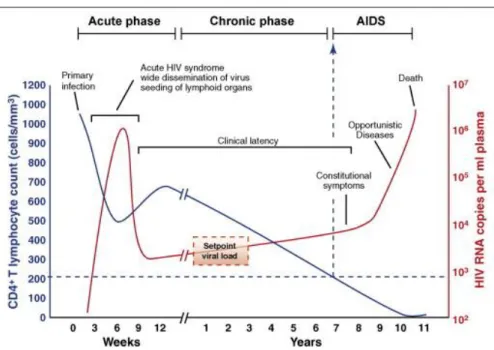
There are three stages of HIV infection: acute, chronic and AIDS (figure 1.4). The acute stage usually lasts for 2-4 weeks and, on the first 10 days (eclipse phase), the virus is not detectable in the plasma. During this phase, mucosal CD4+ T cells, macrophages and DCs are infected, breaching the mucosal barrier and allowing HIV penetration and spread (Perreau, M. [et al.], 2013a). At this point, the virus is detectable in the plasma and there is an exponential infection increase, where there is a decline of CD4+ T cell count, characterized by flu-like symptoms (Kahn, J.O. and Walker, B.D., 1998).
Figure 1.4- HIV course of infection. From (An, P. and Winkler, C.A., 2010)
viral set point. The other component of HIV-immune response is the seroconversion, which begins in the acute phase but it extends to the chronic phase. The seroconversion is a process where plasma cells (mature B cells) secrete HIV-specific antibodies (Hecht, F.M. [et al.], 2011).
The chronic stage (asymptomatic period) is characterized by a clinical latency due to low viral load and it can last for 10 years or more. Despite all the efforts taken by the immune system, infected cells became dysfunctional. Consequently, their responses are insufficient to restrain the infection (lack of effective CD8+ response and inadequate CD4+ T cell help leads to poor antibodies responses), inducing a state of chronic infection (McMichael, A.J. [et al.], 2010). This chronic state of immune activation also damages its responses, increasing the progressive depletion of CD4+ T cells (< 200 cells/µL of plasma) and the proliferation of HIV (Fanales-Belasio, E. [et al.], 2010). Therefore, after years of consistent destruction of the immune system, it culminates with totally loss of immune responses, leaving the body extremely susceptible to opportunistic infections, known as AIDS (Brooks, J.T. [et al.], 2009).
1.1.5 Antiretroviral Therapy
According to the World Health Organization (WHO), more than 70 million people have been infected by HIV and about 35 million people died from it (http://www.who.int/gho/hiv/en/), so finding a way to control or even to totally suppress HIV infection is still a main concern of nowadays society. So far, it was developed six classes of antiretroviral drugs that act on different stages of HIV replication cycle and are usually prescribed in combination:
Entry inhibitors (EIs): target the co-receptor CCR5, interfering with HIV bidding and entry into the host cell (e.g. Maraviroc);
Nucleoside/Nucleotide reverse transcriptase inhibitors (NRTIs): nucleoside/nucleotide analogues that inhibit reverse transcription, by acting as competitive substrate inhibitors (e.g. Zidovine);
Non-nucleoside reverse transcriptase inhibitors (NNRTIs): interfere with reverse transcription by acting as non-competitive substrate inhibitors (e.g. Rilpiverine);
Integrase inhibitors (INs): impair proviral DNA integration into the host genome (e.g. Raltegravir);
Protease inhibitors (PIs): block viral protease, preventing cleavage of Gag/Pol precursors proteins during viral maturation, leading to the production of defective viral particles (e.g. Amprenavir) (Arts, E.J. and Hazuda, D.J., 2012).
The major challenge imposed by HIV is its genetic variability which counteracts both effective antiretroviral therapy (ART) and host immunity. This variability is interconnected with HIV rapid replication (around 1010 virions/day), recombination of different viruses within the same infected individual and high mutation rate by the action of reverse transcriptase (around 0.2 errors/genome/cycle) (Rambaut, A. [et al.], 2004).
1.1.6 Traditional HIV reservoirs
Furthermore, there is another serious obstacle to HIV eradication: latent HIV reservoirs. They act as sanctuaries since they harbor HIV without actively producing it, meaning that these group of immune cells are in a resting state and, for that reason, they are not accessible to antiretroviral drugs. Approximately two weeks upon ART discontinuation, there is viral rebound, meaning that infected individuals require life-long therapy which obviously entails several concerns (virus resistance, cumulative toxicities and economic concerns) (Chun, T.W. [et al.], 2015).
circulating memory CD4+ T cells have been described as the hotspot of HIV-1 reservoir, recent work identified another one as the major source of replication-competent HIV-1: follicular helper CD4+ T cells (T
FH) (Banga, R. [et al.], 2016).
1.2
Immune system
The innate immune system is the first line of host defense comprising multiple mechanisms and cells (macrophages, DCs and natural killer cells (NKs)) that provide immediate protection against infection. It is composed by physical and chemical barriers, such as skin and tears, and by complement system that helps in pathogens clearance (Turvey, S.E. and Broide, D.H., 2010). The main innate immune response is inflammation which is initiated by the recognition of pathogen structural motifs, known as pathogen-associated molecular patterns (PAMPs), by receptors present on some leukocytes (such as macrophages and DCs) – the pattern recognition receptors (PRRs), such as toll-like receptors (TLRs). This recognition allows cell activation and release of inflammatory cytokines, like TNF-α, IL-1 and interferon gamma (IFNγ) (Brubaker, S.W. [et al.], 2015).
Althought innate immune system provides a rapid response, it is not a long-lasting one and, more importantly, it is not pathogen-specific. Hence the evolutionary importance in acquiring a specific yet adaptable response able to prepare the host for future challenges and that generates an immunological memory enhancing the host response to subsequent encounter with the same pathogen – the adaptive immune system (Bonilla, F.A. and Oettgen, H.C., 2010).
(CD8+), MHC class II is present in APCs and is recognized via TCR expressed by helper T cells (CD4+) (Bonilla, F.A. and Oettgen, H.C., 2010). Consequently, cytotoxic T cells are responsible for inducing programmed cell death to target cells (potentially harmful cells) (Berg, R.E. and Forman, J., 2006) and helper T cells have the crucial role of assisting other leukocytes, as it is the case of antibody secretion by B cells (Luckheeram, R.V. [et al.], 2012).
1.3 Follicular helper T cells (TFH)
Follicular helper T cells are a specialized subset of helper T cells that are mainly localized in germinal centers (GCs) within secondary lymphoid organs, as lymph nodes and tonsils. They play an important role in the formation and maintenance of GCs and generating lasting immune memory, which is fundamental for vaccination, considering that TFH cells provide differentiation signals to B cells in order to secrete antibodies (Crotty, S., 2011).
1.3.1 TFH differentiation
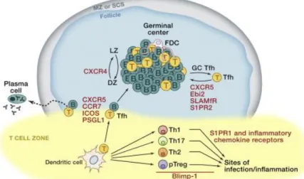
TFH cell differentiation is a multifactorial process and it can be divided into three stages: APC-dependent, B cell interaction and GC TFH cell (figure 1.5) (Crotty, S., 2014).
In the first stage, there is interaction between a naïve CD4+ T cell and a dendritic cell in an antigen-presentation context, priming the CD4+ T cell, in the T cell zone. Upon activation, it can differentiate into TFH cell if it is Bcl6+ (B cell lymphoma 6) which is an antagonist of another transcriptor factor Blimp-1, responsible for CD4+ T cell differentiation into Th1, Th2, Th17 (T helper 1, 2 and 17 cells) (Crotty, S., 2011). Their differentiation also depends on IL-21, IL-6 and inducible co-stimulator (ICOS) (Jogdand, G.M. [et al.], 2016).
In the second stage, early-TFH cell needs to migrate from T cell zone to T-B cell border. B cells express chemokines (C-X-C motif) ligand 13 (CXCL13) which are recognized by CXCR5 present on the early-TFH. In addition to over-expressing CXCR5, early-TFH cells downregulate CCR7 since it is the primary chemotactic receptor for the T-zone expressed chemokines (motif C-C) ligand 19 (CCL19) and 21 (CCL21) (Crotty, S., 2014).
The final stage is the germinal center formation where GC TFH cells drive B cell differentiation into memory B cells and plasma cells and where antibody diversification (somatic hypermutation) occurs. This stage relies mainly on cytokine secretion by GC TFH, such as IL-21 and IL-4, and surface protein CD40L which binds to CD40 expressed by GC B cells (Vinuesa, C.G. [et al.], 2016).
TFH cells can also be found in peripheral blood althought they are less abundant under normal circumstances; in autoimmune disease or in infection context, they migrate from lymphoid tissues to peripheral blood. However, there is still some controversy whether lymphoid tissue TFH cells play the same role than peripheral blood ones. Lymphoid tissue TFH cells are commonly identified as high expressors of CXCR5 and programmed cell death protein 1 (PD-1) - CXCR5highPD-1high (Yu, D. and Vinuesa, C.G., 2010).
1.3.2 TFH cells as HIV reservoir
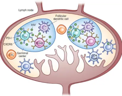
2016, Paiardini, M. and Lichterfeld, M., 2016). Furthermore, these cells exhibit an enhanced susceptibility to HIV infection (about 40 times more than a non-GC cell) (Connick, E. [et al.], 2007) and they constitute 60-75% of HIV-producing cells (Connick, E. [et al.], 2007, Folkvord, J.M. [et al.], 2005). Moreover their lifespan is not affected by HIV infection. Actually, it has been reported that there is an expansion of this CD4+ T cells subset (Lindqvist, M. [et al.], 2012). This GC permissitivity and expansion may be partially explained by the presence of highly infectious virions harbored by follicular dendritic cells (FDCs) that are chronically exposed to TFH cells (figure 1.6) and by alterations in cytokine profile (as IL-6 and IFNγ) (Miles, B. and Connick, E., 2016).
However, there is still much to know about how HIV achieves this seemingly antagonic effects of increased viral replication and TFH cell survival in order to maintain its own replication.
1.4 Objectives
The work presented on this thesis was inserted in the project «HIV-1 replication on follicular sanctuaries», funded by Gilead (#PGG/009/2016) which aims at determining how HIV-1 manipulates TFH microenvironment in order to improve its chances to thrive, without implicating TFH cells survival, becoming a major reservoir. Ultimately, this project intends to provide the necessary tools to the pursuit of a functional cure for HIV-1 infection, by immunotherapy.
In the present work, the main goal is to get an insight of the nature of TFH viral sanctuary by studying ex vivo HIV-1-infected TFH cells isolated from healthy human tonsils. This constitute an advantage comparing with the previous studies that investigate TFH compartment from HIV-1-infected patients as a whole, not allowing to discriminate between HIV-1 infection direct from bystander effects (Buranapraditkun, S. [et al.], 2017, Perreau, M. [et al.], 2013b).
2.
Materials and Methods
2.1
Materials
2.1.1 General reagents and materials
Luria-Bertani (LB) Broth, Miller medium was obtained from Fisher BioReagentsTM, NucleoBond® Xtra Column Filters was from Macherey-Nagel, Dulbecco’s modified Eagle’s (DMEM) medium supplemented with 10% Fetal Bovine Serum (FBS) and 1%
Penicillin-Streptomycin (P/S) was from ThermoFisher as well as Roswell Park Memorial Institute (RPMI) 1640 medium supplemented with 10% FBS and 1% P/S. Fluorescence-activated cell sorting (FACS) buffer is constituted by Phosphate-buffered saline 1X (PBS) from VWR and 2% of FBS from Biochrom. Fluorescence-activated cell sorting-Saponin (FACS-SAP) buffer is constituted by FACS buffer and saponin 0.1%. Biocoll (density 1,077 g/mL) used in density gradient cell separation was from Biochrom. For CD4+ T cell isolation, MojoSortTM Human CD4 T cell isolation Kit from Biolegend was used. The MojoSort buffer is constituted by PBS 1X, 100 mM ethylenediamine tetraacetic acid (EDTA) and 5 mg/mL bovine serum albumin (BSA). For HIV-1 infection, it was used a ThermoMixer C for 15 mL tubes from Eppendorf.
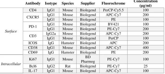
2.1.2 FACS antibodies
Table 2.1 - Primary surface and intracellular antibodies used in FACS.
Antibody Isotype Species Supplier Fluorochrome Concentration (μg/ml )
Surface
CD4 IgG1 Mouse Biolegend PerCP-Cy5.5 100 CXCR5 IgG1 IgG1 Mouse Mouse Biolegend Biolegend APC-Cy7 PE 200 100
PD-1 IgG1 IgG1 Mouse Mouse Biolegend Biolegend PE-Cy7 BV421 100 200
CD3 IgG2a IgG1 Mouse Mouse Biolegend Biolegend APC-Cy7 PerCP 200 100 ICOS IgG Hamster Biolegend APC 200 CD38 IgG1 Mouse Biolegend APC-Cy7 400 CD69 IgG Hamster Biolegend PE 200
Intracellular
IL-21 IgG1 Mouse Biolegend A647 80
IFNγ IgG1 Mouse Biolegend Pacific-Blue 100 IL-10 IgG1 Rat Biolegend A647 100 TLR7 IgG1 Mouse Novus PE 700
2.1.3 FACS dyes
Table 2.2 - Dyes used in FACS
Dye Supplier Fluorochrome
LIVE/DEAD™ Fixable Aqua Invitrogen BV510
MitoTracker® Deep Red FM Invitrogen A647
2.2 Methods
2.2.1 Viral plasmid production
2.2.1.1 Bacterial transformation
1 µL of viral DNA (HIV-NL-4-3 (Silva, J.G. [et al.], 2016) or NLENG1-IRES (Trinite, B. [et al.], 2013), both containing a green fluorescent protein (GFP) reporter gene) was
added to DH5α competent cells and incubated on ice for 20 min. The mix was placed in
a water bath at 42ºC, for 45 sec and placed back in ice for 2 min. 1 mL LB medium was added to the mix, incubated in a shaking incubator at 37ºC, 0,45 x g for 1 h and centrifuged at 3200 x g for 5 min. The mix was incubated overnight (O/N), at 37ºC, in a 10 cm LB-agar plate supplemented with 100 µg/mL ampicillin.
2.2.1.2 Bacterial inoculation
2.2.1.3 Viral DNA purification
Bacterial culture was centrifuged at 800 x g for 30 min. DNA was recovered from
bacterial culture’s pellet with NucleoBond® Xtra Column Filters by anion-exchange
chromatography, according to manufacturer’s instructions (Macherey-Nagel). Viral DNA
was resuspended in 100 µL milliQ water and stored at -20ºC.
2.2.2 Viral particles production
2.2.2.1 Transfection
293T packaging cells were seeded O/N at 37ºC, 5% CO2, at 4x10⁶ cells in a 10 cm plate in 10 mL supplemented DMEM medium. They need to grow until they reach 70-90% confluency before transfection. A mix with 20 µg of packaging plasmid (HIV-NL4-3) and 20 µg of envelope plasmid (ENV-VSV) or 40 µg of whole plasmid (NLENG1-IRES), CaCl2 and milliQ water (to a final volume of 1 mL) was added dropwise to 1 mL Hepes-buffered saline (HBS) 2X, while bubbling it. The final mix was added dropwise to 293T cells (1 mL/10 cm plate). The packaging cells were incubated O/N at 37ºC, 5% CO2. Media was changed at 24 h.
2.2.2.2 Supernatant collection
Viral particles (supernatant) were collected with a 10 mL syringe, filtered through a 0.45 µm filter and stored at -80 ºC, at 48 h post-transfection.
2.2.3 CD4+ T cells isolation from human tonsils
2.2.3.1 Human tonsils processing
removed, the remaining tissue was mechanically disintegrated and was filtered with a 100 µm mesh. Then, it was diluted (1:2) with PBS 1X, added carefully on top of Biocoll (1:3) and centrifuged at 700 x g for 30 min (without brake) for density gradient cell separation. The mononuclear cells (MNCs) ring was carefully collected and washed two times with PBS 1X at 700 x g for 10 min.
2.2.3.2 CD4+ T cell isolation
For CD4+ T cell isolation, it was followed the MojoSort™ Isolation Kits No Wash Protocol. They were culture with supplemented RPMI medium at 2x10⁶ CD4+ T cells/mL, O/N at 37ºC, 5% CO2.
2.2.4 CD4+ T cells HIV-1 infection
On the next day, CD4+ T cells were centrifuged, the supernatant was removed and they were resuspended with HIV-1 (500 μL HIV-NL4-3/million cells or 75 μL NLENG1
-IRES/million cells) and 15 μg/mL of polybrene, in 15 mL tubes. They were placed in a
Thermo Mixer C, for 4 h, at 37ºC with agitation cycles of 300 rpm for 1 min every 5 mins. Then, they were incubated for another 4 h at 37ºC, 5% CO2. After that time, cells were centrifuged at 300 x g for 5 min, resuspended in supplemented RPMI medium at 2x10⁶ CD4+ T cells/mL and cultured for 3 days at 37ºC, 5% CO
2.
2.2.5 TLR7 stimulation
For TLR7 stimulation in non-infected CD4+ T cells, it was used its synthetic agonist Imiquimod (IMQ) (1 mg/mL, from InVivoGen) at 0, 0.5, 1, 2.5, 5 and 10 μg/mL. CD4+ T cells were culture at 2.5x10⁶/mL, for 3 days at 37ºC, 5% CO
2.2.6 Data acquisition and processing
2.2.6.1 Flow cytometry
Uninfected and infected CD4+ T cells were washed with PBS 1X (at 700 x g for 3 min) and they were stained with MitoTracker® Deep Red FM and posteriorly with
LIVE/DEAD™ Fixable Aqua, following manufacturer’s instructions. For surface
staining, cells were washed twice with FACS buffer and incubated for 20 min, at RT (room temperature), in the dark, with the surface antibodies (table 1). This staining was followed with two washes with FACS buffer in order to remove the unbound antibodies. Cells were fixed with paraformaldehyde (PFA) 1%, for 20 min, at RT, in the dark and then washed with FACS buffer. For membrane permeabilization, it was used FACS-SAP
with the same incubation’s condition as cell fixation. For intracellular staining, cells were
centrifuged at 700 x g for 3 min and incubated for 30 min, at RT, in the dark, with the intracellular antibodies (table 1). Finally, cells were washed twice with FACS-SAP and once with FACS buffer and the data was acquired in FACS Canto II from BD Biosciences. The data was analyzed with Flow Jo software (BD Biosciences).
2.2.6.2 Statistical analysis
3.
Results
3.1 GC TFH population identification
After CD4+ T cells isolation fromhuman tonsils, GC TFH population was identified by flow cytometry, following the gating strategy represented on figure 3.1. In addition to this population, that have been described as PD-1highCXCR5high (Yu, D. and Vinuesa, C.G., 2010), it is also possible to identify non-GC TFH (nGC TFH), which are intermediate expressors, and non-TFH cells (nTFH) which are considered as PD-1-CXCR5-.
Figure 3.1 – Gating strategy used for GC TFH identification (CD4+PD-1highCXCR5high T
cells), by flow cytometry. GC TFH (green) represents 10.7%, nGC TFH (blue) represents
15.4% and nTFH (orange) represents 22% of tonsils CD4+ T cells.
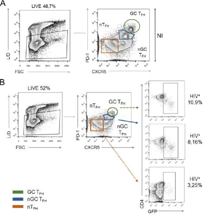
3.2 Tonsils CD4
+T cells populations susceptibility to HIV-1
Figure 3.2 – Tonsils CD4+ T cells populations susceptibility to HIV-1, by flow cytometry, gated on “Lymphocytes” following the gating strategy represented on figure
3.1. (A) Non-infected cells (NI). GC TFH represents 10.7%, nGC TFH represents 15.4%
and nTFH represents 22% of tonsils CD4+ T cells. (B) Infected cells (I). GC TFH represents
6.16%, nGC TFH represents 7.83% and nTFH represents 22% of tonsils CD4+ T cells.
3.3 HIV-1 effects on GC TFH cells
3.3.1 HIV-1 expands TFH population
PD-1, CXCR5 and transcription factor Bcl6 are known TFH markers. Analyzing both mean fluorescence intensity (MFI) of Bcl6 and the percentage of Bcl6-expressors TFH cells (figure 3.3 A), despite the presence of an outlier donor, it is visible a slight increase on this transcription factor upon HIV-1 infection. This virus also seems to increase CXCR5 (figure 3.4) leading to the proposal that HIV-1 drives TFH differentiation (Bcl6 increase) and recruitment (CXCR5 increase). Although there are some evidences that HIV-1 decreases the PD-1 inhibitory signaling (Kohler, S.L. [et al.], 2016); in this study, only half of the donors exhibited that feature, rendering these results inconclusive.
Figure 3.3 – HIV-1 increases the expression of the lineage specific transcription factor
Bcl6 in GC TFH cells. (A) Percentage (left) and MFI (right) of Bcl6+ NI and
HIV-1-infected GC TFH (I). (B) Fold change of the percentage and MFI of Bcl6+ GC TFH cells
Figure 3.4 – HIV-1 seems to increase GC TFH chemotactic receptor CXCR5. (A) MFIs
of PD-1 (left) and CXCR5 (right) from NI and HIV-1-infected GC TFH cells. (B) Fold
change of MFIs of PD-1 and CXCR5 upon HIV-1 infection. Grey bars represent the median. Each symbol represents an individual donor. p values were determined by Wilcoxon matched-pairs test; ns, not significant (p > 0.05).
3.3.2 HIV potentially augments GC TFH cell survival
Since other papers have described a role to bystander HIV-1 effects in expanding GC TFH cells (Buranapraditkun, S. [et al.], 2017, Perreau, M. [et al.], 2013b), it would be interesting to determine if HIV-1 could direct the proliferation of the infected GC TFH cells. Unexpectedly, HIV-1 infection directly increases GC TFH cell proliferation (measured by nuclear protein Ki67), as seen in the percentage of Ki67+ T
FH cells (figure 3.5 A) and in an approximately 6-fold change (figure 3.5 B).
These results raise the intriguing possibility that HIV-1 may promote its replication in GC TFH cells in two ways: one, by increasing the viral production output, and, more unconventionally, by driving the proliferation of the infected cells, turning them into expandable bioreactors.
Figure 3.5 – HIV-1 increases GC TFH cell proliferation. (A) Percentage (left) andMFI
(right) of Ki67+ NI and HIV-1-infected GC TFH cells. (B) Fold change of percentage and
MFI of Ki67+ GC T
FH cells upon HIV-1 infection. Grey bars represent the median. Each
Figure 3.6 –HIV-1 imposes an increased GC TFH metabolism. (A) Percentage (left) and
MFI (right) of MT+ (Mitotracker+) NI and HIV-1-infected GC TFH cells. (B) Fold change
in the percentage and MFI of MT+ GC T
FH cells upon HIV-1 infection. Grey bars represent
the median. Each symbol represents an individual donor. p values were determined by Wilcoxon matched-pairs test; ns, not significant (p > 0.05), * p < 0.05.
Figure 3.7 – HIV-1 potentiates an activation status of GC TFH cells. (A) Percentage (left)
and MFI (right) of CD69+ NI and HIV-1-infected GC T
FH cells. (B) Fold change of
percentage and MFI of CD69+ GC TFH cells upon HIV-1 infection. Grey bars represent
3.3.3 HIV-1 modulates GC TFH cell signaling
Bearing in mind that HIV-1 seemingly enhances TFH metabolism and, consequently, proliferation, the virus is also likely to control TFH signaling. Accordingly, HIV-1 slightly upregulates TCR (CD3) in GC TFH cells (figures 3.8 A and B), possibly to increase TFH activation.
Figure 3.8 – HIV-1 apparently modulates GC TFH signaling. (A) Percentage of CD3+
(top) and ICOS+ (bottom) NI and HIV-1-infected GC TFH cells. (B) MFIs of CD3 (top)
and ICOS (bottom) in NI and HIV-1-infected GC TFH cells. Fold change of percentage
and MFI of (C) CD3+ and (D) ICOS+ GC T
FH cells upon HIV-1 infection. Grey bars
represent the median. Each symbol represents an individual donor. p values were determined by Wilcoxon matched-pairs test; ns, not significant (p > 0.05), * p < 0.05.
3.3.4 TLR7 modulated signaling is HIV-1 specific
TLRs act as sensors of PAMPs expressed on infectious agents, providing a positive co-stimulatory signal that induces pro-inflammatory cytokines and cell proliferation. There are surface and endosomal TLRs, allowing a broad pathogen recognition and immune response. Among several receptors, there is an intracellular one capable of recognize ssRNA, such as HIV-1 – the TLR7.
So far, this work demonstrated that HIV-1 modulates GC TFH cells signaling aiming at improving this cellular reservoir capacities. Therefore, it is imperative to understand the extent of HIV-1 influence on TFH cellular machinery. In fact, HIV-1-infected GC TFH cells present a higher expression of TLR7 (figure 3.9).
Figure 3.9 – HIV-1 increases TLR7 expression in GC TFH cells. (A) Percentage (left) and
MFI (right) of TLR7+ NI and HIV-1-infected GC T
FH cells (B) Fold change of percentage
Each symbol represents an individual donor. p values were determined by Wilcoxon matched-pairs test; ns, not significant (p > 0.05), ** p < 0.01.
In order to determine if the upregulation of TLR7 was a simple response to ligand abundance, a TLR7-synthetic agonist Imiquimod (IMQ) was added to non-infected cells. Strikingly, IMQ induced different outcomes comparing with HIV-1-infected cells. Upon IMQ stimulation, there is a trend for TLR7 expression decrease in NI TFH cells (figure
3.10), more pronounced for the highest IMQ concentration (10 μg/mL), indicating
cellular defensive mechanism (negative feedback) to avoid chronic stimulation. Additionally, IMQ decreases GC TFH cells proliferation (figure 3.11).
Altogether, these data show that HIV-1 may induce an alternative signaling pathway of TLR7 in order to escape a cellular protective response and to allow its cellular reservoir expansion.
Figure 3.10 –IMQ decreases TLR7 expression in NI GC TFH cells. Percentage (left) of
TLR7+ T
FH cells and MFI (right) of TLR7 from NI GC TFH, for each IMQ concentration
Figure 3.11 – IMQ decreases proliferation capacity of NI GC TFH cells. Percentage (left)
andMFI (right) of Ki67+ NI GC TFH cells, for each IMQ concentration used (0, 0.5, 1,
2.5, 5 and 10 μg/mL).
3.3.5 GC TFH cytokine profile may be shaped by HIV-1
A recent work demonstrated that TLR7 engagement by HIV-1, in circulating CD4+ T cells, induces a state of anergy and an inability to secrete cytokines (Dominguez-Villar, M. [et al.], 2015). In the light of the previous results, it was important to check if this hold true to the present work. For that reason, the pro-inflammatory cytokines IL-21, IL-17
and IFNγ were measured in GC TFH, without any stimulation, and compared with
HIV-1-infected ones, knowing that they present an amplified TLR7 expression.
The TFH cytokine profile seems to be affected by HIV-1 infection, since it is visible an expansion of IL-21+, IL-17+ and IFNγ+ T
FH cells (figure 3.10 A and B) and in their expression levels (figure 3.10 C), even though it is more evident in IL-21 and IFNγ considering their major role in directing antibody production. Hence, contrary to circulating CD4+ T cells, HIV-1 does not induce anergy in GC T
FH cells.
Figure 3.12 – HIV-1 interferes with GC TFH cytokine production. Percentage of IL-21+
(left), IL-17+ (middle) and IFNγ+ (right) (A) NI and (B) HIV-1-infected GC TFH cells. (C)
4.
Discussion
The combination antiretroviral therapy, introduced in 1996, has been one of the most outstanding achievements of medical research for the treatment of HIV infection, preventing millions of AIDS-related deaths and improving HIV-infected individual’s life
expectancy. However, it is unable to clear persistent HIV reservoirs which remain the major obstacle to a functional cure and enforces a life time of treatment (Chun, T.W. [et al.], 2015). Such therapy entails a number of serious side effects such as heart and liver diseases and drug interactions (Marzolini, C. [et al.], 2011). Moreover, a life time of ART leads to development of drug resistance, an evasion mechanism in which occurs mutations in ART-targeted viral proteins, resulting in a great genetic variation within HIV populations. This phenomenon poses a real concern since it generates a high-fitness virions population capable of evade treatment (Clavel, F. and Hance, A.J., 2004). In fact, it is estimated that, in the United States of America, 76% of ART-treated patients developed resistance to one or more antiretroviral drugs (Richman, D.D. [et al.], 2004). That is why a therapy targeting the host signaling instead of the virus offers, not only the advantage of minimize the described selective pressure on HIV, but also the opportunity to compile a library of signaling inhibitors that, unlike ART, are capable of permeate lymphoid tissue (the major HIV-persistent reservoir).
Follicular helper T cells constitute the most important cellular reservoir for HIV-1 persistence and replication (Paiardini, M. and Lichterfeld, M., 2016). As described previously in HIV-1-infected patients (Perreau, M. [et al.], 2013b), they exhibited an enhanced susceptibility to HIV-1 infection compared to non-follicular helper T cells or even non-germinal center ones (figure 3.2 B). In addition to that, GC TFH cells display a longer lifespan upon infection (figure 3.2) and an increased differentiation and recruitment, by the upregulation of TFH transcription factor Bcl6 and the chemokine receptor CXCR5, respectively (figures 3.3 and 3.4). These data suggest that HIV-1 preferentially drives GC TFH population expansion.
known that TFH cells rely both on glycolysis and mitochondrial oxidative phosphorylation (Ray, J.P. [et al.], 2015, Zeng, H. [et al.], 2016), being the last one the most efficient
pathway in energy generation but glycolysis enables “building blocks” formation which
is crucial for viral particles production. In a previous study, it was demonstrated that HIV-1-infected circulating CD4+ T cells display an upregulated glucose transporter Glut1 expression meaning that HIV-1 most likely shifts the metabolism towards glycolysis (Loisel-Meyer, S. [et al.], 2012). Nevertheless, whether HIV-1 reprograms GC TFH metabolism towards one or the other metabolic pathway and, if so, which metabolic factors are involved, is still unknown. Combining the data obtained in the present work with the data from Loisel-Meyer, S. [et al.], 2012, it rises the idea that HIV-1 shapes TFH metabolism by activating the oxidative phosphorylation metabolic pathway to fulfill the
energetic needs along with upregulation of the glycolytic pathway to provide “building
blocks” to sustain a highly active HIV-1 replication.
Even though HIV-1 replication also rely on an activated cellular state (Stevenson, M. [et al.], 1990), there is still much to know about how this virus affects TCR signaling in TFH cells. It is described that the increased pathogenicity observed in HIV-1 results in a loss of capacity of Nef protein to downregulate TCR-CD3, as seen in less pathogenic HIV strains (Feldmann, J. [et al.], 2009, Schindler, M. [et al.], 2006) where it act as a host protective measure by ensuring a minimal cellular activation and, consequently, a reduced activation-induced T cell death (Foster, J.L. and Garcia, J.V., 2006). Knowing that GC TFH cells are in an activated and proliferative state, upon HIV-1 infection, it is not surprising that TCR-CD3 is upregulated (figure 3.8 A).
Toll-like receptors represent a critical role in activating an innate immune response, TLR7, in particular, is responsible for ssRNA recognition in intracellular compartments. This study showed that HIV-1 enhances TLR7 expression in GC TFH cells (figure 3.9) indicating a modulated TLR signaling in order to promote HIV-1 expansion. These data raise questions regarding the altered TFH signaling by HIV-1 that need to be answered in the near future.
Although TLR engagement act as a positive co-stimulatory signal to increase pro-inflammatory cytokines secretion and cell proliferation, it was demonstrated that, in circulating HIV-1-infected CD4+ T cells, it induces an anergic state (Dominguez-Villar, M. [et al.], 2015). This does not appear to apply to GC TFH cells considering the increased pro-inflammatory cytokines expression in HIV-1-infected cells (figure 3.12). This data also corroborates the chronic state of inflammation as a HIV-1 infection hallmark.
5.
Conclusions and future perspectives
The present work provided the framework for further investigation in TFH cellular machinery upon HIV-1 infection, by demonstrating an enhanced TFH differentiation, proliferation and survival at the expense of an altered metabolism (increased oxidative phosphorylation metabolic pathway) and signaling (increased TCR, TLR7 and pro-inflammatory cytokines).
Nonetheless, not only there is still serious gaps of knowledge regarding GC TFH cells signaling and metabolism, but also concerning the HIV-1 effects on them. It is imperative to know which signaling events/molecules are altered due to infection as well as the effect of PD-1 inhibitory and ICOS co-stimulatory signals both on TCR activation and on TFH cell function, since it was described an inadequate B cell help upon PD-1 engagement in HIV-1-infected cells (Cubas, R.A. [et al.], 2013). In addition, there is the need to investigate the possible crosstalk between TCR and TLR7, along with cytokine profile, and their consequences on viral and antibody production.
Since it was proved that HIV-1 is able to manipulate TFH metabolism, it is important to check whether it favors a single metabolic pathway and which metabolic molecules are involved in order to selectively inhibit metabolic pathways hijacked by HIV-1 instead of targeting general ones, preventing the harmful of functional cells.
6.
References
Alexaki, A.; Liu, Y. J.; Wigdahl, B. - Cellular Reservoirs of HIV-1 and their Role in Viral Persistence. Current Hiv Research. Vol. 6. n.º 5 (2008). p. 388-400. Disponível em
WWW: <<Go to ISI>://WOS:000260032800001>. ISSN: 1570-162X
An, P.; Winkler, C. A. - Host genes associated with HIV/AIDS: advances in gene discovery. Trends in Genetics. Vol. 26. n.º 3 (2010). p. 119-131. Disponível em WWW:
<<Go to ISI>://WOS:000275272600006>. ISSN: 0168-9525
Arts, E. J.; Hazuda, D. J. - HIV-1 Antiretroviral Drug Therapy. Cold Spring Harbor
Perspectives in Medicine. Vol. 2. n.º 4 (2012). Disponível em WWW: <<Go to
ISI>://WOS:000314277000001>. ISSN: 2157-1422
Banga, R.; Procopio, F. A.; Noto, A.; Pollakis, G.; Cavassini, M.; Ohmiti, K.; Corpataux, J. M.; de Leval, L.; Pantaleo, G.; Perreau, M. - PD-1(+) and follicular helper T cells are responsible for persistent HIV-1 transcription in treated aviremic individuals. Nature
Medicine. Vol. 22. n.º 7 (2016). p. 754-761. Disponível em WWW: <<Go to
ISI>://WOS:000379366900014>. ISSN: 1078-8956
Barresinoussi, F.; Chermann, J. C.; Rey, F.; Nugeyre, M. T.; Chamaret, S.; Gruest, J.; Dauguet, C.; Axlerblin, C.; Vezinetbrun, F.; Rouzioux, C.; Rozenbaum, W.; Montagnier, L. - ISOLATION OF A T-LYMPHOTROPIC RETROVIRUS FROM A PATIENT AT RISK FOR ACQUIRED IMMUNE-DEFICIENCY SYNDROME (AIDS). Science. Vol.
220. n.º 4599 (1983). p. 868-871. Disponível em WWW: <<Go to ISI>://WOS:A1983QP69600068>. ISSN: 0036-8075
Barton, K.; Winckelmann, A.; Palmer, S. - HIV-1 Reservoirs During Suppressive Therapy. Trends in Microbiology. Vol. 24. n.º 5 (2016). p. 345-355. Disponível em
WWW: <<Go to ISI>://WOS:000375811500007>. ISSN: 0966-842X
Berg, R. E.; Forman, J. - The role of CD8 T cells in innate immunity and in antigen non-specific protection. Current Opinion in Immunology. Vol. 18. n.º 3 (2006). p. 338-343.
Disponível em WWW: <<Go to ISI>://WOS:000237735700017>. ISSN: 0952-7915
for HIV-1. Nature. Vol. 391. n.º 6664 (1998). p. 240-240. Disponível em WWW: <<Go
to ISI>://WOS:000071484400033>. ISSN: 0028-0836
Bonilla, F. A.; Oettgen, H. C. - Adaptive immunity. Journal of Allergy and Clinical
Immunology. Vol. 125. n.º 2 (2010). p. S33-S40. Disponível em WWW: <<Go to
ISI>://WOS:000280170600004>. ISSN: 0091-6749
Brooks, J. T.; Kaplan, J. E.; Holmes, K. K.; Benson, C.; Pau, A.; Masur, H. - HIV-Associated Opportunistic Infections-Going, Going, But Not Gone: The Continued Need for Prevention and Treatment Guidelines. Clinical Infectious Diseases. Vol. 48. n.º 5
(2009). p. 609-611. Disponível em WWW: <<Go to ISI>://WOS:000263061700014>. ISSN: 1058-4838
Brubaker, S. W.; Bonham, K. S.; Zanoni, I.; Kagan, J. C. - Innate Immune Pattern Recognition: A Cell Biological Perspective. Annual Review of Immunology Vol 33. Vol.
33. (2015). p. 257-290. Disponível em WWW: <<Go to ISI>://WOS:000352911900010>. ISSN: 0732-0582
Buranapraditkun, S.; Pissani, F.; Teigler, J. E.; Schultz, B. T.; Alter, G.; Marovich, M.; Robb, M. L.; Eller, M. A.; Martin, J.; Deeks, S.; Michael, N. L.; Streeck, H. - Preservation of Peripheral T Follicular Helper Cell Function in HIV Controllers. Journal of Virology.
Vol. 91. n.º 14 (2017). Disponível em WWW: <<Go to ISI>://WOS:000405465700031>. ISSN: 0022-538X
Carvalho, A.; Costa, P.; Triunfante, V.; Branca, F.; Rodrigues, F.; Santos, C. L.; Correia-Neves, M.; Saraiva, M.; Lecour, H.; Castro, A. G.; Pedrosa, J.; Osorio, N. S. - Analysis of a Local HIV-1 Epidemic in Portugal Highlights Established Transmission of Non-B and Non-G Subtypes. Journal of Clinical Microbiology. Vol. 53. n.º 5 (2015). p.
1506-1514. Disponível em WWW: <<Go to ISI>://WOS:000353036500008>. ISSN: 0095-1137
Chun, T. W.; Moir, S.; Fauci, A. S. - HIV reservoirs as obstacles and opportunities for an HIV cure. Nature Immunology. Vol. 16. n.º 6 (2015). p. 584-589. Disponível em WWW:
Clavel, F.; Hance, A. J. - Medical progress: HIV drug resistance. New England Journal
of Medicine. Vol. 350. n.º 10 (2004). p. 1023-1035. Disponível em WWW: <<Go to
ISI>://WOS:000189363600010>. ISSN: 0028-4793
Connick, E.; Mattila, T.; Folkvord, J. M.; Schlichtemeier, R.; Meditz, A. L.; Ray, M. G.; McCarter, M. D.; MaWhinney, S.; Hage, A.; White, C.; Skinner, P. J. - CTL fail to accumulate at sites of HIV-1 replication in lymphoid tissue. Journal of Immunology. Vol.
178. n.º 11 (2007). p. 6975-6983. Disponível em WWW: <<Go to ISI>://WOS:000246896300039>. ISSN: 0022-1767
Crotty, S. - Follicular Helper CD4 T Cells (T-FH). Annual Review of Immunology, Vol
29. Vol. 29. (2011). p. 621-663. Disponível em WWW: <<Go to
ISI>://WOS:000289959200021>. ISSN: 0732-0582
Crotty, S. - T Follicular Helper Cell Differentiation, Function, and Roles in Disease.
Immunity. Vol. 41. n.º 4 (2014). p. 529-542. Disponível em WWW: <<Go to
ISI>://WOS:000345504300008>. ISSN: 1074-7613
Cubas, R. A.; Mudd, J. C.; Savoye, A. L.; Perreau, M.; van Grevenynghe, J.; Metcalf, T.; Connick, E.; Meditz, A.; Freeman, G. J.; Abesada-Terk, G.; Jacobson, J. M.; Brooks, A. D.; Crotty, S.; Estes, J. D.; Pantaleo, G.; Lederman, M. M.; Haddad, E. K. - Inadequate T follicular cell help impairs B cell immunity during HIV infection. Nature Medicine. Vol.
19. n.º 4 (2013). p. 494-+. Disponível em WWW: <<Go to ISI>://WOS:000317109100036>. ISSN: 1078-8956
Dominguez-Villar, M.; Gautron, A. S.; de Marcken, M.; Keller, M. J.; Hafler, D. A. - TLR7 induces anergy in human CD4(+) T cells. Nature Immunology. Vol. 16. n.º 1
(2015). p. 118-+. Disponível em WWW: <<Go to ISI>://WOS:000346494200015>. ISSN: 1529-2908
Emerman, M.; Malim, M. H. - HIV-1 regulatory/accessory genes: Keys to unraveling viral and host cell biology. Science. Vol. 280. n.º 5371 (1998). p. 1880-1884. Disponível
em WWW: <<Go to ISI>://WOS:000074323800045>. ISSN: 0036-8075
Di Sanita. Vol. 46. n.º 1 (2010). p. 5-14. Disponível em WWW: <<Go to
ISI>://WOS:000286606900002>. ISSN: 0021-2571
Faria, N. R.; Rambaut, A.; Suchard, M. A.; Baele, G.; Bedford, T.; Ward, M. J.; Tatem, A. J.; Sousa, J. D.; Arinaminpathy, N.; Pepin, J.; Posada, D.; Peeters, M.; Pybus, O. G.; Lemey, P. - The early spread and epidemic ignition of HIV-1 in human populations.
Science. Vol. 346. n.º 6205 (2014). p. 56-61. Disponível em WWW: <<Go to
ISI>://WOS:000342446900048>. ISSN: 0036-8075
Feldmann, J.; Leligdowicz, A.; Jaye, A.; Dong, T.; Whittle, H.; Rowland-Jones, S. L. - Downregulation of the T-Cell Receptor by Human Immunodeficiency Virus Type 2 Nef Does Not Protect against Disease Progression. Journal of Virology. Vol. 83. n.º 24 (2009).
p. 12968-12972. Disponível em WWW: <<Go to ISI>://WOS:000271932900030>. ISSN: 0022-538X
Folkvord, J. M.; Armon, C.; Connick, E. - Lymphoid follicles are sites of heightened human immunodeficiency virus type 1 (HIV-1) replication and reduced antiretroviral effector mechanisms. Aids Research and Human Retroviruses. Vol. 21. n.º 5 (2005). p.
363-370. Disponível em WWW: <<Go to ISI>://WOS:000229825700004>. ISSN: 0889-2229
Foster, J. L.; Garcia, J. V. - HIV pathogenesis: Nef loses control. Cell. Vol. 125. n.º 6
(2006). p. 1034-1035. Disponível em WWW: <<Go to ISI>://WOS:000238602700009>. ISSN: 0092-8674
Gallo, R. C.; Sarin, P. S.; Gelmann, E. P.; Robertguroff, M.; Richardson, E.; Kalyanaraman, V. S.; Mann, D.; Sidhu, G. D.; Stahl, R. E.; Zollapazner, S.; Leibowitch, J.; Popovic, M. - ISOLATION OF HUMAN T-CELL LEUKEMIA-VIRUS IN ACQUIRED IMMUNE-DEFICIENCY SYNDROME (AIDS). Science. Vol. 220. n.º
4599 (1983). p. 865-867. Disponível em WWW: <<Go to ISI>://WOS:A1983QP69600067>. ISSN: 0036-8075
Greene, W. C. - A history of AIDS: Looking back to see ahead. European Journal of
Immunology. Vol. 37. (2007). p. S94-S102. Disponível em WWW: <<Go to
Gulzar, N.; Copeland, K. F. T. - CD8+T-cells: Function and response to HIV infection.
Current Hiv Research. Vol. 2. n.º 1 (2004). p. 23-37. Disponível em WWW: <<Go to
ISI>://WOS:000189289400004>. ISSN: 1570-162X
Hecht, F. M.; Wellman, R.; Busch, M. P.; Pilcher, C. D.; Norris, P. J.; Margolick, J. B.; Collier, A. C.; Little, S. J.; Markowitz, M.; Routy, J. P.; Holte, S.; Acute Infect Early Dis Res, Program - Identifying the Early Post-HIV Antibody Seroconversion Period. Journal
of Infectious Diseases. Vol. 204. n.º 4 (2011). p. 526-533. Disponível em WWW: <<Go
to ISI>://WOS:000293304400008>. ISSN: 0022-1899
http://www.who.int/gho/hiv/en/ -. ISBN/ISSN:
Jogdand, G. M.; Mohanty, S.; Devadas, S. - Regulators of Tfh Cell Differentiation.
Frontiers in Immunology. Vol. 7. (2016). Disponível em WWW: <<Go to
ISI>://WOS:000388337300001>. ISSN: 1664-3224
Kahn, J. O.; Walker, B. D. - Acute human immunodeficiency virus type 1 infection. New
England Journal of Medicine. Vol. 339. n.º 1 (1998). p. 33-39. Disponível em WWW:
<<Go to ISI>://WOS:000074500000007>. ISSN: 0028-4793
Katsikis, P. D.; Mueller, Y. M.; Villinger, F. - The Cytokine Network of Acute HIV Infection: A Promising Target for Vaccines and Therapy to Reduce Viral Set-Point? Plos
Pathogens. Vol. 7. n.º 8 (2011). Disponível em WWW: <<Go to
ISI>://WOS:000294298100001>. ISSN: 1553-7366
Kohler, S. L.; Pham, M. N.; Folkvord, J. M.; Arends, T.; Miller, S. M.; Miles, B.; Meditz, A. L.; McCarter, M.; Levy, D. N.; Connick, E. - Germinal Center T Follicular Helper Cells Are Highly Permissive to HIV-1 and Alter Their Phenotype during Virus Replication. Journal of Immunology. Vol. 196. n.º 6 (2016). p. 2711-2722. Disponível em
WWW: <<Go to ISI>://WOS:000372338100028>. ISSN: 0022-1767
Lindqvist, M.; van Lunzen, J.; Soghoian, D. Z.; Kuhl, B. D.; Ranasinghe, S.; Kranias, G.; Flanders, M. D.; Cutler, S.; Yudanin, N.; Muller, M. I.; Davis, I.; Farber, D.; Hartjen, P.; Haag, F.; Alter, G.; zur Wiesch, J. S.; Streeck, H. - Expansion of HIV-specific T follicular helper cells in chronic HIV infection. Journal of Clinical Investigation. Vol. 122. n.º 9
Loisel-Meyer, S.; Swainson, L.; Craveiro, M.; Oburoglu, L.; Mongellaz, C.; Costa, C.; Martinez, M.; Cosset, F. L.; Battini, J. L.; Herzenberg, L. A.; Atkuri, K. R.; Sitbon, M.; Kinet, S.; Verhoeyen, E.; Taylor, N. - Glut1-mediated glucose transport regulates HIV infection. Proceedings of the National Academy of Sciences of the United States of
America. Vol. 109. n.º 7 (2012). p. 2549-2554. Disponível em WWW: <<Go to
ISI>://WOS:000300489200077>. ISSN: 0027-8424
Lorenzo-Redondo, R.; Fryer, H. R.; Bedford, T.; Kim, E. Y.; Archer, J.; Pond, S. L. K.; Chung, Y. S.; Penugonda, S.; Chipman, J. G.; Fletcher, C. V.; Schacker, T. W.; Malim, M. H.; Rambaut, A.; Haase, A. T.; McLean, A. R.; Wolinsky, S. M. - Persistent HIV-1 replication maintains the tissue reservoir during therapy. Nature. Vol. 530. n.º 7588
(2016). p. 51-+. Disponível em WWW: <<Go to ISI>://WOS:000369304500030>. ISSN: 0028-0836
Luckheeram, R. V.; Zhou, R.; Verma, A. D.; Xia, B. - CD4(+)T Cells: Differentiation and Functions. Clinical & Developmental Immunology. (2012). Disponível em WWW:
<<Go to ISI>://WOS:000302595400001>. ISSN: 1740-2522
Marzolini, C.; Back, D.; Weber, R.; Furrer, H.; Cavassini, M.; Calmy, A.; Vernazza, P.; Bernasconi, E.; Khoo, S.; Battegay, M.; Elzi, L.; Swiss, H. I. V. Cohort Study - Ageing with HIV: medication use and risk for potential drug-drug interactions. Journal of
Antimicrobial Chemotherapy. Vol. 66. n.º 9 (2011). p. 2107-2111. Disponível em WWW:
<<Go to ISI>://WOS:000293917000029>. ISSN: 0305-7453
McMichael, A. J.; Borrow, P.; Tomaras, G. D.; Goonetilleke, N.; Haynes, B. F. - The immune response during acute HIV-1 infection: clues for vaccine development. Nature
Reviews Immunology. Vol. 10. n.º 1 (2010). p. 11-23. Disponível em WWW: <<Go to
ISI>://WOS:000272992100009>. ISSN: 1474-1733
Miles, B.; Connick, E. - T-FH in HIV Latency and as Sources of Replication-Competent Virus. Trends in Microbiology. Vol. 24. n.º 5 (2016). p. 338-344. Disponível em WWW:
<<Go to ISI>://WOS:000375811500006>. ISSN: 0966-842X
Paiardini, M.; Lichterfeld, M. - Follicular T helper cells: hotspots for HIV-1 persistence.
Nature Medicine. Vol. 22. n.º 7 (2016). p. 711-712. Disponível em WWW: <<Go to
Perreau, M.; Levy, Y.; Pantaleo, G. - Immune response to HIV. Current Opinion in Hiv
and Aids. Vol. 8. n.º 4 (2013a). p. 333-340. Disponível em WWW: <<Go to
ISI>://WOS:000326730100012>. ISSN: 1746-630X
Perreau, M.; Savoye, A. L.; De Crignis, E.; Corpataux, J. M.; Cubas, R.; Haddad, E. K.; De Leval, L.; Graziosi, C.; Pantaleo, G. - Follicular helper T cells serve as the major CD4 T cell compartment for HIV-1 infection, replication, and production. Journal of
Experimental Medicine. Vol. 210. n.º 1 (2013b). p. 143-156. Disponível em WWW:
<<Go to ISI>://WOS:000313560900013>. ISSN: 0022-1007
Rambaut, A.; Posada, D.; Crandall, K. A.; Holmes, E. C. - The causes and consequences of HIV evolution. Nature Reviews Genetics. Vol. 5. n.º 1 (2004). p. 52-61. Disponível em
WWW: <<Go to ISI>://WOS:000187641100016>. ISSN: 1471-0056
Ray, J. P.; Staron, M. M.; Shyer, J. A.; Ho, P. C.; Marshall, H. D.; Gray, S. M.; Laidlaw, B. J.; Araki, K.; Ahmed, R.; Kaech, S. M.; Craft, J. - The Interleukin-2-mTORc1 Kinase Axis Defines the Signaling, Differentiation, and Metabolism of T Helper 1 and Follicular B Helper T Cells. Immunity. Vol. 43. n.º 4 (2015). p. 690-702. Disponível em WWW:
<<Go to ISI>://WOS:000363478700012>. ISSN: 1074-7613
Richman, D. D.; Morton, S. C.; Wrin, T.; Hellmann, N.; Berry, S.; Shapiro, M. F.; Bozzette, S. A. - The prevalence of antiretroviral drug resistance in the United States.
Aids. Vol. 18. n.º 10 (2004). p. 1393-1401. Disponível em WWW: <<Go to
ISI>://WOS:000222641500005>. ISSN: 0269-9370
Rolf, J.; Fairfax, K.; Turner, M. - Signaling Pathways in T Follicular Helper Cells.
Journal of Immunology. Vol. 184. n.º 12 (2010). p. 6563-6568. Disponível em WWW:
<<Go to ISI>://WOS:000278516700001>. ISSN: 0022-1767
Schindler, M.; Munch, J.; Kutsch, O.; Li, H.; Santiago, M. L.; Bibollet-Ruche, F.; Muller-Trutwin, M. C.; Novembre, F. J.; Peeters, M.; Courgnaud, V.; Bailes, E.; Roques, P.; Sodora, D. L.; Silvestri, G.; Sharp, P. M.; Hahn, B. H.; Kirchhoff, F. - Nef-mediated suppression of T cell activation was lost in a lentiviral lineage that gave rise to HIV-1.
Cell. Vol. 125. n.º 6 (2006). p. 1055-1067. Disponível em WWW: <<Go to


![Figure 1.3 - HIV life cycle. From (Fanales-Belasio, E. [et al.], 2010)](https://thumb-eu.123doks.com/thumbv2/123dok_br/15750395.637891/28.892.200.599.126.416/figure-hiv-life-cycle-fanales-belasio-e-et.webp)