Polymerase chain reaction with two molecular targets in mucosal
leishmaniasis' diagnosis: a validation study
Clemencia Ovalle Bracho
+, Luisa Porras de Quintana,
Sandra Muvdi Arenas,
Melania Rios Parra
Tropical Dermatology Research Group, National Institute of Dermatology, Centro Dermatológico Federico Lleras Acosta, E.S.E., Bogotá, Colombia
We validated the polymerase chain reaction (PCR) with a composite reference standard in 61 patients clini-cally suspected of having mucosal leishmaniasis, 36 of which were cases and 25 non-cases according to this reference standard. Patient classification and test application were carried out independently by two blind observers. One pair of primers was used to amplify a fragment of 120 bp in the conserved region of kDNA and another pair was used to amplify the internal transcript spacers (ITS) rDNA. PCR showed 68.6% (95% CI 59.2-72.6) sensitivity and 92% (95% CI 78.9-97.7) specificity; positive likelihood ratio: 8.6 (95% CI 2.8-31.3) and negative likelihood ratio: 0.3 (95% CI 0.3-0.5), when kDNA molecular target was amplified. The test performed better on sensitivity using this target compared to the ITS rDNA molecular target which showed 40% (95% CI 31.5-42.3) sensitivity and 96% (95% CI 84.1-99.3) specificity; positive likelihood ratio: 10 (95% CI 2.0-58.8) and negative likelihood ratio: 0.6 (95% CI 0.6-0.8). The inter-observer agreement was excellent for both tests. Based upon results obtained and due to low performance of conventional methods for diagnosing mucosal leishmaniasis, we consider PCR with kDNA as molecular target is a useful diagnostic test and the ITS rDNA molecular target is useful when the aim is to identify species.
Key words: leishmaniasis mucocutaneous - diagnosis - polymerase chain reaction - ribosomal spacer
Leishmaniasis is a disease found throughout the world: 350 million people are at risk, and the disease burden is 2.4 DALYs (years of life lost due to premature death or disability). It is estimated that there are 12 mil-lion cases in the world: 1.5 to 2 milmil-lion new cases a year of which 1-1.5 million correspond to cutaneous leish-maniasis and 500,000 to visceral leishleish-maniasis (TDR/ WHO 2005). The total leishmaniasis cases reported in Colombia in 2005 were 18,100 showing a clear tendency for an increase in the last decade. This number repre-sents a 21.85% increase with respect to the total cases informed by 2004. The incidence rate estimated for cu-taneous leishmaniasis for 2005 was 14.6 × 10,000, tak-ing as denominator the rural population (Instituto Na-cional de Salud 2006).
Mucosal leishmaniasis is a chronic progressive dis-ease, difficult to treat and diagnose which usually ap-pears months or years after skin inoculation and may lead to destructive lesions of the upper respiratory tract. It can be accompanied by malnutrition when it causes
dys-phagia and can lead to death due to pneumonia or as-phyxia (Marsden 1985).
Clinical diagnosis is difficult because symptoms and signs can be confused with those of other granuloma-tous and malignant conditions (Rodríguez et al. 1994). Parasite identification is difficult and sensitivity reports vary with different techniques, from 0.2% with direct smear to 68.6% when various methods are used (Weigle et al. 1987, Zajtchuk et al. 1989, Schubach et al. 2001, Disch et al. 2005, Oliveira et al. 2005). In cases where there is a strong clinical suspicion and no parasite is iso-lated, treatment is prescribed with pentavalent antimony which is highly effective, but has been associated with fre-quent adverse effects: malaise, headache, muscle pain, joint pain and hepatic, pancreatic, renal, hematologic, and car-diac toxicity that has lead to death in some cases (Chulay et al. 1985, Hepburn et al. 1994, Aronson et al. 1998).
Polymerase chain reaction (PCR) has been used in the last decade for cutaneous and more recently for mu-cosal leishmaniasis. Different authors have reported sen-sitivities that vary from 62 to 97.1% using mostly mini-circle kDNA as target in mucosal leishmaniasis (Pirmez et al. 1999, Oddone et al. 2004, Oliveira et al. 2005, Disch et al. 2005). After carrying out a critical appraisal of these studies, we found that in most of them the mea-surements were not blind, the patient spectrum was not described, and the sample size ranged from 6 to 40 pa-tients. The reference standard was not clearly stated in most, and varied in different articles, some used only clinical suspicion for case definition. There has been no previous study rigorously designed to validate PCR in the diagnosis of mucosal leishmaniasis, which was the main purpose of our research.
Financial support: Centro Dermatológico Federico Lleras Acosta and Instituto Colombiano para el Desarrollo de la Ciencia y la Tecnología "Francisco José de Caldas" (Colciencias) grant 2120-12-16939
Corresponding author: [email protected] Received 19 June 2006
MATERIALS AND METHODS
Study design - Diagnostic test validation study.
Eligibility criteria - Tissue samples used in this study were obtained from the tissue bank from the National Institute of Dermatology Centro Dermatológico Fede-rico Lleras Acosta, Bogotá. The tissue samples used in this study belonged to patients that had a clinical diag-nosis of leishmaniasis with suspicion of being the mu-cosal type. Samples from the tissue bank were used if complete clinical records were available, and if the DNA was of good quality.
Samples - Sixty-one biopsy samples were included: 52 from patients with clinical diagnosis of leishmania-sis with suspicion of being the mucosal type and 9 mu-cosal samples from individuals submitted to rhinoplasty. The nasal samples were collected by an otorhino-laryngologist or a trained dermatologist using the fol-lowing methodology: nasal mucosa was sprayed with lidocaine 1% and then cleaned with a saline solution and clorhexidine solution. After examination with a rhino-scope, the site with evident mucosal infiltration was iden-tified. Then, 2% lidocaine was injected in the selected site and a biopsy sample was taken with a nasal biopsy forceps. The biopsy samples were divided in two: one part was preserved in 10% buffered formalin and pro-cessed for histopathologic examination, and the other part was flash frozen and stored at –70oC until used in the PCR assay.
Reference standard - True positives were consid-ered according to the below reference standard if the criteria 1 to 3 were met and at least one of the criteria 4 to 6. The following criteria were considered for case definition: (1) clinical suspicion - the lesion was con-sidered as suggestive of mucosal leishmaniasis if it had clinical infiltration associated with one or more symp-toms and signs defined according to the anatomical area
involved (Table I); (2) histopathology findings - when either of these two categories were present in the bi-opsy samples: (a) when there was a pattern compatible or suggestive of mucosal leishmaniasis with dense and diffuse inflammatory infiltrate of plasma cells, lympho-cytes, and macrophages, or isolated epithelioid cells and giant cells associated with the described inflammatory infiltrate or well circumscribed granulomas in the ab-sence of amastigotes; (b) when amastigotes were iden-tified associated with one of the inflammatory patterns previously described; (3) therapy test: when symptoms and signs were reduced one month after specific treatment with pentavalent antimony was begun; (4) Montenegro skin test (MST): considered positive if induration of 5 mm or greater was observed 48 h after antigen intradermal ap-plication; (5) scar: the presence or absence of a scar typical of cutaneous leishmaniasis was recorded; (6) his-tory of living or having visited endemic areas before symptoms appeared.
Control patients (non-cases) were defined if one of the following criteria was fulfilled: (1) clinical suspicion of mucosal leishmaniasis, but a confirmed diagnosis of a dif-ferent clinical entity; (2) clinical suspicion of mucosal leishmaniasis, a non specific inflammatory pattern not sug-gestive of mucosal leishmaniasis observed on histology, negative MST and clinical follow-up which showed no dis-ease progression; (3) clinical diagnosis of a different mu-cosal disease confirmed by other diagnostic test.
In order to avoid interobserver variability that could be present if the histopathology reported in the patient's clinical record was used, each one of the histopathology slides was reviewed by one pathologist in order to clas-sify each case in one of the described categories.
Although every case had an immunofluorescence test result, this criteria was not included in the reference stan-dard because false positive results have been reported in Chagas disease, malaria, lupus erythematosus, and toxo-plasmosis (Pappas et al. 1983).
DNA extraction - A positive control DNA was prepared from promastigotes of Leishmania braziliensis
brazi-liensis strain MHOM/BR/75/M2903, and L. mexicana
amazonensis reference strain IFLA/BR/67/PH8 which were grown in Schneider's insect medium. Positive con-trol DNA and biopsy samples were immersed to com-plete lysis with 500 µl of NET 10 solution (NaCl 5M; buffer 10 mM Tris-HCl (pH8.0) and 10 mM EDTA; SDS 1%) and 20 µl of proteinase K (20 mg/ml) and heated at 56°C for 18 h. The phenol-chloroform extraction pro-tocol described by Isaza (2002) was used. The final DNA pellet was dissolved in 50 µl of TE buffer 0.1× and stored at –70°C until used. DNA quality was assessed through electrophoresis in agarose gel 0.8%. DNA was consid-ered to be of good quality if it showed a visible band of high molecular weight and no degradation. Samples that did not present these characteristics were excluded from the study.
Inhibition control - In order to detect reaction in-hibitors, 2 ng of L. b. braziliensis MHOM/BR/75/ M2903 DNA was added to one half of the reaction mix-ture. If this mixture, which contained both the patient's DNA and L. b. braziliensis DNA, did not reveal a visible
TABLE I
Symptoms and signs according to area involved Area involved Symptoms Signs
Nasal Rhynorrea Mucosal infiltration Pruritus Ulcer
Obstruction Septum perforation Epystaxis Cartilaginous septum
destruction
Oropharynx Odynophagia Granulomatous infiltration Dysphagia Uvula destruction
Fibrosis of the tonsilar bed Larynx Dysphonia Granulomatous infiltration
of laryngeal structures Dysnea Hoarse voice Lip Bleeding Infiltration
Pain Ulcer
band, it was interpreted as having inhibitors and a DNA purification protocol was used with 5M ammonium ac-etate (Sambrook et al. 2001). Once this procedure had been carried out, another PCR was performed in order to evidence the inhibitor removal.
PCR - Each sample was analyzed using three differ-ent pairs of PCR primers: for ITS (internal transcript spacers) rDNA (Cupolillo et al. 1995), a 1000 bp sequen-ce was amplified with IR1 (5' - GCT GTA GGT GAA CCT GCA GCA GCT GGA TCA TT 3') and IR2 (5' GCG GGT AGT CCT GCC AAA CAC TCA GGT CTG -3'); for nested ITS PCR, primers ITS 1F (5' - GCA GCT GGA TCA TTT TCC - 3') and ITS 2R (5' - AAC ACT CAG GTC TGT AAA C - 3') were used. In order to amplify a 120 bp sequence from the conserved region of kDNA (Rodgers et al. 1990), a modified 13A (5' - TAG GGG CGT TCT GCG AA - 3') and 13B (5' - ATT TTA CAC CAA CCC CCA GTT - 3') primers were used. A PCR reaction mixture for ITS rDNA was prepared containing 1 × am-plification buffer (20 mM Tris-HCL pH 8,4 and 50 mM KCl), 0,8 mM MgCl2, 0,2 mM of each dNTPs, 1 µM of forward and reverse primer and 1.5 units of Taq poly-merase in a final volume of 50 µl. Amplification condi-tions used for IR1-IR2 and ITS1F- ITS2R primers were as follows: samples were initially denatured at 94°C for 3 min, followed by 40 cycles of 94°C, 59°C, and 72°C for 30 s each, with a final extension of 72°C for 10 min. When using primers 13B and modified 13A, reaction mixture was similar to the previous protocol except for 2 mM MgCl2, 1.0 mM dithiothreitol (DTT) and 1.25 units of Taq DNA polymerase (Invitrogen). Amplification con-ditions used were: samples were initially denatured at 94°C for 3 min, followed by 35 cycles of 1 min each at 94°C, 60°C, and 72°C respectively with a final exten-sion for 10 min at 72°C. Five microliters of DNA sample were added to final volume of 50 µl for each reaction mixture. Each assay contained a negative control in which no DNA had been added to the reaction mixture and two positive controls in which 2 ng of DNA from L. b. braziliensis MHOM/BR/75/M2903 DNA and L. m.
amazonensis reference strain IFLA/BR/67/PH8 DNA
had been included as a template for the PCR.
PCR product analysis - The PCR products were ana-lyzed by electrophoresis in agarose gels of 1 and 2.5% for 1000 pb and 120 pb respectively using Promega G2101 y G3161 size marker. Gels were visualized un-der ultraviolet light and photographed using PolaroidTM film. Interobserver agreement was measured for both molecular targets by two independent and blind observ-ers who evaluated the presence or absence of the respec-tive band on the gel for each sample.
Statistical analysis - Data obtained from clinical records for case definition was registered in a pre-de-signed form and entered in an Excel 2003 database. Sta-tistical analysis was carried out with SPSS 11.5. Point estimates and 95% confidence intervales were estimated for: sensitivity, specificity, likelihood ratios, diagnostic odds ratio, kappa for inter and intra observer agreement using Javastat package.
Ethical considerations - This research was devel-oped in accordance with the ethical standards established by the Institutional Ethical Committee of Centro Der-matológico Federico Lleras Acosta, which are based upon the Helsinki Declaration and was considered as less than minimal risk research.
RESULTS
Population characteristics - Fifty-two patients clini-cally suspected of having mucosal leishmaniasis and nine mucosal samples from individuals submitted to rhino-plasty entered the study. Thirty-six were classified as cases and 25 as non cases according to the reference standard used. The samples derived from individuals sub-mitted to rhinoplasty were included as non cases. One hundred and eight eligible patients were excluded due to lack of information required for case definition in the clinical records or to inadequate biopsy samples or poor DNA quality. The mean age of the study subjects was 40.5 years and 65.6% were men. The range for evolu-tion time was 1 to 25 years with a median of 18 months, and patients came from 10 different regions of the coun-try. Patients classified as non cases were diagnosed with paracoccidioidomycoses, traumatic perforation, chronic cocaine or vasoconstrictor use, lepromatous leprosy, lymphoma, allergic rhinitis, and lupus. Eighteen percent of the non case group showed a positive MST. Among the group of patients classified as cases, 94% were MST positive and in 5% of them parasites were identified in the histopathology. Eighty percent of cases showed a scar suggestive of cutaneous leishmaniasis. Indirect immun-ofluorescence was positive for 80% of cases, and nega-tive for all controls.
Demographic characteristics' comparison between included and excluded individuals showed no significant differences: mean age difference was not statistically significant: p = 0.645. There were no significant differ-ences in gender proportions: p = 0.647, and median time of evolution (months): p = 0.10.
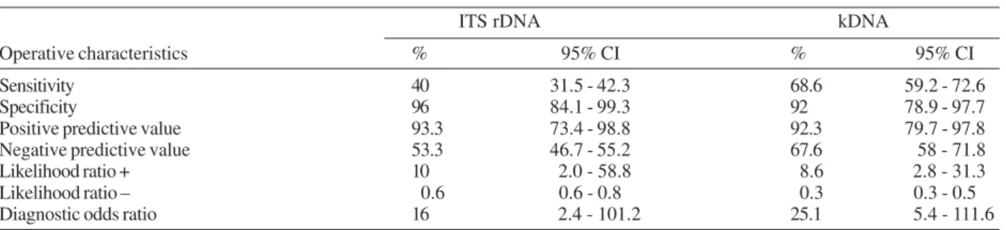
PCR leishmania detection - Twenty percent of the samples studied showed PCR inhibitors. After applying the DNA purification with 5M ammonium acetate only one showed persistent inhibition. DNA amplification with primer IR1-IR2 and ITS1F-ITS2R exhibited a 1000 bp band (Fig. 1) and with primers modified 13A and 13B a 120 bp band was observed (Fig. 2). Table II shows the diagnostic operative test characteristics with a 95% con-fidence interval.
The two biopsy samples in which amastigotes were identified were positive for PCR using primers IR1-IR2, ITS1F-ITS2R, and modified 13A and 13 B.
The inter-observer agreement was excellent; there were no discordant results for either test. The kappa sta-tistic for kDNA was 1.0 (95% CI 0.88-1.0) and for ITS rDNA was 1.0 (95% CI 0.85-1.0)
DISCUSSION
The aim of this study was to analyze the diagnostic operative characteristics of PCR using two molecular targets: conserved region kDNA and ITS rDNA in mu-cosal leishmaniasis. The reference standard used was a sum of clinical, epidemiological, and laboratory crite-ria, defined for this study.
Although the reference standard had not been used previously, we consider it had a good discrimination power. One could question the inclusion of individuals who showed a non-specific inflammatory infiltrate as controls, given the evidence of this type of infiltrate in leishmaniasis reported in the literature. Ridley described a series of over 400 biopsies of mucocutaneous leish-maniasis and found that non-specific inflammation was the most frequent and least effective response; its onset might be delayed, and in this event particularly the inci-dence of metastasis from skin to mucosa was high (Rid-ley 1989). This infiltrate can also be seen in individuals without the disease. In this work we considered individu-als could be included as controls if the mucosal biopsy showed a non-specific inflammatory infiltrate and if they also had a negative MST and a follow up with no pro-gressive course. Those patients included in our study as non cases had to have on the medical record a follow up were no disease progression was noted. If patients were lost, they were called and evaluated by researchers to determine if there was disease progression. We believe if the patient had a non specific infiltrate on mucosal biopsy, had a negative skin test, and was followed up in order to evaluate the disease did not have a progressive course without specific treatment, we could confidently classify this subject as a control and include him (her) in our study.
Studies designed to evaluate test diagnostic perfor-mance can have several biases which should be kept in mind when analyzing the results. The exclusion of a high proportion of eligible patients from the study could have left out a population not representative of the initial one. This could introduce a selection bias which occurs when not every patient presenting clinically relevant condi-tions is included in a consecutive order and when the selection is not random (Lijmer et al. 1999). Demogra-phic characteristics and evolution time of symptoms
TABLE II
Diagnostic operative test characteristics
ITS rDNA kDNA
Operative characteristics % 95% CI % 95% CI
Sensitivity 40 31.5 - 42.3 68.6 59.2 - 72.6
Specificity 96 84.1 - 99.3 92 78.9 - 97.7
Positive predictive value 93.3 73.4 - 98.8 92.3 79.7 - 97.8 Negative predictive value 53.3 46.7 - 55.2 67.6 58 - 71.8 Likelihood ratio + 10 2.0 - 58.8 8.6 2.8 - 31.3 Likelihood ratio – 0.6 0.6 - 0.8 0.3 0.3 - 0.5 Diagnostic odds ratio 16 2.4 - 101.2 25.1 5.4 - 111.6 Sample with inhibition not included.
1000 pb 750 pb 500 pb
1000 pb 1 2 3 4 5 6 7 8 9 10
300 pb 200 pb
100 pb
1 2 3 4 5 6 7 8 9 10 11
400 pb
120 pb
were compared between excluded and included individu-als in order to assess differences that could have intro-duced a selection bias in the study. No statistically sig-nificant differences were found between these groups (p > 0.05). The inclusion of healthy mucosal samples could have introduced a spectrum bias and enhance PCR specificity. We analyzed confidence intervals for diag-nostic operative characteristics including and excluding these samples and did not find any significant differ-ences. Test revision bias, which occurs when the result for the reference standard is known when applying the index test, as well as diagnosis revision bias which oc-curs when the results of the index test are known when interpreting the reference standard, were avoided by blinding the observers to each of the tests. The decision to apply one test was independent from the result of the other test and there was only one reference standard ap-plied to all cases and non cases. Although the ideal would have been prospective data collection, in Colombia eco-nomic and sociopolitical reasons make it difficult for patients from certain rural areas to reach urban centers for follow-up. This is a limitation for retrospective as well as for prospective study designs.
Our result for PCR sensitivity when conserved re-gion kDNA was used, showed a higher value 69.6% (95% CI 59.2-72.6%) than when ITS rDNA was used as mo-lecular target. This difference could be due to differ-ences in the number of copies present in Leishmania: around 10,000 copies for kDNA and near 200 copies for ITS rDNA (Rodgers et al. 1990, Pirmez et al. 1999, Marfurt et al. 2003, Bensoussan et al. 2006). Also, dif-ferences in the detection limits for each molecular tar-get have been reported. Primers 13A and 13B can am-plify 0.1 fg of L. mexicana and 10 fg of kDNA of L. braziliensis (Rodgers et al. 1990). Other authors inform detection limits for 13A and 13B primers of < 0.001 parasite/reaction and 0.2 parasite/reaction for ITS1 (Marfurt et al. 2003, Schonian et al. 2003). It is pos-sible that the small amount of parasites present in the affected mucosa could be under the detection limit for ITS rDNA but could be enough for kDNA.
Previous studies inform sensitivity values that range from 62-97.1% with no confidence intervals reported. The reference standard used by some authors is not well described and varies from one study to another, which makes it difficult to establish comparisons. Most of the studies report case series with PCR made by non blind observers. None of the reports estimate inter-observer agreement (Pirmez et al. 1999, Medeiros et al. 2002, Oddone et al. 2004, Disch et al. 2005).
Sensitivity values in our report may have been af-fected by inhibitors, which were present in 20% of evalu-ated samples. The presence of contaminants in tissue samples that could inhibit Taq polymerase activity and reduce the sensitivity 10 to 100 times when compared with kDNA obtained from cultures has been described (Rodgers et al. 1990). The procedure used for DNA pu-rification can reduce DNA quantity and increase the num-ber of false negatives. Hemoglobin could be an inhibi-tor in these samples, but there may also be elements present in nasal mucus that can inhibit PCR reaction. The
presence of inhibitors in 30% of samples from nasal swabs of leprosy patients has been reported (Pattyn et al.1993). The specificity reported was good for both molecu-lar targets, so if a test is positive one can be highly con-fident that the diagnosis is mucosal leishmaniasis. One of the false positive results we obtained was positive for both molecular targets and we believe could have been misinterpreted by the reference standard. Previous re-ports have informed 100% specificity with a poor de-scription of the population included as non cases.
Likelihood ratios for positive results were good and similar for both tests, although the confidence intervals were wide. For ITS rDNA there would be 10 positive results in mucosal leishmaniasis patients for each posi-tive result in a patient with another condition. Likelihood ratios for negative results were also similar for both tests. The diagnostic odds ratio, which expresses a ratio of the odds of a positive test in diseased and the odds of a positive test in non diseased is a measurement of the discrimination ability of the test and was good for both molecular targets, even though the precision was low due to a small sample size (Lijmer et al. 1999).
We consider our study has some strengths as well as some weaknesses. The strengths were the inclusion of a reasonable spectrum of individuals, the use of a com-posite reference standard, the independency of PCR in relation to the reference standard, and the blind inter-pretation of PCR and the reference standard. Among the weaknesses we can mention that the reference standard had not been used previously in the literature, the retro-spective data collection and the exclusion of a high pro-portion of eligible subjects. We analyzed possible bias the study could have and could not identify any.
Considering the difficulty in identifying parasites from mucosal leishmaniasis lesions, we consider PCR a useful tool in the diagnosis of this clinical entity. When clinically suspected, PCR with kDNA as molecular target should be used due to its higher sensitivity. PCR ITS rDNA remains a useful test as it allows species identification. Researchers should refine this technique in order to remove inhibitors without losing sensitivity. Multicenter prospective studies should be carried out in order to in-clude a larger number of subjects and obtain more pre-cise estimates. Well designed small studies could be useful for meta-analysis which address this subject.
ACKNOWLEDGEMENTS
To Martha Inirida Guerrero who was our scientific advisor and critically reviewed the manuscript, and Diana Alvarez for histopathology slides analysis.
REFERENCES
Aronson N, Glenn W, Johnson S, Jackson J, Gasser R, Magill A 1998. Safety and efficacy of intravenous sodium stiboglu-conate in the treatment of leishmaniasis: recent U.S. military experience. Clin Infect Dis27: 1457-1464.
Bensoussan E, Nasereddin A, Jonas F, Schnur L, Jaffe L 2006. Comparison of PCR assays for diagnosis of cutaneous leish-maniasis. J Clin Microbiol40: 1435-1439.
pentavalent antimony (sodium stibogluconate). Am J Trop Med Hyg34: 702-709.
Cupolillo E, Grimaldi Jr G, Momen H, Beverley S 1995. Intergenic region typing (IRT): a rapid molecular approach to the characterization and evolution of Leishmania. Mol Bioch Parasitol 73: 145-155.
Disch J, Junqueira M, Orsini M, Pirmez C, Oliveira AC, Castro M, Rabello 2005. Leishmania (Viannia) subgenus kDNA amplification for the diagnosis of mucosal leishmaniasis.
Diag Microbiol infect Dis51: 185-190.
Hepburn NC, Siddique I, Howie AF, Beckett GJ, Hayes PC 1994. Hepatotoxicity of sodium stibogluconate therapy for American cutaneous leishmaniasis. Trans R Soc Trop Med Hyg 88: 453-455.
Instituto Nacional de Salud 2006. Informe de enfermedades trans-mitidas por vectores (ETV). Inf Quinc Epidem Nac 11: 40-44. Isaza DM, Arboleda M, Restrepo M, McCann SHE, Barker DC 2002. Validation of the polymerase chain reaction for the di-agnosis of human cutaneous leishmaniasis in north-west Colombia. Trans R Soc Trop Med Hyg 96: 165-168. Lijmer J, Mol BW, Heisterkamp S, Bonsel G, Prins MH, van der
Meulen JHP 1999. Empirical evidence of design-related bias in studies of diagnostic tests. JAMA 282: 1062-1066. Marsden PD 1985. Clinical presentations of Leishmania
bra-ziliensis brabra-ziliensis. Parasitol Today 1: 129-133. Medeiros A, Rodrigues S, Roselino A 2002. Comparison of the
specificity of PCR and the histopathological detection of
Leishmania for the diagnosis of American cutaneous leish-maniasis. Braz J Med Biol Res35: 421-424.
Marfurt J, Nasereddin A, Niederwieser I, Jaffe CL, Beck H-P, Felger I 2003. Identification and differentiation of Leishma-nia species in clinical samples by PCR amplification of the miniexon sequence and subsequent restriction fragment length polymorphism analysis. J Clin Microbiol 41: 3147-3153. Oddone R, Arbo C, Nara E, Velázquez GR, Acosta ME, Poletti
D 2004. Utilidad diagnóstica de los métodos laboratoriales en leishmaniasis mucosa, incluyendo la “PCR”. Noticias Técnicas del Laboratorio 4: 7-9.
Oliveira JG, Novail OF, Oliveira CI, Cruz JA, Campos FL, Rocjha A, Boaventura V, Noroña A, Jackson, ML Jackson, Barral A 2005. Polymerase chain reaction (PCR) is highly sensible for diagnosis of mucosal leishmaniasis. Acta Trop 94: 55-59. Pappas Michael G, McGreevy PB, Hajkowski R 1983. Evalua-tion of promastigote and amastigote antigens in the indirect
fluorescent antibody test for American cutaneous leishma-niasis. Am J Trop Med Hyg 32: 1260-1267.
Pattyn S, Ursi D, Ieven M, Grillone S, Raes V 1993. Detection of Mycobacterium leprae by the polymerase chain reaction in nasal swabs of leprosy patients and their contacts. Int J Lepr 61: 389-393.
Pirmez C, Da Silva TV, Oliveira NMP, Da Cruz AM, Gonçalves CSC, Catanho M 1999. Use of PCR in diagnosis of human American tegumentary leishmaniasis in Rio de Janeiro, Brazil. J Clin Microbiol June: 1819-1823.
Ridley D, Magalhães A, Marsden P 1989. Histological analysis and the pathogenesis of mucocutaneous leishmaniasis.
J Pathol159: 293-299.
Rodgers MR, Popper SJ, Wirth DF1990. Amplification of Kinetoplat ADN as a tool in the detection and diagnosis of
Leishmania. Exp Parasitol 71: 267-275.
Rodriguez N, Guzman B, Rodas A, Takiff H, Bloom BR, Convit J 1994. Diagnosis of cutaneous leishmaniasis and species discrimination of parasites by PCR and hibridization. J Clin Microbiol Sep: 2246-2252.
Sambrook J 2001. Molecular Cloning A LaboratoryManual. Preparation and Analysis ofEukaryotic Genomic DNA, 3rd ed., Cold Spring Harbor Laboratory Press, New York, p. 6.4-6.12
Schonian G, Nasereddin A, Dinse N, Schweynoch C, Schallig H, Presber W 2003. PCR diagnosis and characterization of
Leishmania in local and imported clinical samples. Diag Microbiol and Infect Dis 47: 349-358.
Schubach A, Cuzzi-Maya T, Oliveira AV, Sartori A, De Oliveira-Neto MP, Mattos MS 2001. Leishmanial antigens in the di-agnosis of active lesions and ancient scars of American tegu-mentary leishmaniasis patients. Mem Inst Oswaldo Cruz 96: 987-996.
Weigle KA, De Davalos M, Heredia P, Molineros R, Saravia NG, D'Alessandro A 1987. Diagnosis of cutaneous and mucocu-taneous leishmaniasis in Colombia: a comparison of seven methods. Am J Trop Med Hyg 36: 489-496.
WHO-TDR 2005. Surveillance and control of leishmaniasis. http:/ /www.who.int/tdr/diseases/default.htm. Priorities and stra-tegic emphases for TDR supported research on leishmania-sis. http://www.who.int/tdr/disease/leish/strategy.htm Zajtchuk CJT, Casler JD, Netto EM, Grog M, Neafie RC, Hessel