Parasitology
Comparison of polymerase chain reaction with other laboratory
methods for the diagnosis of American cutaneous leishmaniasis
Diagnosis of cutaneous leishmaniasis by polymerase chain reaction
Marcos J. Marques
a, A
ˆ ngela C. Volpini
b, George L.L. Machado-Coelho
c,
Jackson Machado-Pinto
d, Carlos A. da Costa
a, Wilson Mayrink
a,
Odair Genaro
a,1, Alvaro J. Romanha
e,T
a
Departamento de Parasitologia, Instituto de Cieˆncias Biolo´gicas, Universidade Federal de Minas Gerais, Belo Horizonte 31270-901, MG, Brazil
b
Laborato´rio de Pesquisas em Leishmanioses, Instituto Oswaldo Cruz, FIOCRUZ, Rio de Janeiro 21040-900, Brazil
c
Escola de Farma´cia da Universidade Federal de Ouro Preto, Ouro Preto 35400-000, MG, Brazil
dClı´nica Dermatolo´gica da Santa Casa de Miserico´rdia, Belo Horizonte 30150-221, MG, Brazil
eLaborato´rio de Parasitologia Celular e Molecular, Centro de Pesquisas Rene´ Rachou-FIOCRUZ, CEP 30190-002, Belo Horizonte, MG, Brazil
Received 20 April 2005; accepted 8 August 2005
Abstract
An evaluation of 5 laboratory methods for diagnosing American cutaneous leishmaniasis (ACL) was carried out on patients from an endemic area of Brazil. From 164 patients presenting cutaneous lesions, and suspected to have ACL, 133 (81.1%) were confirmed for the disease by Montenegro skin test (MST) and/or parasitologic examination (PE). In both groups of patients, the positivity of polymerase chain reaction (PCR) was similar to that of immunofluorescence assay and enzyme-linked immunosorbent assay, and higher than that of MST and PE (P b.05). In the group of patients suspected to have ACL, PCR presented the same positivity as PE and MST together. No correlation
between positivity of the laboratory methods and clinical or epidemiologic aspects was observed. Our data confirmed the value of PCR as an alternative laboratory method for diagnosing ACL, especially for those patients with negative PE and MST.
D2006 Elsevier Inc. All rights reserved.
Keywords:American cutaneous leishmaniasis; Polymerase chain reaction; Diagnosis
1. Introduction
American cutaneous leishmaniasis (ACL) is a parasitic disease caused by several species of the protozoa Leish-mania, from the subgeneraVianniaandLeishmania, and is endemic from the Yucata´n peninsula to northern Argentina (WHO, 1990; Dedet, 1999; Lawn et al., 2004). ACL is therefore considered a great public health problem, with social and economic reflexes, and in most cases character-ized as an occupational disease because it affects mainly rural workers. Recently, the disease has been reported to also present some characteristics of domiciliary and
periurban transmissions (Marzochi and Marzochi, 1994; Passos et al., 2000, Weigle et al., 2002; Castro et al., 2005). The annual incidence of cutaneous leishmaniasis in the world is estimated at 1.0 –1.5 million cases (WHO, 1990). More than 90% of the cutaneous cases occur in Afghanistan, Saudi Arabia, Algeria, Brazil, Iran, Iraq, Syria, and Sudan (Singh and Sivakumar, 2003). In Brazil, the advance of ACL can be assessed by the disease detection rate, which increased from 10.45 in 1985 to 21.88/100 000 inhabitants in 2001 (BRASIL/MS/FUNASA/CENEPI, 2002).
The diagnostic methods available at present are mostly based on clinical and epidemiologic features, parasite detection (stained smears, culture, and histopathology), and immunologic methods (Rodrigues et al., 2002). Up to now, no single laboratory method has been accepted as the gold standard for diagnosing ACL. Parasitologic tests of a skin biopsy specimen are not always conclusive in patients
0732-8893/$ – see front matterD2006 Elsevier Inc. All rights reserved. doi:10.1016/j.diagmicrobio.2005.08.003
T Corresponding author. Tel.: 3295-3566x180; fax: +55-31-3295-3115.
E-mail address:[email protected] (A.J. Romanha).
1 In memoriam.
with a clinical diagnosis of cutaneous leishmaniasis (Faber et al., 2003). Therefore, the association of immunologic tests with parasitologic examination has been used in the laboratory routine. The Montenegro skin test (MST) has been the immunologic method of choice followed by immunofluorescence assay (IFA) and enzyme-linked immu-nosorbent assay (ELISA) (Marzochi, 1992; Passos et al., 2000). Although serologic tests for ACL diagnosis present some limitations (Ulrich et al., 1988; Chiaramonte et al., 1999), they may contribute to the early diagnosis of mucosal lesions or extensive and multiple cutaneous lesions (Chiari et al., 1973a, 1973b; Mendonc¸a et al., 1988).
The direct microscopic examination and MST, even when associated, are not sufficient to diagnose all ACL cases (Lopez et al., 1993). Thus, polymerase chain reaction (PCR) has recently been used for ACL diagnosis. PCR has been able to detectLeishmania DNA in human lesions and in other animals suspected to bear the infection (Rodgers et al., 1990; De Brujin et al., 1993; Lopez et al., 1993; Rodriguez et al., 1994; Pirmez et al., 1999; Marques et al., 2001; Weigle et al., 2002; Lawn et al., 2004).
The present study compares the positivity of the 5 most used laboratory methods for diagnosing ACL and reports clinical and epidemiologic aspects of the disease in the endemic region of Minas Gerais, Brazil.
2. Materials and methods
2.1. Patients
From 1997 to 2000, 164 patients with cutaneous but not with mucosal lesions were detected on clinical examination at the Leishmaniasis outpatient clinic in the city of Caratinga, Minas Gerais, Brazil. These patients were from rural regions of Vale do Rio Doce. A study was carried out with the purpose of describing clinical, epidemiologic, and laboratory aspects related to ACL. A questionnaire regarding the subject was submitted to all patients who answered the following questions about clinical and epidemiologic variables: lesion’s time, number, site, and diameter; and patient’s age, sex, and skin color. A differential diagnosis with other similar lesions was considered. Patients who presented typical cutaneous ulcers and a positive parasitologic examination (PE) and/or MST tests were defined as ACL clinical cases. Informed consent for blood collection and biopsies was obtained from all patients. The project was previously approved by the Ethical Committee for Human Research of Santa Casa de Miserico´rdia Hospital in Belo Horizonte, State of Minas Gerais, Brazil (CEPH # 007/98).
2.2. Parasitologic examination
After lesion asepsis, 0.5 – 1.0 mL of 2% xylocaine was subcutaneously injected nearby. A scalpel with a disposable blade was used for the biopsy. A wedged skin fragment (5 mm length) from the border of the lesion of each patient
was obtained. After removing the blood excess from the biopsy fragment, 12 imprints were performed for each of 3 microscopic slides. After air drying, slides were fixed in methanol, stained with Giemsa, and observed via optical microscopy (magnification 1000) for the presence of amastigote forms of the parasite. The results were obtained after a thorough examination of the 3 slides.
2.3. Montenegro skin test
This test was used to assess the patients’ cellular immune response in vivo. A promastigote antigen fromLeishmania (Leishmania) amazonensis (clone PH8-1 IIId) forms was used (da Costa et al., 1996). The antigen was produced by Biobra´s (Montes Claros, Minas Gerais, Brazil) under good manufacturing practice conditions and registered as Mon-tenegro antigen C-40R. MST was carried out as described by Melo et al. (1977). The result was considered positive when an induration of z5 mm in diameter could be observed after 48 h of the antigen injection.
2.4. Polymerase chain reaction
The same tissue biopsy fragments used for slide imprints were further used for extracting Leishmania DNA. The DNA extraction from samples was performed with 100AL buffer solution (10 mmol/L Tris–HCl and 1 mmol/L ethylenediaminetetraacetic acid, pH 8.0) and 100 Ag/mL proteinase K (final concentration), incubated at 568C for 3 h, and homogenized from time to time. The digestion was stopped by proteinase K inactivation by boiling it for 15 min. Samples were centrifuged, and the supernatant was used as the Leishmania template DNA source for the PCR reaction (Belli et al., 1998; Passos et al., 1999). Reaction mixtures contained 200 Amol/L each of dUTP, dATP, dCTP, and dGTP, 1 Amol/L of each primer, buffer (10 mmol/L Tris–HCl, 50 mmol/L KCl, pH 8.0), 1.5 mmol/L MgCl2, 0.75 U of Taq DNA polymerase (PhN, Belo Horizonte, Minas Gerais, Brazil), and 1AL of DNA sample in a final volume of 10 AL. Twenty microliters of mineral oil was poured over the reaction mixture to avoid evaporation. PCR conditions were as follows: initial denaturation at 948C for 5 min, followed by 29 cycles at 94 8C for 1 min, 60 8C for 1 min, 72 8C for 30 s, and a final extension at 72 8C for 5 min. Primers directed to amplify the conserved region of theLeishmania kDNA minicircle were: forward 5V- GGG (G/T)AG GGG CGT TCT (G/C)CG AA-3Vand reverse 5V- (G/C)(G/C)(G/C) (A/T)CT AT(A/T) TTA CAC CAA CCC C-3V, and the PCR product obtained was 120 nucleotide bp long (Degrave et al., 1994; Passos et al., 1996).
tryptose medium and collected at the exponential growth phase. Cultures were washed 3 times in sterile phosphate-buffered saline solution, pH 7.2, at 4 8C, centrifuged at 500g for 15 min, and DNA was extracted as described previously. DNA samples were also obtained from skin biopsies of 26 individuals without lesions or other le-sions than ACL. These patients were diagnosed by clinical and parasitologic examinations at the Clı´nica Derma-tolo´gica (a dermatological clinic) of the Santa Casa de Miserico´rdia Hospital in Belo Horizonte, Brazil, and were used as controls.
A search for inhibitors was performed in samples that showed PCR-negative results to assess possible PCR failures to detectLeishmania DNA. In the same PCR tube, 1AL of DNA preparation from a negative sample and 1AL of Leishmania DNA standard sample at 10 fg/AL were added as template. A positive result means no presence of inhibitors in the tested sample. The biologic materials were coded before the laboratory tests to avoid any bias on the interpretation of the results.
2.5. Serology
The IFA and ELISA were used for detecting anti-Leishmaniatotal IgG in sera of 164 patients with cutaneous lesions who were suspected to have ACL. IFA was evaluated using total anti-IgG conjugates labeled with fluorescein isothiocyanate, specific for humans and diluted at 1:100 (Bio-Manguinhos/FIOCRUZ, Rio de Janeiro, Brazil). The antigen was obtained from promastigote forms from L. (L.) amazonensis MHOM/BR/1960/BH6 strain fixed with 1.0% formalin in saline. Titer z1:40 was considered positive (da Costa et al., 1991). ELISA was performed using total anti-IgG conjugates, labeled with peroxidase, specific for humans, and diluted at 1:3000 (Sigma Chemical Company, St. Louis, MO). Soluble antigen of L. (V.) braziliensis obtained from the MHOM/ BR/1975/M2903 strain was used. Sera from 40 individuals from Caratinga, Minas Gerais, with no history of ACL or cutaneous lesions and negative IFA were diluted at 1:80 and used to determine the cutoff point for ELISA. The cutoff point was established as the average absorbance at 492 nm of those sera plus 2 SD (Abs z0.125).
2.6. Treatment
Patients defined as ACL clinical cases received antimo-nial therapy (GlucantimeR, Rhodia, Brazil) at the dosage of 17 mg Sbv/(kg day) i.m. (Mayrink et al., 1976), for 10 consecutive days, alternated with a 10 day-interval without treatment. Patients were submitted to treatment cycles until the lesions were healed.
2.7. Statistics
A correlation between clinical and epidemiologic param-eters was determined by Pearson’s coefficient. A compar-ative analysis of positivity between the laboratory methods was performed usingv2test.
3. Results
3.1. Casuistic description
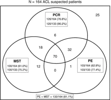
Of 164 suspected patients, 133 (81.1%) were confirmed as ACL clinical cases according to the case history (individuals exposed to risk of acquiring the disease), clinical examination of lesions, MST, and/or PE (Fig. 1). Most ACL patients were white males, aged 2 – 89 years (mean, 29 years; median, 32 years), and 63.9% aged 10.5 – 40.5 years.
3.2. Clinical characteristics of lesions
The main clinical characteristics of ACL patients’ lesions were a single lesion (66.2%), with an average of 14.5 mm, median of 12.0 mm, and diameter range of 5.0 –50.0 mm, on the lower limbs (47.6%) and a developing time ofV60 days (66.9%). Pearson’s index values have shown no correlation between clinical and epidemiologic parameters (lesions’ developing time, number, site, and diameter; and patients’ age, sex, and skin color).
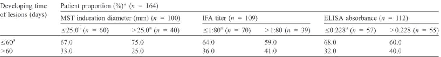
3.3. Quantitative analysis of MST, IFA, and ELISA according to the developing time of lesions
One hundred patients positive to MST, 109 to IFA, and 112 to ELISA were evaluated (Table 1). The induration of MST ranged from 8 to 45 mm, with a mean of 24.3 mm and median of 25.0 mm. The titer of IFA ranged from 1:40 to 1:640, with mean of 1:116 and median of 1:80. The absorbance of ELISA ranged from 0.129 to 0.504, with a mean of 0.248 and median of 0.228. Median was used as cutoff point to separate samples in the laboratory tests. Quantitative analyses show no significant differences for
MST, IFA, and ELISA regarding the developing time of lesions (P N 0.05).
3.4. Comparative analysis of ACL diagnosis by different laboratory methods
Fig. 1 and Table 2 show the positivity of 5 laboratory methods for diagnosing ACL in groups of suspected and confirmed patients. Patients were considered suspected based on suggestive clinical and epidemiologic aspects only or when they were negative for PE and MST. On the other hand, patients were confirmed with ACL when, besides the suggestive clinical and epidemiologic aspects, they were also positive for PE and/or MST. Analysis of positivity for MST, PCR, and PE of patients suspected of ACL (n = 164) shows that 12, 6, and 1 patients were positive exclusively in each one of the methods, respec-tively. Furthermore, 70 patients were positive and 25 neg-ative in the 3 methods. The positivity of the 5 methods was higher in patients of the ACL-confirmed group than those from the suspected one (P b .05). In both groups of
patients, the positivity of PCR was the same as that of IFA and ELISA (Table 2) and higher than that of MST and PE (Fig. 1;P b.05). In the group of patients suspected to have
ACL, PCR presented the same positivity as PE and MST together (Fig. 1). PE and MST were used as standards to calculate the positive predictive value (PPV) and the negative predictive value (NPV) of diagnostic methods (Table 2); therefore, these parameters were not applicable
for both. Thus, the following percentages regarding PCR, IFA, and ELISA, respectively, were found: 95.2%, 98.2%, and 100.0% for PPV, and 65.8%, 52.7%, and 59.6 for NPV. Emphasis should be given to the fact that in the group of ACL-suspected patients, PCR was positive in 6 of 31 individuals (19.3%) who were negative for PE and MST (Table 2). Of those 6 patients, 3 returned to the clinic and were then submitted to new tests (PE and MST), being confirmed as ACL cases. They were treated and their lesions healed.
Samples from patients with a positive PCR result have shown a unique migration band of the expected size for Leishmania (120 bp) after polyacrylamide gel electropho-resis. It was exactly in the same position as for the positive controls at 0.01 and 1.0 pg ofL.(V.)braziliensisDNA (data not shown). The 26 patients without lesions or with skin lesions other than ACL have shown no specific PCR products for Leishmania sp. PCR-negative samples pre-sented no inhibitors in their DNA preparations.
The Pearson’s index values have shown no correlation between positivity of the laboratory methods (PE, MST, IFA, ELISA, and PCR) and clinical and epidemiologic parameters (lesions’ developing time, number, site, and diameter; patients’ age, sex, and skin color).
3.5. Treatment
ACL patients needed an average of 3.54 treatment cycles (71 days) for the complete healing of the lesions. The 133 ACL patients were treated and cured. No side effects requiring treatment discontinuation were observed.
4. Discussion
The advantages of PCR for diagnosing leishmaniasis should be considered in the context of the diagnostic services and according to epidemiologic and clinical characteristics of the disease (Weigle et al. 1993, 2002; Lawn et al., 2004). In the present study, clinical and epidemiologic aspects of ACL in the endemic region of Minas Gerais, Brazil, were reported. Although most ACL patients still come from rural areas, the regular attendance at the Leishmaniasis outpatient clinic, the awareness of the population about the health care facilities and services of the counties around the endemic region have changed the profile of the ACL patients over the last 30 years. A study undertaken byChiari et al. (1973a)in the same region Table 2
Positivity and predictive values of different laboratory methods for diagnosing ACL in groups of suspected and confirmed ACL patients
Group of patients Positivity of the laboratory method,a
n(%)
P
PCR IFA ELISA
PE+ (n = 103) 102 (99.0) 97 (94.2) 98 (95.1) .16 PE /MST+ (n = 30) 18 (60.0) 10 (33.3) 14 (46.7) .11 ACL confirmed
(n = 133)
120 (90.2) 107 (80.4) 112 (84.2) .10
ACL-suspected (n = 164)
126 (76.8) 109 (66.5) 112 (68.4) .08
PE /MST (n = 31) 6 (19.4) 2 (6.5) 0 (0.0) .02T
Predictive value
PPV (95.2) (98.2) (100.0)
NPV (65.8) (52.7) (59.6)
a
The positivity of the methods was higher in the ACL patient confirmed group than in the suspected one (P b.05).
T P b.05, statistically significant. Table 1
Quantitative analysis of positive patients to MST, IFA, and ELISA according to the developing time of lesions
Developing time of lesions (days)
Patient proportion (%)T(n = 164)
MST induration diameter (mm) (n = 100) IFA titer (n = 109) ELISA absorbance (n = 112)
V25.0a(n = 60) N25.0a(n = 40) V1:80a(n = 70) N1:80 (n = 39) V0.228a(n = 57) N0.228 (n = 55)
V60a 67.0 75.0 64.0 59.0 68.0 60.0
N60 33.0 25.0 36.0 41.0 32.0 40.0
a
Cut-off represents the median of the parameters (developing time of lesions, MST diameter, indirect IFA titer, and ELISA absorbance).
showed that 62.2% of the ACL cases presented skin lesions with a developing time of N60 days. At present, the rate
decreased to 33.1% suggesting that the population has acquired more knowledge about the disease and has looked for medical assistance more rapidly.
The ACL transmission pattern has changed in the recent years with outbreaks occurring in long-established rural settlements and urban areas (Lainson, 1989; Gontijo et al., 2002; Castro et al., 2005). However, our data have shown that the ACL patients were from rural regions and that 63.9% were between 10.5 and 40.5 years old. At these ages, individuals are more involved in agricultural activ-ities and, as a consequence, they are more at risk for infection, suggesting that ACL transmission in that region still occurs predominantly outside the dwellings. These data corroborate with those of Machado -Coelho et al. (1999)on the occupational character of ACL in the region of Vale do Rio Doce.
In the present study, it was observed that most patients presented a single lesion (66.2%), with an average of 14.5 mm in diameter, on the lower limbs (47.6%), and a developing time of V60 days (66.9%, P b .05). The
duration of these patients’ lesions differed from that of other regions, which varied between 0.5 and 168 months (Saravia et al., 1989; de Brujin et al., 1993; Aviles et al., 1999; Passos et al., 2000). On the other hand, the predominance of a unique lesion on the lower limbs has been the most frequent finding (Cuba-Cuba et al., 1984; Mendonc¸a et al., 1988; Marzochi, 1992; Convit et al., 1993; Passos et al., 2000). Numerous primary and secondary skin diseases and conditions are frequently misdiagnosed as early lesions of cutaneous leishmaniasis (Singh and Sivakumar, 2003). In that sense, a differential diagnosis with other similar lesions, such as varicose ulcer, impetigo, tuberculosis, leprosy, syphilis, blastomycosis, sporotrichosis, skin cancer, and other cutaneous lesions, was considered. However, other diagnostic methods are required to confirm the clinical suspicion (Herwaldt, 1999).
PE positivity in ACL-suspected patients (62.8%) reached the upper values reported — 33.0% to 63.0% (de Brujin et al., 1993; Rodriguez et al., 1994; Pirmez et al., 1999; Aviles et al., 1999). Along the years, the finding of Leishmania by PE at the leishmaniasis outpatient clinic in Caratinga has improved greatly. PE increased from 19.4% (Mayrink et al., 1979) to 62.8% at present. This improve-ment seems to be due to a better tissue collection, slide preparation, and an exhaustive examination of 12 imprints performed for each of the 3 microscopic slides. No correlation between positivity of the PE and developing time of the lesions could be observed. PE positivity for patients with V60 -day-old lesions was identical to that of patients with N60 -day-old lesions. Nevertheless, there are
studies showing a greater difficulty to detect parasites on
N90 -day-old (Pirmez et al., 1999) and 180 -day-old lesions
(Weigle et al., 1987;Gutierrez et al., 1991; Marzochi, 1992; Singh and Sivakumar, 2003).
The ease of processing and the short time to obtain MST results (48 h) allowed its use as the main indirect test performed in the laboratory for diagnosing ACL cases in endemic areas (Gontijo et al., 2002; Oliveira et al., 2003). In a study of mucocutaneous leishmaniasis in Treˆs Brac¸os, Bahia, Brazil,Cuba-Cuba et al. (1984)found out that 93.0% of patients with cutaneous lesions were MST positive. They also showed that MST could not be positive up to 6 or more weeks after the emergence of cutaneous lesions. The present work shows that MST presented 61.0% positivity for patients suspected and 75.2% for patients confirmed with ACL. It has been described that MST may remain positive after clinical cure (Furtado, 1980; Cuba-Cuba et al., 1984; Kar, 1995). Moreover, MST has been pointed out as a good marker of cell immune response in leishmaniases whose negative results may imply in failure in the treatment (Saravia et al., 1990; Passos et al., 2001). Furthermore, MST is not able to differentiate an active from an inactive infection. In our study, MST was associated with PE for diagnosing ACL infection and confirmed the disease in 81.1% of the patients suspected to have ACL.
Although, at present, no single laboratory technique has been accepted as the gold standard for diagnosing Leishmania infection, the results obtained by PCR are significant. PCR presented 76.8% positivity for the patients suspected to have ACL and 90.2% for the patients confirmed with ACL. The positivity of the 5 methods used in this work was higher in patients of the confirmed group than in those from the suspected one (P b .05). Belli
et al. (1998)described that PCR showed 100.0% sensitivity and specificity when compared with direct microscopy. Pirmez et al. (1999) reported in ACL patients a PCR positivity of 96.9% and a PE positivity of 67.4%. Our results have shown that in both groups of patients the positivity of PCR was higher than that of PE and MST alone. PCR appears to be the most sensitive single diagnostic test for cutaneous leishmaniasis (Weigle et al., 2002; Faber et al., 2003; Lawn et al., 2004). As the parasite finding or the isolation and growth have a lower sensitivity, PCR should be added to the lesion healing as criteria of cure for the ACL treatment (WHO, 1990, Guevara et al., 1993; Delgado et al., 1996; Schubach et al., 1998; Passos et al., 2001, Coutinho et al., 2002).
It is worthwhile to note that PCR was positive in 6 of 31 patients (19.4%) who were negative for PE and MST, thus showing the failure of these 2 methods in diagnosing ACL, even when they were associated. Of the 6 PCR-positive patients, 3 returned to the clinic and were further confirmed for ACL by either PE and/or MST. They were treated and cured, therefore demonstrating the PCR value under those circumstances.
there are reports in the literature about the correlation between quantitative data of IFA and ELISA and the number and time of lesions (Chiari et al., 1973a, 1973b; Mendonc¸a et al., 1988; Kar, 1995), we have not observed such correlations. Maybe because the humoral immune response is time-dependent and most of the lesions of our casuistic were recent (V60 days), given insufficient time for the immune response to be elicited. Moreover, quantitative serologic evaluation has been suggested in the follow-up of the treated patients, as there is a reduction of the titers with the therapy (Chiari et al., 1973b; Walton, 1980; Mendonc¸a et al., 1988; Passos et al., 2001).
PCR should be used selectively for chronic cases or when other methods have not detected ACL case. In conclusion, our data confirm the value of PCR as an alternative laboratory method for diagnosing ACL, partic-ularly in those cases where PE and MST had failed to detect the disease.
Acknowledgments
We thank Mr. Jair Cecı´lio de Paula for technical assis-tance in the Dr. Paulo Araujo Magalha˜es ambulatory.
References
Aviles H, Belli A, Armijos R, Monroy FP, Harris E (1999) PCR detection and identification of Leishmania parasites in clinical specimens in Ecuador: a comparison with classical diagnostic methods.J Parasitol
85:181 – 187.
Belli A, Rodriguez B, Aviles H, Harris E (1998) Simplified polymerase chain reaction detection of new worldLeishmaniain clinical specimens of cutaneous leishmaniasis.Am J Trop Med Hyg58:102 – 109. Brasil, Ministe´rio da Sau´de, Fundac¸a˜o Nacional de Sau´de, Centro Nacional
de Epidemiologia (2002) Guia de Vigilaˆncia Epidemiolo´gica. Quinta Edic¸a˜o. Brası´lia7FUNASA.
Castro EA, Luza E, Telles FQ, Pandeyd A, Bisetob A, Dinaiskib M, Sbalqueiroe I, Soccola VT (2005) Eco-epidemiological survey of
Leishmania(Viannia)braziliensisAmerican cutaneous and mucocuta-neous leishmaniasis in Ribeira Valley River, Parana´ State, Brazil.Acta Trop93:141 – 149.
Chiaramonte MG, Frank FM, Furer GM, Taranto NJ, Margni RA, Malchiodi EL (1999) Polymerase chain reaction revealsTrypanosoma cruziinfection suspected by serology in cutaneous and mucocutaneous leishmaniasis patients.Acta Trop72:295 – 308.
Chiari CA, Magalha˜es PA, Mayrink W (1973) Pesquisa de anticorpos, por imunofluoresceˆncia, em soros de pacientes com Leishmaniose tegu-mentar americana apresentando lesoes cutaˆneas recentes.Rev Inst Med Trop Sa˜o Paulo15:304 – 309.
Chiari CA, Mayrink W, Magalha˜es PA (1973) Reac¸a˜o de imunofluor-esceˆncia indireta no controle de tratamento da Leishmaniose tegumentar americana.Rev Inst Med Trop Sa˜o Paulo15:298 – 303.
Convit J, Ulrich M, Ferna´ndez CT, Tapia FJ, Ca´ceres-Dittmar G, Caste´s M, Rondo´n A (1993) The clinical and immunological spectrum of American cutaneous leishmaniasis. Trans R Soc Trop Med Hyg
87:444 – 448.
Coutinho SG, Pirmez C, Da-Cruz AM (2002) Parasitological and immunological follow-up of American tegumentary leishmaniasis patients.Trans R Soc Trop Med Hyg96(Suppl 1):S173 – S178. Cuba-Cuba C, Llanos-Cuentas EA, Barreto AC, Magalha˜es AV, Lago EL,
Reed SG, Marsden PD (1984) Human mucocutaneous leishmaniasis in
Treˆs Brac¸os, Bahia — Brazil. An area of Leishmania braziliensis braziliensis transmission. I. Laboratory diagnosis.Rev Soc Bras Med Trop17:161 – 167.
da Costa CA, Genaro O, De Lana M, Magalha˜es PA, Dias M, Michalick MSM, Melo MN, Da Costa RT, Magalha˜es-Rocha NM, Mayrink W (1991) Leishmaniose visceral canina: avaliac¸a˜o da metodologia sorolo´gica utilizada em inque´ritos epidemiolo´gicos. Rev Soc Bras Med Trop24:21 – 25.
da Costa CA, Toledo VPCP, Genaro O, Williams P, Mayrink W (1996) Montenegro skin test — evaluation of the composition and stability of the antigen preparation.Mem Inst Oswaldo Cruz91:193 – 194. de Brujin MHL, Labrada LA, Smyth AJ, Santrich C, Barker DC (1993) A
comparative study of diagnosis by the polymerase chain reaction and by current clinical methods using biopsies from Colombian patients with suspected leishmaniasis.Trop Med Parasitol44:201 – 207.
Dedet JP (1999) E´ pide´miologie des leishmanioses du nouveau monde. In
Les leishmanioses. Ed, JP Dedet. Les leishmanioses. Parı´s7ELLIPSES AUPELF/UREF, pp 147 – 160.
Degrave W, Fernandes O, Campbell D, Bozza M, Lopes U (1994) Use of molecular probes and PCR for detection and typing ofLeishmania— a mini-review.Mem Inst Oswaldo Cruz89:463 – 469.
Delgado O, Guevara P, Silva S, Belfort E, Ramirez JL (1996) Follow-up of a human accidental infection by Leishmania (Viannia) braziliensis
using conventional immunologic techniques and polymerase chain reaction.Am J Trop Med Hyg55:267 – 272.
Faber WR, Oskam L, van Gool T, Kroon NC, Knegt-Junk KJ, Hofwegen H, van der Wal AC, Kager PA (2003) Value of diagnostic techniques for cutaneous leishmaniasis.J Am Acad Dermatol49:70 – 74.
Furtado TA (1980) Crite´rios para o diagno´stico da leishmaniose tegumentar americana.An Bras Dermatol55:81 – 86.
Garcia-Miss MR, Andrade-Narvaez FJ, Esquivel-Vin˜as RE, Simmonds-Diaz EB, Canto-Lara SB, Cruz-Ruiz AL (1990) Localized cutaneous leishmaniasis (chiclero’s ulcer) in Mexico: sensitivity and specificity of ELISA for IgG antibodies toLeishmania mexicana mexicana.Trans R Soc Trop Med Hyg84:356 – 358.
Gontijo CMF, da Silva ES, de Fuccio MB, de Sousa MCA, Pacheco RS, Dias ES, Andrade Filho JD, Brazil RP, Melo MN (2002) Epidemiological studies of an outbreak of cutaneous leishmaniasis in the Rio Jequitinhonha Valley, Minas Gerais, Brazil. Acta Trop
81:143 – 150.
Guevara P, Ramirez JL, Rojas E, Scorza JV, Gonza´lez N, An˜ez N (1993)
Leishmania braziliensisin blood 30 years after cure.Lancet341:1341. Gutierrez Y, Salinas GH, Palma G, Valderrama LB, Santrich CV, Saravia NG (1991) Correlation between histopathology, immune response, clinical presentation, and evolution inLeishmania braziliensis infec-tion.Am J Trop Med Hyg45:281 – 289.
Herwaldt BL (1999) Leishmaniasis.Lancet354:1191 – 1199.
Kar K (1995) Serodiagnosis of leishmaniasis. Crit Rev Microbiol
21:123 – 152.
Lainson R (1989) Demographic changes and their influence on the epidemiology of the American leishmaniasis. In Demography of vector-borne diseases. Ed, MW. Service Demography of vector-borne diseases. Boca Raton (FL)7CRC Press, pp 85 – 106.
Lawn SD, Whetham J, Chiodini PL, Kanagalingam J, Watson J, Behrens RH, Lockwood DNJ (2004) New world mucosal and cutaneous leishmaniasis: an emerging health problem among British travelers.
Q J Med97:781 – 788.
Lopez M, Inga R, Cangalaya M, Echevarria J, Llanos-Cuentas A, Orrego C, Arevalo J (1993) Diagnosis ofLeishmaniausing the polymerase chain reaction: a simplified procedure for field work.Am J Trop Med Hyg
49:348 – 356.
Machado-Coelho GLL, Assunc¸a˜o R, Mayrink W, Caiaffa WT (1999) American cutaneous leishmaniasis in southeast Brazil: space–time clustering.Int J Epidemiol28:982 – 989.
leishmaniasis via polymerase chain reaction. Am J Trop Med Hyg
65:902 – 906.
Marzochi MCA (1992) Leishmanioses no Brasil — as leishmanioses tegumentares.J Bras Med63:82 – 102.
Marzochi MCA, Marzochi KBF (1994) Tegumentary and visceral leishmaniases in Brazil—emerging anthropozoonosis and possibilities for their control.Cad Sau´de Pu´blica10:359 – 375.
Mayrink W, Melo MN, da Costa CA, Magalha˜es PA, Dias M, Coelho MV, Araujo FG, Williams P, Figueiredo YP, Batista SM (1976) Intradermorreac¸a˜o de Montenegro na leishmaniose tegumentar amer-icana apo´s terapeˆutica antimonial. Rev Inst Med Trop Sa˜o Paulo
18:182 – 185.
Mayrink W, Williams P, Coelho MV, Dias M, Martins AV, Magalha˜es PA, da Costa CA, Falca˜o AR, Melo MN, Falca˜o AL (1979) Epidemiology of dermal leishmaniasis in the Rio Doce Valley, State of Minas Gerais, Brazil.Ann Trop Med Parasitol73:123 – 137.
Melo MN, Mayrink W, da Costa CA, Magalha˜es PA, Dias M, Williams P, Araujo FG, Coelho MV, Batista SM (1977) Padronizac¸a˜o do antı´geno de Montenegro.Rev Inst Med Trop Sa˜o Paulo19:161 – 164. Mendonc¸a SCF, Souza WJS, Nunes MP, Marzochi MCA, Coutinho SG
(1988) Indirect immunofluorescence test in new world leishmaniasis: serological and clinical relationship. Mem Inst Oswaldo Cruz 83: 347 – 355.
Oliveira CI, Ba´fica A, Oliveira F, Favali CBF, Correa T, Freitas LAR, Nascimento E, Costa JM, Barral A (2003) Clinical utility of polymerase chain reaction-based detection of Leishmania in the diagnosis of American cutaneous leishmaniasis.Clin Infect Dis37:149 – 153. Passos VMA, Lasmar EB, Gontijo CMF, Fernandes O, Degrave W (1996)
Natural infection of a domestic cat (Felis domesticus) withLeishmania
(Viannia) in the metropolitan region of Belo Horizonte, state of Minas Gerais, Brazil.Mem Inst Oswaldo Cruz91:19 – 20.
Passos VMA, Fernandes O, Lacerda PAF, Volpini AC, Gontijo CMF, Degrave W, Romanha AJ (1999)Leishmania(Viannia)braziliensis is the predominant species infecting patients with American cutaneous leishmaniasis in the state of Minas Gerais, Southeast Brazil.Acta Trop
72:251 – 258.
Passos VMA, Barreto SM, Romanha AJ, Krettli AU, Volpini AC, Costa MFFL (2000) American cutaneous leishmaniasis: use of a skin test as a predictor of relapse after treatment.Bull WHO78:968 – 974. Passos VM, Barreto SM, Romanha AJ, Krettli AU, Volpini AC, Gontijo
CM, Falca˜o AL, Lima-Costa MF (2001) Cutaneous leishmaniasis in the Metropolitan Region of Belo Horizonte: clinical, laboratorial, thera-peutic and prognosis features (1989–1995).Rev Soc Bras Med Trop
34:5 – 12.
Pirmez C, Trajano VS, Oliveira-Neto MP, Da-Cruz AM, Goncalves-da-Costa SC, Catanho M, Degrave W, Fernandes O (1999) Use of PCR in
diagnosis of human American tegumentary leishmaniasis in Rio de Janeiro, Brazil.J Clin Microbiol37:1819 – 1823.
Rodgers MR, Popper SJ, Wirth DF (1990) Amplification of kinetoplat DNA as a tool in the detection and diagnosis of Leishmania. Exp Parasitol71:267 – 275.
Rodrigues EHG, Brito MEF, Mendonc¸a MG, Werkh7user RP, Coutinho EM, Souza WV, Albuquerque MFPM, Jardim ML, Abath FGC (2002) Evaluation of PCR for diagnosis of American cutaneous leishmaniasis in an area of endemicity in Northeastern Brazil. J Clin Microbiol
40:3572 – 3576.
Rodriguez N, Guzman B, Rodas A, Takiff H, Bloom BR, Convit J (1994) Diagnosis of cutaneous leishmaniasis and species discrimination of parasites by PCR and hybridization.J Clin Microbiol32:2246 – 2252. Saravia NG, Valderrama L, Labrada M, Holguı´n AF, Navas C, Palma G,
Weigle KA (1989) The relationship of Leishmania braziliensis
subspecies and immune response to disease expression in new world leishmaniasis.J Infect Dis159:725 – 735.
Saravia NG, Weigle K, Segura I, Giannini SH, Pacheco R, Labrada LA, Goncalves A (1990) Recurrent lesions in human Leishmania brazil-iensisinfection–reactivation or reinfection.Lancet336:398 – 402. Schubach A, Haddad F, Neto MP, Degrave W, Pirmez C, Grimaldi Jr G,
Fernandes O (1998) Detection of Leishmania DNA by polymerase chain reaction in scars of treated human patients. J Infect Dis
178:911 – 914.
Singh SS, Sivakumar RR (2003) Recent advances in the diagnosis of leishmaniasis.J Postgrad Med49:55 – 60.
Ulrich M, Centeno M, Mattout Z, Convit J (1988) Serological patterns and specificity in American cutaneous leishmaniasis.Am J Trop Med Hyg
39:179 – 184.
Walton BC (1980) Evaluation of chemotherapy of American leishman-iasis by the indirect fluorescent antibody test. Am J Trop Med Hyg
29:747 – 752.
Weigle KA, de Davalos M, Heredia P, Molineros R, Saravia NG, D’Alessandro A (1987) Diagnosis of cutaneous and mucocutaneous leishmaniasis in Colombia: a comparison of seven methods.Am J Trop Med Hyg36:489 – 496.
Weigle KA, Santrich C, Martinez F, Valderrama L, Saravia NG (1993) Epidemiology of cutaneous leishmaniasis in Colombia: a longitudinal study of the natural history, prevalence and incidence of infection and clinical manifestations.J Infect Dis168:699 – 708.
Weigle KA, Labrada LA, Lozano C, Santrich C, Barker DC (2002) PCR-based diagnosis of acute and chronic cutaneous leishmaniasis caused by
Leishmania(Viannia).J Clin Microbiol40:601 – 606.