906 Mem Inst Oswaldo Cruz, Rio de Janeiro, Vol. 110(7): 906-913, November 2015
online | memorias.ioc.fiocruz.br
Aspidosperma
(Apocynaceae) plant cytotoxicity and activity towards
malaria parasites. Part II: experimental studies with
Aspidosperma ramiflorum
in vivo and in vitro
Anna CC Aguiar1,2, Ananda C Cunha3, Isabela Penna Ceravolo1, Regina A Correia Gonçalves3, Arildo JB Oliveira3, Antoniana Ursine Krettli1,2/+
1Fundação Oswaldo Cruz, Centro de Pesquisas René Rachou, Belo Horizonte, MG, Brasil 2Universidade Federal de Minas Gerais, Faculdade de Medicina, Belo Horizonte, MG, Brasil
3Universidade Estadual de Maringá, Departamento de Farmácia, Programa de Pós-Graduação em Ciências Farmacêuticas, Maringá, PR, Brasil
Several species of Aspidosperma plants are used to treat diseases in the tropics, including Aspidosperma ramiflo-rum, which acts against leishmaniasis, an activity that is experimentally confirmed. The species, known as guatambu -yellow, yellow peroba, coffee-peroba and matiambu, grows in the Atlantic Forest of Brazil in the South to the Southeast regions. Through a guided biofractionation of A. ramiflorum extracts, the plant activity against Plasmodium falci-parum was evaluated in vitro for toxicity towards human hepatoma G2 cells, normal monkey kidney cells and nonim-mortalised human monocytes isolated from peripheral blood. Six of the seven extracts tested were active at low doses (half-maximal drug inhibitory concentration< 3.8 µg/mL); the aqueous extract was inactive. Overall, the plant ex-tracts and the purified compounds displayed low toxicity in vitro. A nonsoluble extract fraction and one purified alka-loid isositsirikine (compound 5) displayed high selectivity indexes (SI) (= 56 and 113, respectively), whereas compounds 2 and 3 were toxic (SI < 10).The structure, activity and low toxicity of isositsirikine in vitro are described here for the first time in A. ramiflorum, but only the neutral and precipitate plant fractions were tested for activity, which caused up to 53% parasitaemia inhibition of Plasmodium berghei in mice with blood-induced malaria. This plant species is likely to be useful in the further development of an antimalarial drug, but its pharmacological evaluation is still required.
Key words: Aspidosperma ramiflorum - ethnopharmacology - antimalarials - P. falciparum and medicinal plants
doi: 10.1590/0074-02760150188
Financial support: CNPq/FAPEMIG, Ministry of Health (MCT/CNPq 09/2009 PRONEX Rede de Malária), CNPq (MCT/CNPq/CT-Saúde/ MS/SCTIE/DECIT, 034/2008) (fellowships to ACCA and AUK) + Corresponding author: [email protected]
Received 14 May 2015 Accepted 31 August 2015
Malaria, one of the most prevalent parasitic diseases in the world, still causes a high morbidity and is respon-sible for approximately 600,000 deaths yearly world-wide mainly due to Plasmodium falciparum (WHO 2014). This increasing global importance is a result of the spread of drug-resistant parasites, the current limita-tions of vector control and the absence of an effective vaccine. Treatment remains the main strategy for ma-laria control and new drugs are required (Ridley 2002, de Ridder et al. 2008, WHO 2014).
Popular medicine remains an important source for ma-laria treatment using plant remedies in the endemic areas of African sub-Saharan countries (Willcox & Bodeker 2004, Bourdy et al. 2007, Adebayo & Krettli 2011) and in Latin America (Oliveira et al. 2009, 2015). The use of plants in the search for new antimalarials is also influenced by the fact that two important antimalarials, quinine and
artemis-inin, originated from the Cinchona species native to the Peruvian Amazon (White 2008) and the Artemisia annua
native to China (Cui & Su 2009), respectively.
The activities of several plant species used against fe-ver and malaria in Brazil have been tested against malar-ia parasites after fractionation and chemical characterisa-tion (Krettli et al. 2001, 2009, Frausin et al. 2015). Plants of the Aspidosperma genus tested in vitro and in vivo were active, like the compounds from the barks of Arte-misia vargasii, Artemisia ulei and Artemisia desmanthum
(de Andrade-Neto et al. 2007, Rocha e Silva et al. 2012, Torres et al. 2013), as well as Artemisia nitidum, which is used in the Amazon against fever and malaria (Coutinho et al. 2013), and Artemisia olivaceum, a plant from South Brazil (Aguiar et al. 2012).
The extracts and compounds purified from Aspi-dosperma ramiflorum (Apocynaceae), known as gua- tambu-yellow and yellow peroba, matiambu, matambu,
MATERIALS AND METHODS
Ethics- The use of laboratory animals was approved by the Oswaldo Cruz Foundation (Fiocruz) Ethical Committee for Animal Use (CEUA LW23/13).
A. ramiflorum (Müll. Arg.) plant material - The plant materials were collected in July 2010 in Maringá (PR) at the forest garden (51º30’54ºW 22º30’S) at an altitude of 556 m by Dr Luís Teixeira Mendes. Permission for the collection was provided by the System Authorisation and Information on Biodiversity, Brazilian Institute of Environment and Renewable Natural Resources (reg-istration 5003641). The species was identified by Prof Washington Marcondes Ferreira, Department of Biolo-gy, University of Campinas (state of São Paulo, Brazil), and a voucher (HUEM 20501) was deposited at the State University Herbarium, Maringá. An adult specimen may reach 20-30 m in height (sementesdopantanal.dbi. ufms.br/entrada.php?inf= 1&opcao= 1&id= 1115).
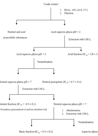
Extraction of plant materials - The collected A. ra- miflorum materials (stem bark 2 kg and leaves 0.340 g) were dried in a circulating air oven, ground and subject-ed to extraction by maceration with methanol for seven days. The organic solvent was evaporated at 40°C under rotation and reduced pressure. The crude extract was lyophilised and subjected to acid base fractionation, as previously described (Marques et al. 1996, Cunha et al. 2012). After a complete partition of the plant stem bark methanolic crude extract (138 g), four alkaloid rich fractions were obtained: the acid, the neutral precipitate (NP), the neutral and the basic fractions, which were all concentrated under reduced pressure at 40ºC, lyoph-ilised and provided for biological testing at the René Ra-chou Research Centre, Fiocruz (Belo Horizonte, MG).
A crude extract of the plant leaves (20.4 g) was sub-jected to a simplified acid-base partition employing the same solvents (Tanaka et al. 2007). Two alkaloid rich fractions were obtained (the acid fraction and the basic fraction) and were concentrated under reduced pressure at 40ºC for solvent evaporation and then lyophilised (Cunha et al. 2012) (Fig. 2).
The alkaloid extracts from the stem barks and the leaves were analysed by thin-layer chromatography (TLC) on a silica gel 60 GF254 and developed with CHCl3/ MeOH 92:8 in an NH3 atmosphere with the p -anisalde-hyde reagent followed by heating to 105ºC for 2-4 min.
Electrospray ionisation mass spectrometric anal-yses (ESI-MS) - Samples from the extracts of the A. ramiflorum barks and leaves were submitted to off-line ESI-MS analysis of alkaloids. The lyophilised extracts were dissolved in methanol:water (1:1) and 100 mL of trifluoroacetic acid (1.0 mg/mL), filtered through a 0.2 mm size pore membrane and introduced into the mass spectrometer using a syringe pump. The spectra were obtained in the positive ionisation mode after using a tri-ple quadrutri-ple Micromass® Liquid Chromatograph Mass
Spectrometer model Quattro MicroTM API setting the
capillary voltage at 2,300 V, the cone voltage at 60 V and the source at 100ºC. Each spectrum was produced by accumulation data over 1 min.
Isolation of alkaloids from the stem bark extracts - The neutral (0.69 g) and basic (0.36 g) fractions from the methanolic crude extract were subjected to a silica gel column chromatography, eluted with CHCl3 and fol-lowed by CHCI3 with increasing amounts of methanol. Further purification by preparative TLC was performed using a silica gel 60 GF254 as the stationary phase and CHCl3:CH2Cl2:ethyl acetate:MeOH 4.5:4:1:0.5 (v/v) as
908 Mem Inst Oswaldo Cruz, Rio de Janeiro, Vol. 110(7), November 2015
98.8, was previously described in the stem barks of A. ramiflorum and its structure was established by uni and bidimensional nuclear magnetic resonance (NMR). MS and comparison with the literature values confirmed compound 3 as 10-methoxygeissoschizol (Marques et al. 1996, Tanaka et al. 2007).
Compound 5 [α]25
D 0.0° (c = 1.0 mg/mL, EtOH) was
identified by infrared (IR) (KBr,νmax cm
-1), 3345 (N-H
and O-H), 1731 (COOCH3), ultraviolet (UV) λ (EtOH,
Max) nm (log ε), 224 (4.7), 280 (4.14), 289 (4.08), elec
-tron impact (EI)/MS 70 eV, m/z (%), 354 [M+., 98], 323 (30), 251 (100), 249 (30), 169 (50), ESI-MS at m/z 355.3 [M+ H] and daughter ions at m/z 353.0 and m/z 279, 1H
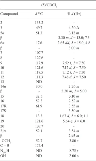
and 13C NMR (Table I). Compound 5 has not been
pre-viously described in A. ramiflorum and its structure was established by uni and bidimensional NMR and MS. By comparison with literature values compound 5 was (±-(E)-isositsirikine (Kan et al. 1981).
Isolation of alkaloids from the leave extracts - The basic alkaloid fraction (0.35 g) was placed on a silica gel column, eluted with CHCl3 followed by CHCI3 with increasing amounts of MeOH and was further purified using a preparative TLC silica gel 60 GF254 as the sta-tionary phase and then eluted with CHCl3:CH2Cl2:ethyl acetate:MeOH 4.5:4:1:0.5 (v/v) in an NH4OH atmosphere (40 mg of extract/per plate). The steps permitted the iso-lation of compound 2, an indole alkaloid.
Compound 2 [α]25
D + 58.5º (c = 1.0 mg/mL, EtOH)
was identified by ESI-MS at m/z 467.3 [M+ H] with daughter ions at m/z 281.3 and m/z 174.0 as described before in stem barks (Marques et al. 1996). Its structure was established by uni and bidimensional NMR and MS data that confirmed its identity (2) as ramiflorine B by comparison with previous data (Tanaka et al. 2007).
Tests with plant materials against P. falciparum blood parasites in vitro - The activity of the A. rami-florum extracts and fractions was evaluated against
P. falciparum blood parasites [clone W2, chloroquine (CQ)-resistant], which were cultured as previously
de-Fig. 2: flowchart fractionation of Aspidosperma ramiflorum using acid base crude extract from stem bark of the plant. IC50: half-maxi-mal drug inhibitory concentration.
TABLE I
Antimalarial activity of Aspidosperma ramiflorum extracts and fractions in mice infected with Plasmodium berghei treated during three consecutive days by gavage
Oral treatment with
Dose (mg/kg)
Parasitaemia reduction at day 10a
Mice survival in days (increase)b
Plant extracts
Neutral precipitate 250 66 26 ± 6 (4)
500 53 29 ± 1 (7)c
Nonsoluble 250 16 29 ± 1 (7)c
500 22 22 ± 1(0)
Chloroquine 20 100 > 30 (> 8)c
Control 0 0 22 ± 2
a: parasitaemia reduction in relation to untreated mice. Compounds inhibiting ≤ 30% were considered inactive, 30-50% as
partially active and ≥ 50% as active; b: mice survival increase in days after drug treatment compared to survival of nontreated controls; c:significant differences in animal survival (p ≤ 0.05) by Mann-Whitney U test.
eluents in an ammonium hydroxide (NH4OH) atmos-phere (40 mg of catechin equivalents per plate). Two in-dole alkaloids, compounds 3 and 5, were isolated.
scribed (Trager & Jensen 1976) with modifications (de Andrade-Neto et al. 2004). The freshly sorbitol synchro-nised ring stages (Lambros & Vanderberg 1979) were immediately incubated with the test compounds at var-ious concentrations that were prevvar-iously solubilised in 0.05% dimethyl sulfoxide (DMSO) (v/v). Each test was performed in triplicate and the results were compared with the control cultures in complete medium with no drugs. CQ was used in each experiment as an antima-larial control. The compounds’ effects were measured through the [3H]-hypoxanthine incorporation assay
(Desjardins et al. 1979) and by the immunoenzymatic test, using specific monoclonal antibodies to a parasite protein histidine and alanine-rich protein (HRPII) that were commercially acquired (MPFM ICLLAB-55A®
and MPFG55P ICLLAB®, USA) (Noedl et al. 2002).
For the [3H]-hypoxanthine assay, the parasites were
maintained at least four days in medium without hypoxan-thine and adjusted to 1% parasitaemia and 1% haematocrit. The levels of [3H]-hypoxanthine incorporated into the
par-asites were measured using a beta counter (PerkinElmer, EUA). For the anti-HRPII test, parasitaemia was adjusted
to 0.05% and the haematocrit to 1.5%; binding of the HRPII
antibodies was quantified at 450 nm using a spectropho-tometer (SpectraMax340PC384, Molecular Devices).
The half-maximal drug inhibitory concentration (IC50) was estimated by curve fitting using software from the OriginLab Corporation (USA) and comparing to the parasite growth in the drug-free medium.
Cytotoxicity tests using immortalised or primary cells
- The cytotoxicity of the plant extracts and fractions was evaluated in a human hepatoma cell line (HepG2) and a monkey kidney cell line (BGM) using cells cultured in 75-cm2 sterile flasks containing RPMI-1640 medium
(supple-mented with 10% heat-inactivated foetal calf serum and 40 mg/L gentamicin) under a 5% CO2 atmosphere at 37ºC. When confluent, the cell monolayer was washed with cul-ture medium, trypsinised, distributed in a flat-bottomed 96-well plate (5 × 10³ cells/well) and incubated for 18 h
at 37ºC for cell adherence (Denizot & Lang 1986). The compounds (20 µL), at various concentrations (1,000-1 µg/mL), were placed in the 96-well plates, incubated with the cultured cells for 24 h under a 5% CO2 atmosphere at 37ºC and then the
3-(4,5-dimethylthiazol-2-yl)-2,5-di-phenyltetrazolium bromide (MTT) solution (5 mg/mL;
20 µL/ well for 3 h) was used to evaluate the mitochon-drial viability. The supernatants were carefully removed and 100 µL DMSO was added to each well and mixed to solubilise the formazan crystals. The optical density was determined at 570 nm and 630 nm (background) (Spec-traMax340PC384). The cell viability was expressed as the
percentage of the control absorbance in the untreated cells after subtracting the appropriate background.
Cytotoxicity was also tested using normal peripheral blood mononuclear cells (PBMC) isolated from healthy individuals by Ficoll-Histopaque (Sigma-Aldrich) gra-dient centrifugation (Panda & Ravindran 2013). The cells were washed twice at 100 g in RPMI-1640 (Sig-ma-Aldrich) and resuspended in 2 mL of RPMI supple-mented with 10% heat-inactivated foetal bovine serum.
The viable PBMCs cells were counted using the Trypan blue exclusion test, plated in 96-well cell culture plates at a final concentration of 3 × 10⁶ cells/mL and incubated
with the compounds (20 µL) at various concentrations (≤ 1,000 µg/mL) for 24 h at 37ºC. The MTT solution (5
mg/mL; 20 µL/ well) was added to evaluate the cell mi
-tochondrial viability as described above. The minimum lethal dose for 50% (MLD50) of the cells was determined as previously described (Madureira et al. 2002). The se-lectivity index (SI) was calculated as the ratio between the activity and the cytotoxicity.
Antimalarial tests against P. berghei in mice - The drug suppressive test was performed as previously de-scribed (Peters 1965) with some modifications (Carva- lho et al. 1991). Adult Swiss outbred mice (20 ± 2 g weigh) were inoculated by the intraperitoneal route with 1 × 10⁵ red blood cells infected with P. berghei, the NK65
strain. Groups of 20-30 infected mice were kept together for 2-24 h, then were divided randomly in groups of up to six animals per cage and were treated by gavage for three consecutive days with 25 mg/Kg or 50 mg/Kg of the test compounds diluted in DMSO 3% (v/v). Two con-trol groups were used and either received CQ (20 mg/kg, diluted in water) or the drug vehicle. Smears prepared from the tail blood of the mice on days 5-10 post-in-fection were methanol-fixed, stained with Giemsa and examined microscopically. Parasitaemia was evaluated and the percent inhibition of the parasite growth was cal-culated in relation to the untreated control group, which was considered 100% growth.
RESULTS
The results of the in vitro activity of the extracts from the stem barks and the leaves of A. ramiflorum against the blood cultures of P. falciparum (W2 clone, CQ-resistant) are shown in Table II. The compounds with an IC50 below 10 µg/mL were considered active. All but the aqueous ex-tracts were active, particularly the acid, the NP and the basic bark crude extracts, which showed IC50 values be-tween 0.5-3.8 µg/mL. Both the hypoxanthine and the anti-HRPII assays provided similar results. The methanolic and acetone extracts from the leaves showed IC50 values of 1.4 and 4.0 µg/mL, respectively. The pure substances (5, 3 and 2) isolated from the bark fractions (neutral and basic) were active. Compound 5 had the lowest IC50 (0.3 µg/mL). All of these extracts were rich in indole alkaloids.
The plant showed no toxicity towards the HepG2 and BGM cell lines and showed high MLD50 values for most of the extracts and fractions. The acid and nonsoluble ex-tracts were toxic to the normal cells (BGM) (Table II). When evaluated against freshly isolated human PBMC, the MLD50 values were lower for most of the extracts and fractions tested, except for the acid, the aqueous and the ac-etone extracts, which had similar MLD50 values in relation to the immortalised cell line. The SI of compound 5 was the highest (SI = 100), but substances 3 and 2 had SI values
be-low 10, which were indicative of toxicity; the other purified
compounds had SI values above 22 (Table II).
910 Mem Inst Oswaldo Cruz, Rio de Janeiro, Vol. 110(7), November 2015
NP fraction reduced the P. berghei parasitaemia to 53% on day 10 in relation to the nontreated control mice. However, both of the extracts increased the survival of the mice (Table I). The other extracts and purified com-pounds were not tested in vivo due to the insufficient amounts available.
The mass peaks observed in the chemical charac-terisation of the crude extract from the barks of A. rami-florum used in the ESI-MS analysis are shown in Fig. 3. A new molecule was identified, isositsirikine (5) at
m/z 355 [M + H]+ (Table III). The other isolated com-pounds were ramiflorine A (1) and B (2) at m/z 467 [M + H]+, 10-methoxy-geissoschizol (3) at 327 [M + H]+
and β-yohimbine (4) and these compounds correspond to
compounds previously described in the species (Marques et al. 1996, Tanaka et al. 2007). All of the fractions from the stem bark had similar mass spectra profiles.
DISCUSSION
The chemical analysis of the A. ramiflorum crude extract showed the mass spectra profiles of two com-pounds that were similar to what had been described be-fore in the plant leaves (Marques et al. 1996). Their mass peaks corresponded to the lead alkaloid compounds ramiflorine A (1) and ramiflorine B (2) at m/z 467 [M + H]+. Using preparative TLC, the bioactive fractions from the stem bark (neutral and basic) and the leaves (basic) were chromatographed on a silica gel column to generate purified fractions and two previously described
compounds in A. ramiflorum were identified, the indole alkaloids ramiflorine B (2) and 10-methoxy-geisso-schizol (3), whereas the compound isositsirikine (5) was reported here for the first time in the species.
The structures of 2, 3 and 5 were established in this study using uni and bidimensional NMR and the values of the MS analysis in comparison with previous literature data (Kan et al. 1981, Marques et al. 1996, Tanaka et al. 2007).
The mass spectrum of 5 gave a molecular ion at m/z 354 (M+, EI/MS 70 eV) as well as peaks at m/z 323, 251, 249 and 169, which are typical of the corynanthine skeleton. The IR and UV spectra showed absorption bands charac-teristic of a 10-methoxyindole chromophore (Marques et al. 1996). The 1H and 13C NMR spectra showed downfield
signals at δH 7.52 (1H, t, J= 7.5 Hz), 7.12 (1H, d, J = 7.5 Hz),
7.12 (1H, t, J = 7.5 Hz) and 7.48 (1H, d, J = 7.4 Hz), which
correlate with the respective carbons at δC 117.9 (C-9), 121.6
(C-10), 119.5 (C-11) and 111.3 (C-12) (heteronuclear single quantum coherence). Additional signals were detected from four quaternary carbons, one with downfield signals
at δC 175.4 (COOCH3) and the other four at δC 133.2 (C-2),
107.7 (C-7), 127.6 (C-8) and 137.7 C-20). 1H NMR
spec-trum also showed the presence of an ethylidene side chain
methyl group (C-19) at δH 1.67 (3H, d, J = 6.0 Hz) and an
olefinic proton (C-18) at δH 5.64 (1H, q, J = 6.0 Hz) and at δH
4.30 (1H, sl) that correlated with δC 49.7. The H-3 α-config
-uration was confirmed by comparison with the chemical shift of the protons of similar compounds described in the literature (Kan et al. 1981). The 1H and 13C NMR data of
alkaloid 5 were similar to those reported for a compound
TABLE II
In vitro activity of Aspidosperma ramiflorum compounds tested against blood forms of Plasmodium falciparum [W2 clone, chloroquine (CQ)-resistant parasites], cytotoxicity [minimum lethal dose for 50% (MLD50)] to a human hepatoma cell line (HepG2), a monkey kidney cell line (BGM) and human peripheral blood mononuclear cells (PBMC) and selectivity indexes (SI)
Compoundsa
MLD50 (µg/mL) IC50 (µg/mL) SI
BGM HepG2 PBMC Hypoxanthine Anti-HRPII BGM HepG2 PBMC
Bark extracts
Acid 388 ± 100 155 ± 2 512 ± 34 3.8 ± 1.2 2.5 ± 1.2 138 41 135
Neutral 31 ± 2 35 ± 5 NT 0.9 ± 0.1 0.7 ± 0.5 34 39 NT
Basic 32 ± 0 31 ± 0 11 ± 4 0.5 ± 0.2 0.8 ± 0.5 64 62 22
Neutral precipitate 30 ± 1 45 ± 0 13 ± 4 0.7 ± 0.4 0.7 ± 0.4 43 64 19
Methanolic residue 88 ± 16 80 ± 43 25 ± 6 1.7 ± 1.6 1.5 ± 0.9 52 47 15
Nonsoluble 621 ± 120 173 ± 7 149 ± 36 3.1 ± 0.6 3.1 ± 1.8 200 56 48
Aqueous > 1,000 > 1,000 > 1,000 > 50 > 50 Inative Inative Inative
Pure substances
5 (isositsirikine) 28 ± 7 34 ± 1 NT 0.3 ± 0.1 0.2 ± 0.0 93 113 NT
3 (10-MG) 8 ± 3 5 ± 1 NT 1.0 ± 0.9 0.4 ± 0.3 Toxic Toxic NT
2 (ramiflorine B) 5 ± 2 5 ± 1.7 NT 1.2 ± 0.4 0.9 ± 0.9 Toxic Toxic NT
Leave extract
Methanolic 98 ± 22 137 ± 18 64 ± 3 3.6 ± 1.3 1.4 ± 0.7 27 38 18
Acetone 37 ± 6 67 ± 12 32 ± 8 1.7 ± 1.7 1.4 ± 0.4 22 39 19
CQ 457 ± 22 398 ± 12 222 ± 73 0.100 ± 0.21 0.0 7± 0.10 4,570 3,980 2,220
Fig. 3: off-line ESI-MS of crude extract from stem bark of Aspidosperma ramiflorum. Ion at m/z 467 corresponding to ramiflorine A (1) and rami-florine B (2), ion at m/z 327 corresponding to 10-methoxy-geissoschizol (3) and ion at m/z 355 corresponding to β-yohimbine (4) and isositsikine (5).
known as isositsirikine (Kan et al. 1981). We suggest that the presence of the indole alkaloid skeleton (3 and 5) ex-plains the decrease in the cytotoxicity of the compounds
and the consequent increase in the SI values; indeed, the
dimeric ramiflorine B (2) was more cytotoxic than 3 and 5. The results of the values of the MLD50 in the PBMC nonimmortalised cells showed a slightly higher cytotox-icity in comparison with the immortalised cells for some samples, which can be attributed to the high multiplica-tion rates of the other cell lines.
It was only possible to test the in vivo antimalarial activity of the neutral and precipitate extracts from the stem barks in mice and both extracts increased survival of the mice, but whether this was due to the presence of
the alkaloids in A. ramiflorum having anti-inflammato-ry and antioxidant activities (Barbosa-Filho et al. 2006) needs to be clarified. It is also necessary to study the in vivo activity of isositsirikine (compound 5), the most active compound against P. falciparum in vitro, which was demonstrated for the first time in the present study.
912 Mem Inst Oswaldo Cruz, Rio de Janeiro, Vol. 110(7), November 2015
TABLE III
1H (300 MHz) and 13C (75 MHz) nuclear magnetic resonance
(NMR) data of compound (5) in CDCl3 as solvent and tetra-methylsilane used as internal reference, chemical shifts
(d, ppm) and coupling constants (J, Hz)
(5)/CDCl3
Compound δ13C 1H J (Hz)
2 133.2
-3 49.7 4.30 ls
5α
β 51.3
-3.12 m
3.30 m, J = 13.0; 7.3
6α 17.6 2.65 dd, J = 15.0; 4.8
β 7
-107.7
3.00 m
-8 127.6
-9 10 11 12 13 14α β 117.9 121.6 119.5 111.3 ND 30.0
-7.52 t, J = 7.50 7.12 d, J = 7.50 7.12 t, J = 7.50 7.48 d, J = 7.50
-2.26 m
2.20 m, J = 5.00 15
16
32.5 52.3
3.10 m
2.52 m
17R 17S
61.9
-3.55 m
3.50 m
18 13.3 1,67 d, J = 6.0; 1.1
19 121.6 5.64 q, J = 6.0
20 21α β 137.7 52.1 -3.54 m
2.93 m
-OCH3 C = 0
52.7 175.4
3.80 s
-N(1)-H ND 8.75 s
OH ND 2.00 s
ND: not determined.
ACKNOWLEDGEMENTS
To Fiocruz-MG, for logistics and facilities, and to Prof Washington Marcondes Ferreira, for plant identification.
REFERENCES
Adebayo JO, Krettli AU 2011. Potential antimalarials from Nigerian plants: a review. J Ethnopharmacol 133: 289-302.
Aguiar ACC, da Rocha EMM, de Souza NB, França TCC, Krettli AU 2012. New approaches in antimalarial drug discovery and devel-opment - A Review. Mem Inst Oswaldo Cruz 107: 831-845.
Alavijeh MS, Chishty M, Qaiser MZ, Palmer AM 2005. Drug metab-olism and pharmacokinetics, the blood-brain barrier and central nervous system drug discovery. Neuro Rx 2: 554-571.
Barbosa-Filho JM, Piuvezam MR, Moura MD, Silva MS, Lima KVB, da Cunha EVL, Fechine I, Takemura O 2006. Anti-inflammatory activity of alkaloids: a twenty-century review. Rev Bras Farma-cogn 16: 109-139.
Bourdy G, Willcox ML, Ginsburg H, Rasoanaivo P, Graz B, Deharo E 2007. Ethnopharmacology and malaria: new hypothetical leads or old efficient antimalarials? Int J Parasitol 38: 33-41.
Carvalho L, Brandão M, Santos-Filho D, Lopes J, Krettli A 1991. Anti-malarial activity of crude extracts from Brazilian plants studied in vivo in Plasmodium berghei-infected mice and in vitro against Plas-modium falciparum in culture. Braz J Med Biol Res24: 1113-1123.
Coutinho JP, Aguiar ACC, dos Santos PA, Lima JC, Rocha MGL, Santa-na AEG, Pereira MM, Krettli AU 2013. Aspidosperma (Apocynace-ae) plant cytotoxicity and activity towards malaria parasites. Part I: Aspidosperma nitidum (Benth) used as a remedy to treat fever and malaria in the Amazon. Mem Inst Oswaldo Cruz 108: 974-982.
Cui L, Su XZ 2009. Discovery, mechanisms of action and combination therapy of artemisinin. Expert Rev Anti Infect Ther8: 999-1013.
Cunha AC, Chierrito TP, Machado GM, Leon LL, da Silva CC, Tanaka JC, de Souza LM, Gonçalves RA, de Oliveira AJ 2012. Anti-leish-manial activity of alkaloidal extracts obtained from different or-gans of Aspidosperma ramiflorum. Phytomedicine19: 413-417.
de Andrade-Neto VF, Brandão MGL, Oliveira FQ, Casali VWD, Njaine B, Zalis MG, Oliveira LA, Krettli AU 2004. Antimalarial activity of Bidens pilosa L. (Asteraceae) ethanol extracts from wild plants collected in various localities or plants cultivated in humus soil. Phytother Res 18: 634-639.
de Andrade-Neto VF, Pohlit AM, Pinto ACS, Silva ECC, Nogueira KL, Melo MRS, Henrique MC, Amorim RCN, Silva LFR, Cos-ta MRF, Nunomura RCS, Nunomura SM, Alecrim WD, Alec-rim MGC, Chaves FCM, Vieira PPR 2007. In vitro inhibition of Plasmodium falciparum by substances isolated from Amazonian antimalarial plants. Mem Inst Oswaldo Cruz 102: 359-365.
de Ridder S, van der Kooy F, Verpoorte R 2008. Artemisia annua as a self-reliant treatment for malaria in developing countries. J Ethnopharmacol120: 302-314.
Denizot F, Lang R 1986. Rapid colorimetric assay for cell growth and survival. Modifications to the tetrazolium dye procedure giving im-proved sensitivity and reliability. J Immunol Methods89: 271-277.
Desjardins RE, Canfield CJ, Haynes JD, Chulay JD 1979. Quantitative assessment of anti-malarial activity in vitro by a semiautomatated microdilution technique. Antimicrob Agents Chemother16: 710-718.
Frausin G, Hidalgo AF, Lima RB, Kinupp VF, Ming LC, Pohlit AM, Milliken W 2015. An ethnobotanical study of anti-malarial plants among indigenous people on the upper Negro River in the Brazil-ian Amazon. J Ethnopharmacol15: 30050-30057.
Kan C, Kan SK, Lounasmaa M, Husson HP 1981. Trapping of in-termediates in the interconversion of heteroyohimbine alkaloids. Acta Chem Scand35: 269-272.
Krettli AU, Adebayo JO, Krettli LG 2009. Testing of natural products and synthetic molecules aiming at new antimalarials. Curr Drug Targets10: 261-270.
Krettli AU, Andrade-Neto VF, Brandão MGL, Ferrari WMS 2001. The search for new antimalarial drugs from plants used to treat fever and malaria or plants randomly selected: a Review. Mem Inst Oswaldo Cruz96: 1033-1042.
Lambros C, Vanderberg JP 1979. Synchronization of Plasmodium-falciparum erythrocytic stages in culture. J Parasitol65: 418-420.
Madureira MC, Martins AP, Gomes M, Paiva J, da Cunha AP, Rosário V 2002. Antimalarial activity of medicinal plants used in traditional medicine in S. Tomé and Príncipe islands. J Ethno-pharmacol8: 23-29.
Noedl H, Wongsrichanalai C, Miller R, Myint K, Looareesuwan S, Sukthana Y, Wongchotigul V, Kollaritsch H, Wiedermann G, Wernsdorfer W 2002. Plasmodium falciparum: effect of anti-ma-larial drugs on the production and secretion characteristics of his-tidine-rich protein II. Exp Parasitol102: 157-163.
Oliveira AB, Dolabela MF, Braga FC, Jácome RL, Varotti FP, Póvoa MM 2009. Plant-derived antimalarial agents: new leads and efficient phythomedicines. Part I. Alkaloids. An Acad Bras Cienc81: 715-740.
Oliveira DR, Krettli AU, Aguiar AC, Leitão GG, Vieira MN, Mar-tins KS, Leitão SG 2015. Ethnopharmacological evaluation of medicinal plants used against malaria by Quilombola commu-nities from Oriximiná, Brazil. J Ethnopharmacol doi: 10.1016/j. jep.2015.07.035.
Panda SK, Ravindran B 2013. In vitro culture of human PBMCs. Bio Protoc 3: e322.
Peters W 1965. Drug resistance in Plasmodium berghei Vincke and Lips, 1948. I. Chloroquine resistance. Exp Parasitol17: 80-89.
Ridley RG 2002. Medical need, scientific opportunity and the drive for antimalarial drugs. Nature 415: 686-693.
Rocha e Silva LF, Montoia A, Amorim RC, Melo MR, Henrique MC, Nunomura SM, Costa MR, Andrade Neto VF, Costa DS, Dantas
G, Lavrado J, Moreira R, Paulo A, Pinto AC, Tadei WP, Zacardi RS, Eberlin MN, Pohlit AMLF 2012. Comparative in vitro and in vivo antimalarial activity of the indole alkaloids ellipticine, oli-vacine, cryptolepine and a synthetic cryptolepine analog. Phyto-medicine20: 71-76.
Tanaka JCA, Silva CC, Ferreira ICP, Machado GMC, Leon LL, Ol-iveira AJB 2007. Antileishmanial activity of indole alkaloids from Aspidosperma ramiflorum. Phytomedicine 14: 377-380.
Torres ZES, Silveira ER, Silva LFR, Lima ES, de Vasconcellos MC, Uchoa DEA, Braz Filho R, Pohlit AM 2013. Chemical composi-tion of Aspidosperma ulei Markgr. and antiplasmodial activity of selected indole alkaloids. Molecules 18: 6281-6297.
Trager W, Jensen JB 1976. Human malaria parasites in continuous culture. Science193: 673-675.
White NJ 2008. Qinghaosu (artemisinin): the price of success. Sci-ence320: 330-334.
WHO - World Health Organization 2014. Malaria. Available from: who.int/mediacentre/factsheets/fs094/en/.