Neuroprotective changes in degeneration-related gene expression in the
substantia nigra following acupuncture in an MPTP mouse model of
Parkinsonism: Microarray analysis
Sujung Yeo
1,2, Keon Sang An
2, Yeon-Mi Hong
1,2, Yeong-Gon Choi
1,2, Bruce Rosen
2,3,
Sung-Hoon Kim
2and Sabina Lim
1,21
Research Group of Pain and Neuroscience, WHO Collaborating Center for Traditional Medicine,
East-West Medical Research Institute, Kyung Hee University, Seoul, Republic of Korea.
2
Department of Basic Korean Medical Science, College of Korean Medicine, Kyung Hee University,
Seoul, Republic of Korea.
3
Department of Radiology, Athinoula A. Martinos Center for Biomedical Imaging,
Massachusetts General Hospital, Harvard Medical School, Boston, USA.
Abstract
Parkinson’s disease (PD) is a neurodegenerative disorder characterized by the death of dopamine-generating cells in the substantia nigra (SN). Acupuncture stimulation results in an enhanced survival of dopaminergic neurons in the SN in Parkinsonism animal models. The present study investigated changes in gene expression profiles measured using whole transcript array in the SN region related to the inhibitory effects of acupuncture in a chronic 1-methyl-4-phenyl-1,2,3,6-tetrahydropyridine (MPTP) Parkinsonism model. In this model, acupuncture stimulation at GB34 and LR3 attenuated the decrease in tyrosine hydroxylase in the SN region; stimulation at non-acupoints did not sup-press this decrease. Gene array analysis revealed that 22 (10 annotated genes:Cdh1, Itih2, Mpzl2, Rdh9, Serping1, Slc6a13, Slc6a20a, Slc6a4, Tph2, and Ucma) probes that were up-regulated in MPTP animals relative to controls were exclusively down-regulated by acupuncture stimulation. In addition, 17 (two annotated genes:4921530L21Rik andGm13931) probes that were down-regulated in MPTP animals compared to controls were exclusively up-regulated by acupuncture stimulation. These findings indicate that the 39 probes (12 annotated genes) affected by MPTP and acupuncture may be responsible for the inhibitory effects of acupuncture on degeneration-related gene expression in the SN following damage induced by MPTP intoxication.
Keywords: Parkinson’s disease, gene array, differentially expressed genes, neurodegeneration, acupoints.
Received: May 8, 2014; Accepted: September 17, 2014.
Introduction
Parkinson’s disease (PD) is a neurodegenerative dis-order characterized by major behavioral symptoms, which include tremor, akinesia, bradykinesia, and stiffness (Jan-kovic, 2008). PD results from the death of dopaminergic neurons in the substantia nigra (SN; (Goto et al., 1989; Chauhanet al., 2001; Kimet al., 2003; Fernandez-Espejo 2004), which plays an important role in several brain func-tions, particularly, motor planning, eye movement, learn-ing, reward-seeking and addiction. Not surprisingly, gross movement dysfunction caused by damage to this area is a symptom of PD.
Using a 6-hydroxydopamine (6-OHDA)-induced Parkinsonism model, a previous study found that acupunc-ture stimulation at acupoints GB34 and LR3 results in the enhanced survival of dopaminergic neurons in the SN and a ~87.7% improvement in motor dysfunction (Park et al., 2003). Similar finding were found following the adminis-tration of 1-methyl-4-phenyl-1,2,3,6-tetrahydropyridine (MPTP), where the acupuncture-induced enhancement of synaptic dopamine availability may play a critical role in the observed motor function improvement (Kim et al., 2011). Acupuncture stimulation at the same acupoints also attenuates the decrease in tyrosine hydroxylase (TH) immunoreactivity (IR) and generates neuroprotective ef-fects in the SN in an MPTP-induced mouse model of Parkinsonism (Kanget al., 2007). The mechanisms under-lying the neuroprotective effects of acupuncture on the SN are thought to be in part related to decreased microglial ac-tivation and reduced inflammatory responses (Kanget al.,
Send correspondence to Sabina Lim. Research Group of Pain and Neuroscience, WHO Collaborating Center for Traditional Medicine, East-West Medical Research Institute, Kyung Hee University, 130-701 Seoul, Republic of Korea. E-mail: [email protected].
2007); these, in turn, inhibit ferric iron and ferritin heavy chain deposition (Choiet al., 2009).
Several genes that are affected by MPTP and stimula-tion at acupoints may be responsible for the inhibitory ef-fects of acupuncture on MPTP-induced degeneration in striatal regions (Choiet al., 2011b). Additionally, gene ex-pression in thalamic regions damaged by MPTP intoxica-tion may be affected, at least in part, by acupuncture at specific acupoints (Yeoet al., 2013). Changes in gene ex-pression profiles in the SN following acupuncture stimula-tion were identified in an acute Parkinsonism model over three days (Honget al., 2010); however, no such changes have been demonstrated in a chronic Parkinsonism animal model. Because PD is a chronic neurodegenerative disor-der, changes in gene expression in the SN in a chronic Parkinsonism animal model should be investigated to iden-tify the neuroprotective mechanisms underlying acupunc-ture treatment.
Thus, we investigated changes in gene expression profiles in the SN region in a relatively chronic Parkin-sonism model and identified the genes related to the inhibi-tory effect of acupuncture on alterations in gene expression induced by MPTP intoxication.
Materials and Methods
MPTP model of Parkinsonism
Six-week-old male inbred C57BL/6 mice (20-22 g; Samtaco, Korea) were divided into the following four groups: control (CTL); MPTP-treatment only (MPTP); MPTP and acupuncture treatment at GB34 and LR3 (MPTP-A); and MPTP and acupuncture treatment at non-acupoints (MPTP-NA). Mice in the control group (n = 9) were injected intraperitoneally with saline 0.9% (100mL) once daily for four weeks, while mice in the MPTP group (n = 9) were injected intraperitoneally with MPTP-HCl (20 mg/kg of free base) in saline 0.9% (100mL) at 24-h in-tervals over four weeks to produce a sustained chronic model of Parkinsonism (Bezardet al., 1997; Kuhnet al., 2003; Choiet al., 2011a; Yeoet al., 2013). On the day fol-lowing the final MPTP treatment, mice were anesthetized using 16.5% urethane and perfused transcardially with cold 0.05 M sodium-phosphate buffer to enable immunohisto-chemical evaluation. The Kyung Hee University Animal Experimentation Committee approved all animal protocols used in this study. Reagents used but not mentioned were purchased from Sigma (USA).
Acupuncture administration
Acupuncture was performed manually 2 h after the first MPTP injection and at 48-h intervals thereafter for a total of 14 sessions. The acupuncture procedure (acupoints: GB34 [Yanglingquan] and LR3 [Taichong]; non-acu-points: both sides of the hips) was performed as reported previously (Parket al., 2003; Kanget al., 2007; Choiet al.,
2011b). Briefly, mice in the acupoints group were immobi-lized by hand 2 h following MPTP administration, and the acupuncture needles were inserted bilaterally to depths of 1 mm at acupoint LR3 and 3 mm at acupoint GB34 before being turned at a rate of two spins per second for 15 s, as re-ported previously (Kanget al., 2007). In the non-acupoints group, the needles were inserted to a depth of 3 mm on both sides of the hips, and a procedure identical to that described above was performed.
Immunohistochemistry
After four weeks, the brains were removed, post-fixed in 0.05 M sodium-phosphate buffer containing 4% paraformaldehyde for 12 h at 4 °C, rinsed with 0.05 M so-dium-phosphate buffer, dehydrated with sucrose for 12 h at 4°C, and then cryosectioned. Coronal sections of the brains (30-mm thickness) were cut using a cryomicrotome. Immu-nohistochemical analyses were carried out using an ABC kit and a Mouse on Mouse (M.O.M) immunodetection kit (Vector Laboratories, CA) using a modification of the avidin-biotin-peroxidase method. Briefly, sections encom-passing the entire striatal and SN regions were incubated in phosphate-buffered saline (PBS; pH 7.4) with 3% H2O2, exposed to 3% bovine serum albumin (BSA) and 0.3% Tri-ton X-100 in PBS for 1 h, and then treated with an avidin-biotin blocking kit (Vector Laboratories). When using the mouse anti-TH antibody, sections were treated with an M.O.M. mouse Ig-blocking reagent (Vector Laboratories) at room temperature for 1 h prior to incubation with the pri-mary antibody. Thereafter, each section was stained over-night at 4 °C with a mouse anti-TH antibody (1:1,500; Santa Cruz Biotechnology, USA) to identify dopaminergic neurons in the striatal and SN regions. The sections were then sequentially treated with a biotinylated anti-mouse IgG followed by an avidin-biotin-peroxidase complex and developed using a diaminobenzidine-hydrogen peroxide solution (0.003% 3,3-diaminobenzidine and 0.03% hydro-gen peroxide in 0.05 M Tris, pH 7.0).
Western blotting
The bilateral striatal and SN regions were homoge-nized in 20 mM HEPES-KOH buffer (pH 7.5) with 150 mM NaCl, 0.5% sodium deoxycholate, 0.1% SDS, and a protease inhibitor cocktail. After centrifugation at 15,000 xgfor 10 min, soluble supernatant samples of equal protein concentration (30mg total protein) were separated by 12% sodium dodecyl sulfate-polyacrylamide gel elec-trophoresis (SDS-PAGE) and then transferred to polyviny-lidene difluoride (PVDF) membranes (Bio-Rad; USA). The membranes were blocked with 5% skim milk in 0.1% Tris-buffered saline (TBS; 20 mM Tris-HCl [pH 7.5] and 150 mM NaCl containing 0.1% Tween-20; TBST) at room temperature for 1 h and then incubated with mouse anti-TH (1:2,000) and mouse anti-actin (1:5,000, Santa Cruz Bio-technology) antibodies. After being washed with 0.1%
TBST, the membranes were incubated with an anti-mouse IgG-peroxidase antibody (1:2,000, Bio-Rad), and the anti-gen-antibody complexes were visualized using the Pierce ECL western blotting substrate (Thermo Scientific, USA).
RNA extraction and microarray analysis
Total RNA was extracted from the bilateral SN tissue of each group (n = 2; both groups) using an RNeasy Plus Mini kit (QIAGEN, USA) according to the manufacturer’s instructions. Isolated RNA quality was estimated and quan-tified using a NanoDrop ND-1000 spectrophotometer (NanoDrop Technologies, USA). An aliquot (300 ng) of to-tal RNA was subjected to an Affymetrix GeneChip®Mouse Gene 1.0 ST Array (genome-wide expression profiling chip; 28,853 genes of 35,557 probes; Affymetrix, USA) ac-cording to the GeneChip Whole Transcript (WT) Sense Target Labeling Assay manual, as reported previously (Choiet al., 2011a; Hwanget al., 2009; Linet al., 2010). Briefly, the procedures were carried out as follows: mixing of T7-(N)6 primer and poly-A RNA controls with 300 ng of total RNA isolated from the SN of each group; synthesis of first-cycle, first-strand complementary DNA (cDNA) and then second-strand cDNA; synthesis of first-cycle cRNA and cleanup of cRNA; synthesis of second-cycle, single-strand cDNA (ss cDNA) and cleanup of ss cDNA; frag-mentation of ss cDNA and labeling of the fragmented ss cDNA; hybridization of labeled ss cDNA to the GeneChip; and finally, the staining, washing, and scanning of the GeneChip using a Fluidics 450 station and the GeneChip Operating Software (GCOS, Affymetrix).
Microarray data analysis
The usefulness of the signal intensities was first eval-uated by visual examination of the scanned images. Quality control of the scanned data was then conducted by confirm-ing the order of the signal intensities of poly-A controls and hybridization controls using the Expression Console soft-ware (Affymetrix). Microarray data were analyzed using GenPlex ver. 3.0 (ISTECH, Korea, (Anet al., 2009; Choiet al., 2011a; Hwanget al., 2009; Yeoet al., 2013). A total of eight CEL files (two CEL files generated from each group x four experimental groups) were uploaded and normalized under the following conditions: perfect match (PM)-only as a PM intensity adjustment; the Robust Multichip Analysis (RMA) quantification method as a probe set summarization algorithm for log transformation with base 2 (log2); and the quantile normalization method for evaluation of prelimi-nary data quality in the preprocessing module, which func-tions as a data quality control, using the Expression Console software (Affymetrix). The mean signal intensities of the 28,853 genes were obtained from two chips from each group. Following normalization, the differentially ex-pressed genes (DEGs) in the GeneChip that satisfied the conditions of the fold change cutoff (1.3) as well as the Stu-dent’st-test significance criterion (p < 0.05) were identified
using the DEG-finding module;i.e., log1.3/log2 = 0.379 < average log2 fold-change value (X) for the identification of up-regulated genes and - log1.3/log2 = -0.379 > average log2 fold-change value (X) for the identification of down-regulated genes. A pathway analysis of all metabolic and Kyoto Encyclopedia of Genes and Genomes (KEGG) path-ways was performed using the pathway module, as de-scribed previously (Hwanget al., 2009; Choiet al., 2011a; Yeoet al., 2013). A National Center for Biotechnology In-formation Gene Expression Omnibus (NCBI GEO) acces-sion number was assigned to the raw microarray data generated in this study.
Real time RT-PCR
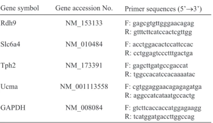
For real-time reverse transcription polymerase chain reaction (RT-PCR) analyses, the total RNA (500 ng) of the samples (CTL, MPTP, and MPTP-A) was subjected to the reaction using a SuperScript First-strand Synthesis Kit (Invitrogen Life Technologies, USA) according to the man-ufacturer’s instructions. The advanced relative expression levels of representative DEGs were monitored with a LightCycler 480 II Real Time PCR Instrument and the LightCycler 480 software ver. 1.5.0.39 (Roche Diagnos-tics; Germany) using a LightCycler 480 SYBR Green I Master (Roche Diagnostics). Mean crossing point (CP) val-ues were obtained and the expression levels of the target genes in the various groups were then compared to those of the reference gene, glyceraldehyde-3-phosphate dehydro-genase (GAPDH), using an advanced relative quantifica-tion method. The ratios of the concentraquantifica-tions of target genes to that of the reference gene were obtained; the primer sequences for each gene are shown in Table 1. The specificity of each primer set was confirmed by determin-ing the meltdetermin-ing temperature and size of each product by gel electrophoresis.
Statistical analysis
Statistical analyses were performed using the statisti-cal package for the social sciences (SPSS) software, ver. 18. Values are means±SE of three independent determina-tions.
Table 1- The sequences of primers.
Gene symbol Gene accession No. Primer sequences (5’®3’)
Rdh9 NM_153133 F: gagcgtgttgggaacagag
R: gtttcttcatccactcgttgg
Slc6a4 NM_010484 F: acctggacactccattccac
R: cctggagtccctttgactga
Tph2 NM_173391 F: gagcttgatgccgaccat
R: tggccacatccacaaaatac
Ucma NM_001113558 F: cgtggaggaacagagagatga
R: aggccatcataatgccactg
GAPDH NM_008084 F: gtcttcaccaccatggagaagg
Results and Discussion
Chronic MPTP-induced Parkinsonism mouse model and the preventive effects of acupuncture
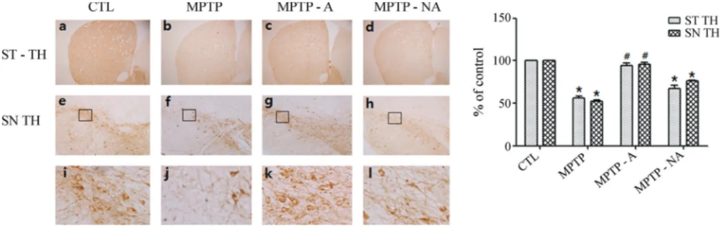
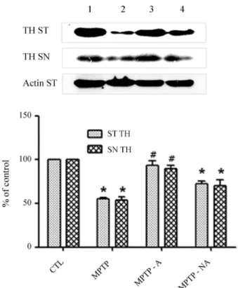
Because TH expression is significantly attenuated in the brains of PD patients (Pardridge, 2005) and in Par-kinsonism animal models (Parket al., 2003; Kanget al., 2007; Choiet al., 2009), we evaluated TH levels to confirm the establishment of a chronic MPTP-induced Parkinso-nism mouse model and the mediating effects of acupunc-ture at GB34 and LR4. In the SN pars compacta (SNpc) and striatal regions, TH levels were noticeably reduced in the MPTP group (MPTP) relative to the control group (CTL; Figures 1a, b, e, f, i, and j). Acupuncture stimulation at GB34 and LR3 (MPTP-A) attenuated the decrease in TH in both areas (Figures 1c, g, and k), but acupuncture at non-acupoints (MPTP-NA) did not similarly influence TH (Fig-ures 1d, h, and l). Immunohistochemical analyses using Western blots confirmed that the decrease in TH levels in the striatal and SNpc regions induced by MPTP intoxica-tion was significantly inhibited by acupuncture at acupoints (MPTP-A) but not at non-acupoints (MPTP-NA; Figure 2). These results confirm that acupuncture stimulation sup-presses the pathological change in TH levels induced by MPTP intoxication in the striatal and SNpc regions, of the mice in our study.
Changes in gene expression in the SN region following chronic MPTP intoxication
As shown in the box plot graph (Figure S1) normal-ization through the preprocessing module was successful. Of the 28,835 genes represented in the oligonucleotide ar-ray, genes were selected and evaluated if they displayed log2-transformed mean signal intensities greater than 0.379 (1.3 fold change) and p < 0.05 by Student’st-test between the control and MPTP groups. Compared to the control, 244 up-regulated (Table S1) and 255 down-regulated
(Ta-ble S2) DEGs were identified in the SN region following chronic MPTP intoxication.
Changes in gene expression in the SN region following acupuncture
Of the 28,853 genes represented in the oligonucleo-tide array, those with log2-transformed mean signal inten-sities greater than 0.379 (1.3-fold change) and a p < 0.05 by Student’st-test between the MPTP-A and MPTP groups or the MPTP-NA and MPTP groups were selected and evalu-ated. Compared to the MPTP group, genes in the SN region were regarded as DEGs following acupuncture stimulation at acupoints and non-acupoints, respectively.
Up-down-regulated genes following MPTP intoxication and acupuncture
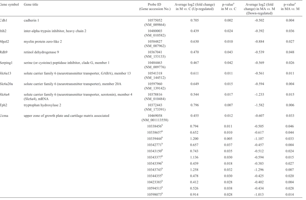
Compared to the control, 22 (10 annotated genes) of the 244 probes were up-regulated in the SN region follow-ing MPTP exposure and exclusively down-regulated by acupuncture at acupoints but not non-acupoints (“Up-down”; Table 2).
Cadherin 1 (Cdh1) is a classic member of the cadherin superfamily that is thought to contribute to the control of synapse formation and synaptic transmission and plasticity (Angstet al., 2001). Cdh1 signaling is an impor-tant component of the regulation of GABAergic synapses in brain neurons (Fiederlinget al., 2011); indeed, elevated GABA levels have been detected in PD (Emiret al., 2012). In the current study,Cdh1was up-regulated in the MPTP group compared to controls. However, acupuncture stimu-lation at GB34 and LR3 maintainedCdh1expression simi-lar to levels in control animals, suggesting that the down-regulation ofCdh1expression by acupuncture may have a neuroprotective effect.
Solute carrier family 6 (neurotransmitter transporter, GABA), member 13 (Slc6a13) is a sodium- and chlo-ride-dependent member of solute carrier family 6 (SLC6)
118 Yeoet al.
that mediates the rapid removal of GABA and maintains low extracellular levels of this neurotransmitter (Chenet al., 2004; Kanner, 2006). Evidence suggests that alteration of GABA transporters is linked to PD (Wanget al., 2007). Accordingly, the overstimulation of GABAAreceptors can potentiate neuronal cell damage; GABA transport blockers are neuroprotective in these situations (Zeevalk and Nicklas, 1996, 1997), suggesting that overexpression of the GABA transporter can damage neurons. In the current study,Slc6a13was up-regulated in the MPTP group com-pared to the control group; therefore this may be the under-lying cause of neuronal intoxication. However, acupunc-ture stimulation at GB34 and LR3 maintainedSlc6a13gene expression at levels similar to those in control animals, sug-gesting that acupuncture has a neuroprotective effect.
Tryptophan hydroxylase 2 (Tph2), with a molecular weight of 56 kDa, is the predominant form of this enzyme present in brain extracts from the mesencephalic tegmen-tum, striategmen-tum, and hippocampus (Sakowskiet al., 2006). Tph2 initiates serotonin synthesis in mammals with Tph1 (Aleninaet al., 2009), and plays a critical role in the main-tenance of brain serotonin homeostasis (Beaulieu et al., 2008). Tph2-derived serotonin is involved in the regulation of behavior and autonomic pathways but is not essential for adult life (Aleninaet al., 2009). However, Tph2 is known to
be highly labile to oxidation (Kuhnet al., 1980). Therefore, the oxidative processes that prevail in PD may cause a misfolding of Tph2 and result in the modification of seroto-nin function such as is seen in dopamine neurons. Tph2 oxi-dation inhibits its activity and leads to the formation of high-molecular-weight aggregates in a dithiothreitol-rever-sible manner (Kuhnet al., 2011). Alteration of serotonin levels by Tph2 hyperinnervation affects the wiring of the brain and can produce long-lasting changes leading to the development of neurodevelopmental disorders such as PD (Migliariniet al., 2012).
Myelin protein zero-like 2 (Mpzl2) is also known as Eva and Eva1. In human choroid plexus epithelial cells and a subset of CD4 T lymphocytes, Mpzl2 is expressed at high levels. Mpzl2 expressed in choroid plexus cells may regu-late the permeability of the blood-cerebrospinal fluid (CSF) barrier (Chatterjeeet al., 2008), suggesting a novel mecha-nism of CNS immune surveillance regulation (Wojciket al., 2011). In the current study, Mpzl2 was up-regulated in the MPTP group compared to controls; however, acupunc-ture stimulation at GB34 and LR3 maintained Mpzl2 gene expression at levels similar to those in control animals. These results also suggest that acupuncture suppresses the Mpzl2 overexpression that can be caused by MPTP neu-ronal loss.
The serine (or cysteine) peptidase inhibitor, clade G, member 1 (Serping1) gene encodes the serine protease in-hibitor (serpin) known as C1 inin-hibitor. The C1 inin-hibitor is important for regulation of several processes, including in-flammation, that are involved in maintenance of blood ves-sels (Shagdarsurenet al., 2008). Serping1 is up-regulated in the microglial pathway upon stimulation by interferon-gamma (IFN-interferon-gamma; (Moranet al., 2007) or lethal lipo-polysaccharide-induced endotoxic shock (Liuet al., 2007).
Inter-alpha trypsin inhibitor, heavy chain 2 (Itih2) has also been identified during inflammation (Scaveniuset al., 2011), and may contribute to the cascade of events leading to neuronal degeneration (Hirsch and Hunot 2009; Tufekci
et al., 2012). Therapeutic strategies aim to down-regulate these inflammatory processes and may slow the progres-sion of PD (Hirsch and Hunot 2009). Therefore, the mainte-nance by acupuncture stimulation of expression levels of Itih2 and Serping1, which are involved in neuroinflam-mation, similar to those in control animals suggests that acupuncture may have a neuroprotective effect.
SLC6 (neurotransmitter transporter), member 20A (Slc6a20a) is also known as Xt3s1, Xtrp3s1, AU022428, and A730081N20Rik. Neurotransmitter transporters of the SLC6 family play an important role in the removal of neurotransmitters in brain tissue and in amino acid trans-port in epithelial cells (Kowalczuket al., 2005). Either the IMINO system or the imino acid carrier supports the main load of proline transport (Munck and Munck, 1994); this gene is expressed at high levels in the brain. More specifi-cally, the IMINO system is present in the microglial cells of Figure 2- Inhibition of the decrease in MPTP-induced TH expression by
120
Yeo
et
al.
Table 2- List of (up-down) substantia nigral genes which were up-regulated in MPTPvs.control and down-regulated in MPTP-Avs.MPTP.
Gene symbol Gene title Probe ID
(Gene accession No.)
Average log2 (fold change) in Mvs.C (Up-regulated)
p-valuea in Mvs.C
Average log2 (fold change) in MAvs.M
(Down-regulated)
p-valuea in MAvs.M
Cdh1 cadherin 1 10575052
(NM_009864)
0.705 0.002 -0.502 0.004
Itih2 inter-alpha trypsin inhibitor, heavy chain 2 10480003 (NM_010582)
0.439 0.024 -0.392 0.036
Mpzl2 myelin protein zero-like 2 10584827 (NM_007962)
0.630 0.010 -0.884 0.027
Rdh9 retinol dehydrogenase 9 10367041
(NM_153133)
0.470 0.043 -0.539 0.048
Serping1 serine (or cysteine) peptidase inhibitor, clade G, member 1 10484463 (NM_009776)
0.467 0.042 -0.569 0.026
Slc6a13 solute carrier family 6 (neurotransmitter transporter, GABA), member 13 10541318 (NM_144512)
0.611 0.011 -0.561 0.011
Slc6a20a solute carrier family 6 (neurotransmitter transporter), member 20A 10597960 (NM_139142)
0.649 0.015 -0.594 0.004
Slc6a4 solute carrier family 6 (neurotransmitter transporter, serotonin), member 4 (Slc6a4), mRNA
10378816 (NM_010484)
0.544 0.017 -1.233 0.015
Tph2 tryptophan hydroxylase 2 10372443
(NM_173391)
0.796 0.007 -1.582 0.006
Ucma upper zone of growth plate and cartilage matrix associated 10469058 (NM_001113558)
0.455 0.012 -0.607 0.033
10338456b 0.794 0.011 -0.505 0.046
10338657b 0.652 0.010 -0.617 0.044
10339444b 1.200 0.005 -1.107 0.033
10342771b 0.657 0.037 -0.457 0.004
10343150b 0.743 0.035 -0.512 0.024
10343377b 1.136 0.030 -0.594 0.015
10343396b 0.439 0.018 -0.383 0.027
10343743b 1.258 0.032 -1.296 0.007
10344355b 0.478 0.030 -0.425 0.020
10423383b 0.412 0.028 -0.402 0.004
10594513b 0.526 0.038 -0.434 0.028
10598073b 0.914 0.028 -1.013 0.014
aDetermined using Student’st-test. b
the brain (Kowalczuk et al., 2005), by which proline is taken up (Hubscher and Berkley, 1992; Mikulska and Lisowski, 2003). This, in turn, influences the proline level in extracellular fluid (ECF). Proline itself has been postu-lated to be neurotoxic when its extracellular concentration is increased in the brain (Cohen and Nadler 1997; Nadleret al., 1988). In the current study, Slc6a20a expression in the SN was up-regulated in the MPTP-induced mouse model of PD. Thus, a disturbance in the uptake of proline influences the extracellular concentration of proline. Increasing pro-line levels in ECF may induce neuronal cell death, suggest-ing that down-regulation of Slc6a20a gene expression following acupuncture may have a neuroprotective effect.
SLC6 (neurotransmitter transporter, serotonin), member 4 (Slc6a4) is also known as Htt, Sert, 5-HTT, or AI323329. Recently, it was reported that MPTP-induced dopaminergic denervation is followed by serotoninergic hyperinnervation (Hebertet al., 2005). In a 6-OHDA ani-mal model of PD, the effects of destroying ascending dopa-mine pathways on extracellular levels of serotonin and se-rotonin innervation in the rat striatum were examined (Balciogluet al., 2003). Specifically, the Slc6a4 promoter may govern the genetic risk of PD (Albaniet al., 2009). In the current study, Slc6a4 was up-regulated in the MPTP group compared to controls, and acupuncture stimulation at GB34 and LR3 maintained Slc6a4 gene expression at lev-els similar to those in control animals. These results indi-cate that acupuncture may suppress the Slc6a4 overexpres-sion caused by MPTP intoxication.
Upper zone of growth plate and cartilage matrix asso-ciated (Ucma) is a highly conserved tyrosine-sulphated se-creted protein with a molecular weight of 17 kDa that is expressed by juvenile chondrocytes (Tagariello et al., 2008). Ucma may be involved in the negative control of the osteogenic differentiation of osteochondrogenic precursor cells in peripheral zones of fetal cartilage and at the carti-lage-bone interface (Surmann-Schmittet al., 2008). An im-balance in isoform expression may, therefore, be involved in skeletal pathology (Le Jeuneet al., 2010). PD patients have been reported to have a significantly increased risk of fracture (Satoet al., 2001; Pouwelset al., 2013), which may be caused by an imbalance inUcmaexpression. In the cur-rent study, Ucma expression was up-regulated in the MPTP group compared to the control group, and acupuncture stimulation at GB34 and LR3 maintained Ucma expression at levels similar to those in control animals.
Retinol dehydrogenase 9 (Rdh9) is a short-chain dehydrogenase/reductase and converts cis-retinol into 9-cis-retinal and 3alpha-androstanediol into dihydrotestos-terone (Zhuanget al., 2002). Widespread Rdh9 function in steroid or retinoid metabolism begins mid-embryogenesis (Huet al., 2007), and may be an important link between neuroactive steroids and neurodegenerative disorders (Melcangiet al., 2012). Moreover, testosterone has been reported to be neurotoxic to dopaminergic neurons
(Cun-ningham et al., 2009). The up-regulation of Rdh9 in the MPTP-treated group relative to the control group is thought to be one aspect of this consequence. However, further evi-dence is needed to confirm this, and the maintenance of
Rdh9expression following acupuncture may have neuro-protective effects.
Down-up regulated genes by MPTP and acupuncture
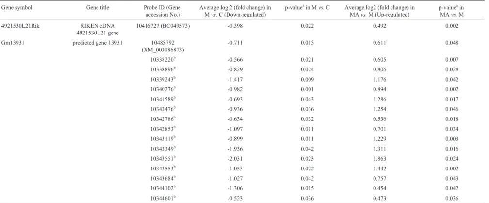
Of the 255 probes down-regulated in the SN region compared to controls, 17 (two annotated genes) were exclu-sively up-regulated following acupuncture at acupoints but not at non-acupoints (“Down-up”; Table 3).
The function of the RIKEN cDNA 4921530L21 gene (4921530L21Rik) has yet to be characterized. The function of the predicted gene 13931 (Gm13931) is known as olfac-tory receptor-related genes. The olfacolfac-tory system may be particularly suitable route for the penetration of xenobiotic agents into the CNS, as was shown in an intranasal MPTP model (Predigeret al., 2009, 2010). Thus, olfactory recep-tor-related genes may be related to the pathological mecha-nisms of MPTP neurotoxicity.
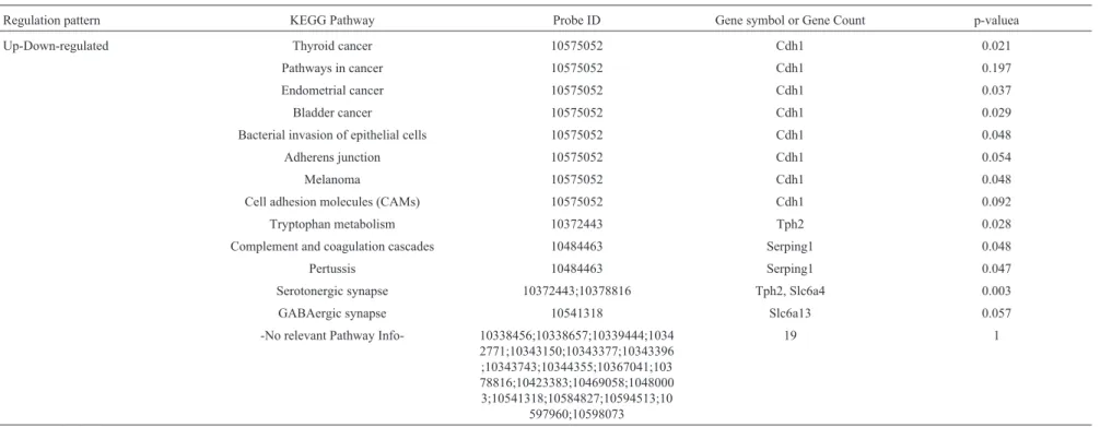
Signal pathway analysis
Pathway analyses for the 22 probes that were up-re-gulated by MPTP intoxication (vs. CTL) and down-regu-lated by acupuncture at acupoints (vs.MPTP), as well as for the 17 probes down-regulated by MPTP intoxication (vs.
CTL) and up-regulated by acupuncture at acupoints (vs.
MPTP; Table 4) were performed. Cdh1, which is “down-up” regulated by MPTP and acupuncture stimulation at acupoints, was involved in “thyroid, endometrial, bladder cancer”, “bacterial invasion of epithelial cells”, and “mela-noma” at significant levels according to the over-repre-sentation analysis (ORA). However, “pathways in cancer”, “adherens junction” and “cell adhesion molecules (CAMs)” were not significant. Tph2 was involved in “tryptophan metabolism” and “serotonergic synapse” at significant levels. Slc6a4 was involved in “serotonergic synapse” at a significant level.Serping1 was involved in “complement and coagulation cascades” and “pertussis” at significant levels.Slc6a13 was involved in “GABAergic synapse”, albeit not significantly so. Gm13931 was in-volved in “olfactory transduction” and was “down-up” reg-ulated by MPTP and acupuncture stimulation at acupoints, even if not significant according to the ORA.
These findings demonstrate that both MPTP and acu-puncture at acupoints influence Cdh1, Tph2, Slc6a4,
Serping1,Slc6a13, andGm13931 expression in the indi-cated pathways in the SN region.
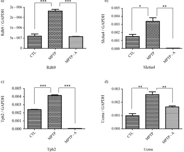
Validation of gene expression changes in the SN region following MPTP and acupuncture at acupoints
122
Yeo
et
al.
Table 3- List of (down-up) substantia nigral genes which were down-regulated in MPTPvs.control and up-regulated in MPTP-Avs.MPTP.
Gene symbol Gene title Probe ID (Gene accession No.)
Average log 2 (fold change) in Mvs.C (Down-regulated)
p-valueain Mvs.C Average log2 (fold change) in MAvs.M (Up-regulated)
p-valueain MAvs.M
4921530L21Rik RIKEN cDNA 4921530L21 gene
10416727 (BC049573) -0.398 0.022 0.492 0.002
Gm13931 predicted gene 13931 10485792 (XM_003086873)
-0.711 0.015 0.611 0.048
10338220b -0.566 0.021 0.605 0.007
10338896b -0.829 0.024 0.806 0.028
10339243b -1.417 0.009 1.176 0.042
10340276b -0.982 0.001 0.894 0.002
10341589b -0.693 0.043 1.286 0.017
10342476b -0.936 0.036 1.254 0.046
10342786b -0.634 0.032 0.536 0.018
10342853b -1.097 0.011 0.701 0.034
10343119b -0.899 0.011 1.229 0.003
10343349b -1.936 0.042 1.311 0.016
10343551b -2.031 0.023 1.863 0.024
10343553b -1.053 0.022 1.442 0.002
10343684b -1.027 0.042 0.757 0.043
10344102b -1.306 0.015 0.454 0.042
10344601b -0.523 0.036 0.473 0.036
a
Determined using Studentt-test.
were evaluated by real time RT-PCR (Figure 3). Expres-sion levels ofRdh9,Slc6a4,Tph2andUcma, which were “up-down” regulated in the microarray analysis, also showed “up-down” regulation in real time RT-PCR (Fig-ure 3). These findings demonstrate that the regulation pat-terns in the SN region regulated by MPTP and acupuncture at acupoints determined by real-time RT-PCR correlate with those determined using microarrays.
Acupuncture stimulation attenuates the reduction of TH induced by 6-OHDA intoxication (Parket al., 2003) and MPTP (Kang et al., 2007; Choi et al., 2011b) in nigrostriatal dopaminergic neurons. Behavioral tests dem-onstrated that acupuncture stimulation improves motor dysfunction in a 6-OHDA Parkinsonism model by ~87.7% (Parket al., 2003). The SN is a crucial aspect of the motor circuit (Brazhniket al., 2012; Gaugleret al., 2012) and is one of the areas damaged most markedly in PD (Castroet al., 2013; Fahimet al., 2013), particularly relative to dys-tonia (Truonget al., 2009).
The neuroprotective effects of acupuncture have been confirmed at a genetic level. The microarray results indi-cate thatSlc6a4andTph2, which are involved in PD, and
Mpzl2andSerping1, which are involved in inflammation,
are primary causes of neurodegenerative disorders (Drou-in-Ouellet and Cicchetti, 2012; Pradhan and Andreasson 2013), and their expression can be maintained at similar to normal levels by acupuncture. Therefore, the effect of MPTP intoxication on gene expression in the SN region may be ameliorated by acupuncture at acupoints. This sug-gests an attenuating effect of acupuncture on the degenera-tion of dopaminergic neuron-like cells by MPTP in the SN. Previous research employing a mouse model of acute MPTP Parkinsonism has demonstrated that acupuncture treatment suppressed genes related to cytokine-cytokine re-ceptor interaction and oxidative phosphorylation pathways (Honget al., 2010). Cellular responses to deleterious events like oxidative stress and cytokine receptor-mediated apoptosis might eventually lead to dopaminergic cell death and hence disease progression. Oxidative stress is an early event that may directly kill dopaminergic neurons (Honget al., 2010). Moreover, the current study showed that at the chronic stage, the expression of genes involved in PD was modulated by acupuncture stimulation. These results sug-gest that a variety of genes may be related to the effect of acupuncture on the treatment of PD.
124
Yeo
et
al.
Table 4- Substantia nigral KEGG pathway list of the 22 probes which were up-regulated in MPTPvs.control and down-regulated only in MPTP-Avs.MPTP, and of the 17 probes which were down-regulated in MPTPvs.control and up-regulated only in MPTP-Avs.MPTP.
Regulation pattern KEGG Pathway Probe ID Gene symbol or Gene Count p-valuea
Up-Down-regulated Thyroid cancer 10575052 Cdh1 0.021
Pathways in cancer 10575052 Cdh1 0.197
Endometrial cancer 10575052 Cdh1 0.037
Bladder cancer 10575052 Cdh1 0.029
Bacterial invasion of epithelial cells 10575052 Cdh1 0.048
Adherens junction 10575052 Cdh1 0.054
Melanoma 10575052 Cdh1 0.048
Cell adhesion molecules (CAMs) 10575052 Cdh1 0.092
Tryptophan metabolism 10372443 Tph2 0.028
Complement and coagulation cascades 10484463 Serping1 0.048
Pertussis 10484463 Serping1 0.047
Serotonergic synapse 10372443;10378816 Tph2, Slc6a4 0.003
GABAergic synapse 10541318 Slc6a13 0.057
-No relevant Pathway Info- 10338456;10338657;10339444;1034 2771;10343150;10343377;10343396 ;10343743;10344355;10367041;103 78816;10423383;10469058;1048000 3;10541318;10584827;10594513;10
597960;10598073
19 1
a
Further studies of the association between gene ex-pression and motor function following acupuncture are warranted, as the investigated genes may mediate the pro-tective effects of acupuncture on SN-mediated motor func-tion. Moreover, the regulation of these genes following acupuncture and their influence on the SN via the involve-ment of abnormal motor circuits should be clarified.
Conclusions
We investigated gene expression changes in the SN using a whole-transcript GeneChip microarray following acupuncture stimulation at acupoints GB34 and LR3 in an MPTP-induced Parkinsonism model. Our data suggest that acupuncture at these acupoints attenuates the decrease in TH in the SN region, while acupuncture at non-acupoints did not suppress this decrease. Compared to the control group, 22 probes (10 annotated genes:Cdh1,Itih2,Mpzl2,
Rdh9, Serping1, Slc6a13, Slc6a20a, Slc6a4, Tph2, and
Ucma) were up-regulated in the MPTP group and were ex-clusively down-regulated by acupuncture at acupoints but not at non-acupoints. Additionally, compared to the control group, 17 probes (two annotated genes; 4921530L21Rik
andGm13931) were down-regulated in the MPTP group and were exclusively up-regulated after acupuncture at acupoints but not at non-acupoints. Therefore, these 39 probes (12 annotated genes) may be responsible for the pro-tective effect of acupuncture in the SN following MPTP-induced impairment.
Acknowledgments
This work was supported by a National Research Foundation of Korea grant funded by the Korean Government [MEST] (No. 2007-0054931, NRF-2014R1A1A1004100). The funders had no role in the study design, data collection and analysis, decision to publish, or preparation of the manuscript.
References
Albani D, Vittori A, Batelli S, Polito L, De Mauro S, Galimberti D, Scarpini E, Lovati C, Mariani C and Forloni G (2009) Se-rotonin transporter gene polymorphic element 5-httlpr in-creases the risk of sporadic Parkinson’s disease in Italy. Eur Neurol 62:120-123.
Alenina N, Kikic D, Todiras M, Mosienko V, Qadri F, Plehm R, Boye P, Vilianovitch L, Sohr R, Tenner K, et al.(2009) Growth retardation and altered autonomic control in mice lacking brain serotonin. Proc Natl Acad Sci USA 106:10332-10337.
An JH, Lee SY, Jeon JY, Cho KG, Kim SU and Lee MA (2009) Identification of gliotropic factors that induce human stem cell migration to malignant tumor. J Proteome Res 8:2873-2881.
Angst BD, Marcozzi C and Magee AI (2001) The cadherin super-family: Diversity in form and function. J Cell Sci 114:629-641.
Balcioglu A, Zhang K and Tarazi FI (2003) Dopamine depletion abolishes apomorphine- and amphetamine-induced in-creases in extracellular serotonin levels in the striatum of conscious rats: A microdialysis study. Neuroscience 119:1045-1053.
Beaulieu JM, Zhang X, Rodriguiz RM, Sotnikova TD, Cools MJ, Wetsel WC, Gainetdinov RR and Caron MG (2008) Role of GSK3 beta in behavioral abnormalities induced by serotonin deficiency. Proc Natl Acad Sci U S A 105:1333-1338. Bezard E, Dovero S, Bioulac B and Gross C (1997) Effects of
dif-ferent schedules of MPTP administration on dopaminergic neurodegeneration in mice. Exp Neurol 148:288-292. Brazhnik E, Cruz AV, Avila I, Wahba MI, Novikov N, Ilieva NM,
McCoy AJ, Gerber C and Walters JR (2012) State-dependent spike and local field synchronization between motor cortex and substantia nigra in hemiparkinsonian rats. J Neurosci 32:7869-7880.
Castro AA, Wiemes BP, Matheus FC, Lapa FR, Viola GG, Santos AR, Tasca CI and Prediger RD (2013) Atorvastatin im-proves cognitive, emotional and motor impairments induced by intranasal 1-methyl-4-phenyl-1,2,3,6-tetrahydropyridine (MPTP) administration in rats, an experimental model of Parkinson’s disease. Brain Res 1513:103-116.
Chatterjee G, Carrithers LM and Carrithers MD (2008) Epithelial v-like antigen regulates permeability of the blood-csf bar-rier. Biochem Biophys Res Commun 372:412-417. Chauhan NB, Siegel GJ and Lee JM (2001) Depletion of glial cell
line-derived neurotrophic factor in substantia nigra neurons of Parkinson’s disease brain. J Chem Neuroanat 21:277-288. Chen NH, Reith ME and Quick MW (2004) Synaptic uptake and beyond: The sodium- and chloride-dependent neurotrans-mitter transporter family SLC6. Pflügers Arch 447:519-531. Choi YG, Park JH and Lim S (2009) Acupuncture inhibits ferric
iron deposition and ferritin-heavy chain reduction in an mptp-induced Parkinsonism model. Neurosci Lett 450:92-96.
Choi YG, Yeo S, Hong YM, Kim SH and Lim S (2011a) Changes of gene expression profiles in the cervical spinal cord by acupuncture in an MPTP-intoxicated mouse model: Microarray analysis. Gene 481:7-16.
Choi YG, Yeo S, Hong YM and Lim S (2011b) Neuroprotective changes of striatal degeneration-related gene expression by acupuncture in an mptp mouse model of Parkinsonism: Microarray analysis. Cell Mol Neurobiol 31:377-391. Cohen SM and Nadler JV (1997) Proline-induced potentiation of
glutamate transmission. Brain Res 761:271-282.
Cunningham RL, Giuffrida A and Roberts JL (2009) Androgens induce dopaminergic neurotoxicity via caspase-3-dependent
activation of protein kinase Cdelta. Endocrinology
150:5539-5548.
Drouin-Ouellet J and Cicchetti F (2012) Inflammation and neuro-degeneration: The story ‘retolled’. Trends Pharmacol Sci 33:542-551.
Emir UE, Tuite PJ and Oz G (2012) Elevated pontine and puta-menal gaba levels in mild-moderate Parkinson disease de-tected by 7 tesla proton MRS. PLoS One 7:e30918. Fahim MA, Shehab S, Nemmar A, Adem A, Dhanasekaran S and
Fernandez-Espejo E (2004) Pathogenesis of Parkinson’s disease: Prospects of neuroprotective and restorative therapies. Mol Neurobiol 29:15-30.
Fiederling A, Ewert R, Andreyeva A, Jungling K and Gottmann K (2011) E-cadherin is required at gabaergic synapses in cul-tured cortical neurons. Neurosci Lett 501:167-172. Gaugler MN, Genc O, Bobela W, Mohanna S, Ardah MT,
El-Agnaf OM, Cantoni M, Bensadoun JC, Schneggenburger R, Knott GW,et al.(2012) Nigrostriatal overabundance of alpha-synuclein leads to decreased vesicle density and defi-cits in dopamine release that correlate with reduced motor activity. Acta Neuropathol 123:653-669.
Goto S, Hirano A and Matsumoto S (1989) Subdivisional involve-ment of nigrostriatal loop in idiopathic Parkinson’s disease and striatonigral degeneration. Ann Neurol 26:766-770. Hebert G, Mingam R, Arsaut J, Dantzer R and Demotes-Mainard
J (2005) A role of IL-1 in MPTP-induced changes in striatal dopaminergic and serotoninergic transporter binding: Clues from interleukin-1 type i receptor-deficient mice. Brain Res Mol Brain Res 136:267-270.
Hirsch EC and Hunot S (2009) Neuroinflammation in Parkinson’s disease: A target for neuroprotection? Lancet Neurol 8:382-397.
Hong MS, Park HK, Yang JS, Park HJ, Kim ST, Kim SN, Park JY, Song JY, Jo DJ, Park SW,et al.(2010) Gene expression pro-file of acupuncture treatment in 1-methyl-4-phenyl-1,2,3,6-tetrahydropyridine-induced Parkinson’s disease model. Neurol Res 32(Suppl 1):74-78.
Hu P, Zhang M and Napoli JL (2007) Ontogeny of rdh9 (crad3) expression: Ablation causes changes in retinoid and steroid metabolizing enzymes, but RXR and androgen signaling seem normal. Biochim Biophys Acta 1770:694-705. Hubscher CH and Berkley KJ (1992) Glial cells: Possible
deter-minants of neuronal uptake of tritiated proline. Neurosci-ence 47:737-743.
Hwang SH, Choi YG, Jeong MY, Hong YM, Lee JH and Lim S (2009) Microarray analysis of gene expression profile by treatment of cinnamomi ramulus in lipopolysaccharide-stimulated BV-2 cells. Gene 443:83-90.
Jankovic J (2008) Parkinson’s disease: Clinical features and diag-nosis. J Neurol Neurosurg Psychiatry 79:68-376.
Kang JM, Park HJ, Choi YG, Choe IH, Park JH, Kim YS and Lim S (2007) Acupuncture inhibits microglial activation and in-flammatory events in the mptp-induced mouse model. Brain Res 1131:211-219.
Kanner BI (2006) Structure and function of sodium-coupled GABA and glutamate transporters. J Membr Biol 213:89-100.
Kim SJ, Sung JY, Um JW, Hattori N, Mizuno Y, Tanaka K, Paik SR, Kim J and Chung KC (2003) Parkin cleaves intracellular alpha-synuclein inclusions via the activation of calpain. J Biol Chem 278:41890-41899.
Kim SN, Doo AR, Park JY, Bae H, Chae Y, Shim I, Lee H, Moon W and Park HJ (2011) Acupuncture enhances the synaptic dopamine availability to improve motor function in a mouse model of Parkinson’s disease. PLoS One 6:e27566. Kowalczuk S, Broer A, Munzinger M, Tietze N, Klingel K and
Broer S (2005) Molecular cloning of the mouse imino sys-tem: An Na+- and Cl—dependent proline transporter. Bio-chem J 386:417-422.
Kuhn DM, Ruskin B and Lovenberg W (1980) Tryptophan hy-droxylase. the role of oxygen, iron, and sulfhydryl groups as determinants of stability and catalytic activity. J Biol Chem 255:4137-4143.
Kuhn DM, Sykes CE, Geddes TJ, Jaunarajs KL and Bishop C (2011) Tryptophan hydroxylase 2 aggregates through disul-fide cross-linking upon oxidation: Possible link to serotonin deficits and non-motor symptoms in Parkinson’s disease. J Neurochem 116:426-437.
Kuhn K, Wellen J, Link N, Maskri L, Lubbert H and Stichel CC (2003) The mouse MPTP model: Gene expression changes in dopaminergic neurons. Eur J Neurosci 17:1-12.
Le Jeune M, Tomavo N, Tian TV, Flourens A, Marchand N, Camuzeaux B, Mallein-Gerin F and Duterque-Coquillaud M (2010) Identification of four alternatively spliced tran-scripts of the ucma/grp gene, encoding a new gla-containing protein. Exp Cell Res 316:203-215.
Lin HQ, Choi R, Chan KL, Ip D, Tsim KW and Wan DC (2010) Differential gene expression profiling on the muscle of acetylcholinesterase knockout mice: A preliminary analysis. Chem Biol Interact 187:120-123.
Liu D, Lu F, Qin G, Fernandes SM, Li J and Davis 3rd AE (2007) C1 inhibitor-mediated protection from sepsis. J Immunol 179:3966-3972.
Melcangi RC, Caruso D, Levandis G, Abbiati F, Armentero MT and Blandini F (2012) Modifications of neuroactive steroid levels in an experimental model of nigrostriatal degenera-tion: Potential relevance to the pathophysiology of Parkin-son’s disease. J Mol Neurosci 46:177-183.
Migliarini S, Pacini G, Pelosi B, Lunardi G and Pasqualetti M (2012) Lack of brain serotonin affects postnatal develop-ment and serotonergic neuronal circuitry formation. Mol Psychiatry 18:1106-1118.
Mikulska JE and Lisowski J (2003) A proline-rich polypeptide complex (prp) from ovine colostrum. Studies on the effect of prp on nitric oxide (NO) production induced by LPS in Thp-1 cells. Immunopharmacol Immunotoxicol 25:645-654.
Moran LB, Duke DC and Graeber MB (2007) The microglial gene regulatory network activated by interferon-gamma. J Neu-roimmunol 183:1-6.
Munck LK and Munck BG (1994) Chloride-dependent intestinal transport of imino and beta-amino acids in the guinea pig and rat. Am J Physiol 266:R997-1007.
Nadler JV, Wang A and Hakim A (1988) Toxicity of l-proline to-ward rat hippocampal neurons. Brain Res. 456:168-172. Pardridge WM (2005) Tyrosine hydroxylase replacement in
ex-perimental Parkinson’s disease with transvascular gene ther-apy. NeuroRx 2:129-138.
Park HJ, Lim S, Joo WS, Yin CS, Lee HS, Lee HJ, Seo JC, Leem K, Son YS, Kim YJ, et al.(2003) Acupuncture prevents 6-hydroxydopamine-induced neuronal death in the nigros-triatal dopaminergic system in the rat Parkinson’s disease model. Exp Neurol 180:93-98.
Pouwels S, Bazelier MT, de Boer A, Weber WE, Neef C, Cooper C and de Vries F (2013) Risk of fracture in patients with Par-kinson’s disease. Osteoporos Int 24:2283-2290.
Pradhan S and Andreasson K (2013) Commentary: Progressive inflammation as a contributing factor to early development of Parkinson’s disease. Exp Neurol 241:148-155.
Prediger RD, Rial D, Medeiros R, Figueiredo CP, Doty RL and Takahashi RN (2009) Risk is in the air: An intranasal MPTP (1-methyl-4-phenyl-1,2,3,6-tetrahydropyridine) rat model of Parkinson’s disease. Ann N Y Acad Sci 1170:629-636. Prediger RD, Aguiar Jr. AS, Rojas-Mayorquin AE, Figueiredo
CP, Matheus FC, Ginestet L, Chevarin C, Bel ED, Mongeau R, Hamon M,et al.(2010) Single intranasal administration
of 1-methyl-4-phenyl-1,2,3,6-tetrahydropyridine in
C57BL/6 mice models early preclinical phase of Parkin-son’s disease. Neurotox Res 17:114-129.
Sakowski SA, Geddes TJ, Thomas DM, Levi E, Hatfield JS and Kuhn DM (2006) Differential tissue distribution of trypto-phan hydroxylase isoforms 1 and 2 as revealed with monospecific antibodies. Brain Res 1085:11-18.
Sato Y, Kaji M, Tsuru T and Oizumi K (2001) Risk factors for hip fracture among elderly patients with Parkinson’s disease. J Neurol Sci 182:89-93.
Scavenius C, Sanggaard KW, Nikolajsen CL, Bak S, Valnickova Z, Thogersen IB, Jensen ON, Hojrup P and Enghild JJ (2011) Human inter-alpha-inhibitor is a substrate for Factor XIIIa and tissue transglutaminase. Biochim Biophys Acta 1814:1624-1630.
Shagdarsuren E, Bidzhekov K, Djalali-Talab Y, Liehn EA, Hristov M, Matthijsen RA, Buurman WA, Zernecke A and Weber C (2008) C1-esterase inhibitor protects against neo-intima formation after arterial injury in atherosclero-sis-prone mice. Circulation 117:70-78.
Surmann-Schmitt C, Dietz U, Kireva T, Adam N, Park J, Taga-riello A, Onnerfjord P, Heinegard D, Schlotzer-Schrehardt U, Deutzmann R,et al.(2008) Ucma, a novel secreted carti-lage-specific protein with implications in osteogenesis. J Biol Chem 283:7082-7093.
Tagariello A, Luther J, Streiter M, Didt-Koziel L, Wuelling M, Surmann-Schmitt C, Stock M, Adam N, Vortkamp A and Winterpacht A (2008) Ucma - a novel secreted factor repre-sents a highly specific marker for distal chondrocytes. Ma-trix Biol 27:3-11.
Truong L, Brooks D, Amaral F, Henderson JM and Halliday GM (2009) Relative preservation of thalamic centromedian nu-cleus in parkinsonian patients with dystonia. Mov Disord 24:2128-2135.
Tufekci KU, Meuwissen R, Genc S and Genc K (2012) Inflamma-tion in Parkinson’s disease. Adv Protein Chem Struct Biol 88:69-132.
Wang H, Katz J, Dagostino P and Soghomonian JJ (2007) Unilat-eral 6-hydroxydopamine lesion of dopamine neurons and subchronic l-dopa administration in the adult rat alters the expression of the vesicular gaba transporter in different sub-sets of striatal neurons and in the substantia nigra, pars reticulata. Neuroscience 145:727-737.
Wojcik E, Carrithers LM and Carrithers MD (2011) Characteriza-tion of epithelial v-like antigen in human choroid plexus epi-thelial cells: Potential role in CNS immune surveillance. Neurosci Lett 495:115-120.
Yeo S, Choi YG, Hong YM and Lim S (2013) Neuroprotective changes of thalamic degeneration-related gene expression by acupuncture in an MTPT mouse model of Parkinsonism: Microarray analysis. Gene 515:329-338.
Zeevalk GD and Nicklas WJ (1996) Attenuation of excitotoxic cell swelling and GABA release by the GABA transport in-hibitor SKF 89976a. Mol Chem Neuropathol 29:27-36. Zeevalk GD and Nicklas WJ (1997) Activity at the GABA
trans-porter contributes to acute cellular swelling produced by metabolic impairment in retina. Vision Res 37:3463-3470. Zhuang R, Lin M and Napoli JL (2002) Cis-retinol/androgen
dehydrogenase, isozyme 3 (crad3): Ashort-chain dehydro-genase active in a reconstituted path of 9-cis-retinoic acid biosynthesis in intact cells. Biochemistry 41:3477-3483.
Supplementary Material
The following online material is available for this article: Table S1 - Up-regulated genes in MPTP-treated substantia nigral region.
Table S2 - Down-regulated genes in MPTP-treated sub-stantia nigral region.
Figure S1 - Box plot before and after normalization. This material is available as part of the online version of this article from http://www.scielo.br/gmb.
Associate Editor: Adriana S. Hemerly