Abstract
Submitted: December 30, 2016 0RGL¿FDWLRQ0DUFK Accepted: April 19, 2017
Effects of triethylene glycol
dimethacrylate (TEGDMA) on the
odontoclastic differentiation ability of
human dental pulp cells
Objectives: The primary purpose of this study was to examine the effects of triethylene glycol dimethacrylate (TEGDMA) on odontoclastic differentiation in the dental pulp tissue. Material and Methods: The effects of different TEGDMA dosages on the odontoclastic differentiation capability of dental pulp cells were analyzed in vit ro using the following methodologies:
ii) apoptotic effects using Annexin V staining; iii) mRNA expression of osteoprotegerin (OPG) and receptor activator of nuclear factor (NF)-kB ligand (RANKL) genes by quantitative Real-time PCR (qRT-PCR); and iv) OPG and RANKL protein expression by enzyme-linked immunosorbent assay (ELISA). Results: TEGDMA caused relatively less odontoclastic differentiation in comparison with the control group; however, odontoclastic differentiation augmented with increasing doses of TEGDMA (p<0.05). The mRNA and protein expression of OPG was lower in TEGDMA treated pulp cells than in the control group (p<0.05). While the mRNA expression of RANKL remained unchanged compared to the control group (p>0.05), its protein expression was higher than the control group (p<0.05). In addition, TEGDMA increased the apoptosis of dental pulp cells dose dependently. Conclusions: TEGDMA reduced the odontoclastic differentiation ability of human dental pulp cells. However, odontoclastic differentiation ratios increased proportionally with the increasing dose of TEGDMA.
Ke yw or ds: Human dental pulp cell. Odontoclast. OPG. RANKL. TEGDMA.
Zeynep ÖNCEL TORUN1
Deniz TORUN2
%DUÕú%$<.$/3
Ali ÖZTUNA2
)DWLK<(ùø/'$/4
Ferit AVCU5
http://dx.doi.org/10.1590/1678-7757-2016-0626
1Balgat Oral and Dental Health Center, Ankara, Turkey.
2University of Health Sciences, Gulhane Faculty of Medicine, Department of Medical Genetics,
Ankara, Turkey.
3University of Health Sciences, Gulhane Faculty of Medicine, Department of Histology and
Embryology, Ankara, Turkey.
4'L\DUEDNÕU6HODKDGGLQ(\\XEL3XEOLF+RVSLWDO'HSDUWPHQWRI0HGLFDO%LRFKHPLVWU\'L\DUEDNÕU
Turkey.
5Memorial Ankara Hospital, Ankara, Turkey.
Corresponding address: Zeynep Öncel Torun Balgat Oral and Dental Health Center, 06520. Ankara - Turkey. Phone: +90 312 2842679 - Fax: +90 312 2841631.
Introduction
Triethylene glycol dimethacrylate (TEGDMA) is
a resin monomer widely used in the composition
of dentin bonding agents and composite resins to
restore teeth structures impaired by caries and/or
fractures. However, resin monomers can be released
into the oral environment and can trigger hazardous
biological effects on oral tissues2. The release of the
resin monomers due to degradation and incomplete
polymerization can occur hours or days after the
treatment7. Due to its hydrophilic nature, hydrolysis
plays an important role in the degradation processes
of TEGDMA24
of resin monomers26. Direct contact or diffusion of
resin monomers through the dentinal tubules creates
ways of interaction between dental pulp tissue and
resin monomers. Dentin thickness and the severity
of caries lesions are important factors in determining
the amount of resin monomers interacting with
dental pulp tissue6,12. TEGDMA has been reported to
cause cytotoxicity, impaired cellular functions, pulpal
system2,6,14,19,27. In addition, TEGDMA may reduce
the mineralization capacity of dental pulp cells by
decreasing the expression of the mineralization related
genes11
pathways and causing adverse effects.
Odontoclasts are key regulators controlling
the eruption of deciduous teeth, and they show
similar biological features to osteoclasts22,25. Besides,
odontoclasts have important functions in pathological
resorption of permanent teeth32. OPG/RANK/RANKL
signaling pathway is known to play a key role in
the differentiation of osteoclasts, which is strictly
controlled by osteoblasts29. Osteoblasts/stromal cells
express the receptor activator of nuclear factor (NF)-kB
dihydroxyvitamin D3 2D3), parathyroid
hormone (PTH), IL-6, and IL-11. The activation of
RANKL with the receptor activator of NF-kB (RANK),
present on the surface of osteoclastic precursor
cells, stimulates the osteoclastic differentiation from
monocyte or macrophage progenitors. Osteoprotegerin
(OPG) is a member of the tumor necrosis factor (TNF)
receptor superfamily, which prevents RANK mediated
activation of osteoclastic differentiation by binding to
RANKL29.
Periodontal ligament fibroblasts are known to
contribute to osteoclast formation and promote
destructive inflammatory periodontal diseases28.
Interactions with bacteria or mechanical loading play
a key role during this process. Moreover, Uchiyama, et
al.31 (2009) has shown that dental pulp and periodontal
ligament cells support the differentiation and function
of osteoclasts. Recently, Inamitsu, et al.16 (2017)
reported that 2-hydroxyethyl methacrylate (HEMA) and
TEGDMA inhibited the osteoclast differentiation through
different signaling pathways in bone marrow-derived
macrophages (BMMs) and murine monocytic cell line
RAW-D. The role of periodontal ligament and pulp cells
on osteoclastogenesis and the effects of different resin
monomers on osteoclast formation have been shown
in various studies. However, there are no studies so
far on the odontoclastogenic activity of pulp cells in
the presence of TEGDMA. The null hypothesis is that
differentiation ability of human dental pulp cells.
Therefore, this study was carried out to determine if
TEGDMA promotes the odontoclastic differentiation
ability of human dental pulp cells (hDPCs). Thus,
we evaluated the effects of TEGDMA on the ability
of hDPCs to produce odontoclasts in the presence of
CD14+ odontoclastic precursor cells derived from human
peripheral blood.
Material and Methods
This study was conducted in full accordance with
the World Medical Association Declaration of Helsinki
and was approved by the Local Ethics Committee of
the Gulhane Military Medical Academy (32/2014). All
participants provided informed consent forms.
Cell culture
Dental pulp tissues were obtained from the molars of
The extracted molars were kept in phosphate-buffered
saline solution (PBS, Biological Industries, Kibbutz Beit
Haemek, Israel) containing 100 U/mL penicillin, and
100 μg/mL streptomycin (Biological Industries, Kibbutz
Beit Haemek, Israel). After they were transferred to the
laboratory, the extracted molars were cut horizontally
at 1 mm below the cementoenamel junction. The
and root and placed in a 100-mm Petri dish. The pulp
tissues were cut into small pieces with a sterile blade
Grand Island, NY, USA) containing 10% fetal bovine
serum (FBS; Biological Industries, Kibbutz Beit
Haemek, Israel), 100 U/mL penicillin, and 100 μg/mL
streptomycin (Biological Industries). Tissue cultures
CO2
for the experiments.
Generation of CD14
+cells
Histopaque–1077 (Sigma-Aldrich, St. Louis,
MO, USA) was used to separate peripheral blood
mononuclear cells (PBMC) from human peripheral
blood by density-gradient centrifugation at 1800 rpm
for 20 min. CD14+ cells were obtained from the PBMC
with CD14 Microbeads (Miltenyi Biotec GmbH, Bergisch
Gladbach, Germany) and SuperMACSTM II Separator
(Miltenyi Biotec GmbH, Bergisch Gladbach, Germany),
in accordance with the manufacturer instructions.
Generation of odontoclasts and designation of
study groups
The following study groups were designed to analyze
odontoclastic differentiation:
Human dental pulp cells (hDPCs);
hDPCs + CD14+ cells;
hDPCs + 1D,25(OH)2D3;
hDPCs + CD14+
2D3;
hDPCs + CD14+ cells + TEGDMA;
2D3 + TEGDMA; and
hDPCs + CD14+
2D3, +TEGDMA.
Each experimental group consisted of nine samples.
hDPCs were seeded into 6-well plates at a concentration
4
Technologies, Grand Island, NY, USA) containing 10%
FBS (Biological Industries, Kibbutz Beit Haemek, Israel),
100 U/mL penicillin, and 100 μg/mL streptomycin
(Biological Industries, Kibbutz Beit Haemek, Israel).
+ cells
were used as odontoclast precursors and co-cultured
5 cells/well. The co-culture was
performed in the presence or absence of 1D,25(OH)2D3
(10–8 M) (Sigma-Aldrich, St. Louis, MO, USA) and
TEGDMA (0.1, 0.3, 1, and 3 mM). The concentration
2D3 was chosen from a previously
reported study21. Culture conditions were maintained
2 at
37°C, with medium change every 3 days.
Flow cytometry
Flow cytometry was used to evaluate odontoclastic
differentiation after 14 days of incubation. After
day 14, cells were suspended in 100 μL PBS, and
stained with 10 μL anti-human CD51/CD61 mouse
monoclonal antibody (BD Biosciences, USA), which
bind selectively to odontoclasts, and incubated for 30
min at room temperature. After incubation, samples
were centrifuged at 300 g for 5 min. Pellets were
re-μ
cytometry (FACSDiva software, FACSAria, USA).
TRAP staining
solution (Sigma-Aldrich, St. Louis, MO, USA). After
serial washing with PBS three times, odontoclasts
were detected by staining for tartrate-resistant
acid phosphatase (TRAP), a marker enzyme for
odontoclasts, using a kit following the manufacturer’s
recommendations (Cosmo Bio Co., Ltd., Tokyo, Japan).
TRAP activity was observed under an optical microscope
Germany) and TRAP-positive cells were counted.
Apoptosis assay
5 cells/
well density and exposed to serial dilutions of TEGDMA
(0.1, 0.3, 1, and 3 mM) for 24 h. Apoptosis in hDPCs was
Staining Kit (Roche, Mannheim, Germany) following the
manufacturer’s instructions. Each experimental group
consisted of nine samples.
Quantitative Real-time PCR (qRT-PCR) analysis
6 cells/of 5% CO2 at 37°C (n=3 per experimental group). The cell cultures were exposed to serial dilutions of TEGDMA
(0.1, 0.3, and 1 mM) for seven days. Total RNA was
extracted from cultured hDPCs using an RNA isolation
kit (High Pure RNA Isolation Kit, Roche, Mannheim,
Germany), and cDNA was synthesized from 100 ng
of total RNA using the Transcriptor High Fidelity cDNA
Synthesis Kit (Roche, Mannheim, Germany). The cDNA
obtained was used as a template for PCR.
Human RANKL and OPG mRNA levels were analyzed
to evaluate the effects of TEGDMA on odontoclastic
differentiation capability of hDPCs.
Glucose-6-phosphate dehydrogenase (G6PD) was used as a
the relative expression levels of RANKL and OPG.
Probe–primer pairs for target genes were purchased
from Roche Diagnostic as RealTime ready assays, i.e.
RANKL (Cat. no. 144633), OPG(Cat. no.142904), and
G6PD (Cat. no.102098). Real-time PCR was performed
using the FastStart Essential DNA Probes Master
(Roche, Mannheim, Germany) and a LightCycler® 480
Instrument II (Roche, Mannheim, Germany). Real-time
PCR conditions were as follows: denaturation at 95°C
for 10 min, followed by 45 cycles of 95°C for 10 s, 60°C
for 30 s, 72°C for 1 s, and cooling at 40°C. The mRNA
method. Each experiment was performed in triplicate.
Enzyme-linked immunosorbent assay (ELISA)
6 cells/of 5% CO2 at 37°C (n=9 per experimental group).
The cell cultures were exposed to serial dilutions of
TEGDMA (0.1, 0.3, and 1 mM) for seven days. Protein
concentrations of OPG and RANKL in the cell culture
supernatant were analyzed by ELISA. The ELISA kits
(MyBioSource Inc., San Diego, CA, USA) employed
a sandwich ELISA technique containing a capture
antibody and label antibody. All steps in the manuals
were followed during the analysis and results were
read on an ELISA plate reader (Reader ELx 800, Biokit,
they were stated as <15% for OPG in the kit manual).
Statistical analysis
The Kolmogorov-Smirnov test of distribution
indicated that data for anti-CD51/CD61 positivity,
TRAP positivity, apoptosis ratio, mRNA, and protein
data for RANKL and OPG were skewed. Thus, results
were summarized as medians and interquartile ranges.
The Kruskal-Wallis test was used to compare three
was established at 0.05. SPSS Statistical Package
Version 20.0 (IBM, Chicago, IL, USA) was used for all
calculations.
Results
Analysis of odontoclastic differentiation
Odontoclastic differentiation ratios in the different
groups were analyzed after 14 days of incubation
CD14+ odontoclast precursor cells did not generate
odontoclasts, as expected (data not shown). Co-culture
of hDPCs and CD14+ cells (group ii) revealed the
highest odontoclastic differentiation ratio. Addition of
2D3 decreased the odontoclastic differentiation
ratio, particularly in hDPCs + CD14+ cells + 0.3 mM
TEGDMA and hDPCs + CD14+ cells + 1 mM TEGDMA
groups (p<0.05). No such changes were seen in hDPCs
+ CD14+ and hDPCs + CD14+ cells + 0.1 mM TEGDMA
groups (p>0.05). In general, the addition of TEGDMA
lowered odontoclastic differentiation relative to groups
without TEGDMA (p<0.05). Notably, 1 mM TEGDMA
caused higher odontoclastic differentiation relative to
0.1 and 0.3 mM TEGDMA (p<0.05). No cell viability/
odontoclastic differentiation was studied in the 3 mM
TEGDMA group due to cytotoxicity (data not shown).
Odontoclast cell formation was confirmed by
counting TRAP-positive cells, which phenotypically
assessed odontoclastic differentiation (Figure 2).
1). However, 95±2% of TRAP-positive cells were
mononuclear, and we did not detect any TRAP-positive
cells in CD14+ negative groups.
Effect of TEGDMA on the apoptosis of dental
pulp cells
hDPCs were exposed to 0.1, 0.3, 1, and 3 mM
TEGDMA for 24 h, and it was found that TEGDMA
dose-dependently induced apoptosis (Figure 3).
Except for the lowest dose of 0.1 mM TEGDMA, the
higher doses of 0.3, 1, and 3 mM TEGDMA increased
apoptosis in comparison to the control group (p<0.001).
Furthermore, there was a dose-dependent effect of
TEDGMA on apoptosis induction in hDPCs.
Expression of RANKL and OPG mRNA in dental
pulp cells
No differences were observed among the TEGDMA
containing groups (0.1, 0.3, 1) in terms of RANKL mRNA
levels after 7 days of incubation (p>0.05) (Figure 4a).
However, OPG mRNA levels were lower in the TEGDMA
containing groups relative to the control group, and
TEGDMA treatments demonstrated a dose-dependent
decrease (p<0.05) (Figure 4b). RANKL and OPG mRNA
levels could not be detected in the 3 mM TEGDMA group
RANKL and OPG protein levels in dental pulp
cells
RANKL protein levels were higher in the TEGDMA
treated hDPCs regarding the control group (p<0.05)
(Figure 5a), and TEGDMA treatments demonstrated
a dose-dependent increase. There was a statistically
and 3 mM TEGDMA containing groups (p<0.05).
OPG protein levels were lower in all TEGDMA treated
groups regarding the control group (p<0.05) (Figure
5b). In particular, the 0.1 mM TEGDMA group had
relatively lower OPG protein levels compared to 0.3
and 1 mM TEGDMA groups (p<0.05). There were no
Study Groups (n=9) Median (Min-max) *(p<0.05) 1 hDPCs + CD14 2465 (1065-3100) 1-2, 1-3, 1-4, 1-5, 1-6, 1-7, 1-8 2 hDPCs + CD14 + Vit D3 1043 (958-3742) 2-3, 2-4, 2-5, 2-6, 2-8
3 hDPCs + CD14 + 0.1 mM TEGDMA 260 (101-369) 3-6, 3-7, 3-8 4 hDPCs + CD14 + 0.1 mM TEGDMA + Vit D3 297 (98-456) 4-6, 4-7, 4-8
5 hDPCs + CD14 + 0.3 mM TEGDMA 276 (101-395) 5-6, 5-7, 5-8 6 hDPCs + CD14 + 0.3 mM TEGDMA + Vit D3 144 (108-165) 6-7, 6-8
7 hDPCs + CD14 + 1 mM TEGDMA 905 (760-1010) 7-8 8 hDPCs + CD14 + 1 mM TEGDMA + Vit D3 640 (586-750)
6WXG\JURXSVWKDWUHYHDOVWDWLVWLFDOGLIIHUHQFHVIURPHDFKRWKHUDFFRUGLQJWR.UXVNDO:DOOLVWHVW
Table 1- Number of TRAP-positive cells generated by the co-colturing of human CD14+ cells and hDPCs in the presence or absence of Vit D3 and TEGDMA
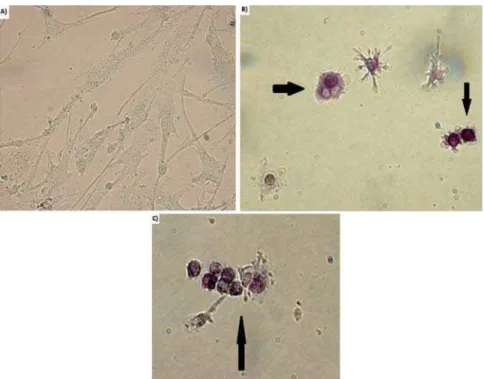
Figure 2-$QDO\VLVRIRGRQWRFODVWLFGLIIHUHQWLDWLRQE\75$3VWDLQLQJXQGHUDQRSWLFDOPLFURVFRSHZLWKîPDJQL¿FDWLRQ75$3SRVLWLYH cells were observed through the co-culture of CD14+ cells and hDPCs at the end of 14 days incubation period. a) TRAP-negative b, c)
TRAP-positive cells
(p>0.05).
Discussion
Resin monomers cause adverse biological effects by
modulating different regulatory cellular mechanisms.
Previous studies have shown that TEGDMA can disrupt
mineralization capacity leading to cytotoxicity in
hDPCs1,6,11,18,20. Recently, it has been shown that dental
pulp cells support the differentiation and function of
odontoclasts31. However, the effect of TEGDMA on
the ability of dental pulp cells to induce odontoclastic
differentiation has not yet been determined. In this
study, our aim was to evaluate the cellular processes
related to odontoclastic differentiation in
TEGDMA-treated hDPCs. We anticipated that any information
about the effects of TEGDMA on odontoclastic
differentiation would provide a better understanding
on the adverse effects of TEGDMA.
In this study, we attempted to determine the
effects of TEGDMA on the generation of odontoclasts.
This was carried out by co-culturing hDPCs and CD14+
monocytes derived from human peripheral blood. We
conducted experiments in the presence or absence
of 1D,25(OH)2D3,followed by incubating with serial
cytometry analysis using anti-human CD51/CD61
mouse monoclonal antibody was consistent with
TRAP staining. However, TRAP-positive multinuclear
cells constituted only 5±2% of all TRAP-positive
cells. Previously, Domon, et al.10 (1997) reported that
both mononuclear and multinucleated odontoclasts
participate in tooth eruption, and further suggested
that not all odontoclasts are multinucleated cells.
Further, Hattersley & Chambers13 (1989) suggested that
osteoclasts are initially mononuclear and might remain
so, but later they become multinucleated. Moreover,
odontoclastic cells can become multinucleated by
cell fusion, and the formation of multinucleated
odontoclasts has been described as an instantaneous
event10. However, this study failed to observe a high
proportion of multinuclear TRAP-positive cells and most
of the odontoclasts were mononuclear. Although we do
not have any evidence, we speculate that short culture
time and higher biological activity of TEGDMA may
have contributed to the high proportion of mononuclear
TRAP-positive cells. Further, extending the culture
period may increase the fusion of mononuclear cells.
The study groups without CD14+ cells did not reveal
any odontoclastic differentiation. This observation
was consistent with a previously reported study and
reinforces the fact that odontoclastic precursor cells are
required for odontoclastic differentiation31. Co-culture of
hDPCs and CD14+ cells led to the highest odontoclastic
differentiation ratio, and an addition of TEGDMA to the
culture medium reduced odontoclastic differentiation
ratios. Notably, the observed suppressive effect of
TEGDMA was attenuated at higher concentrations. A
reduction in the odontoclastic differentiation in the
presence of lower doses of TEGDMA indicates that
treatment with dental materials containing resin
monomers may diminish the potential to induce
odontoclastic resorption. However, a dose-dependent
increase in the odontoclastic differentiation suggests
that circumstances that lead to an increase in the
release of resin monomers into oral environment, such
as mechanical/chemical degradation or inadequate
polymerization, may increase the possibility of
odontoclastic resorption. Based on our observations,
further studies are essential to explore the clinical
effects of TEGDMA related odontoclastic differentiation.
It is known that 1D,25(OH)2D3 plays a key role in
both odontoblastic and odontoclastic differentiation4.
Normal levels of vitamin D and its active metabolite
1D,25(OH)2D3 are important for bone mineralization.
However, increased 1D,25(OH)2D3 enhances bone
degradation by acting on osteoblasts, causing
them to release RANKL, which in turn activates
osteoclasts. Besides that, Kim, et al.17 (2013)
reported that 1D,25(OH)2D3 inhibits the osteoclast
differentiation by suppressing the expression of RANK
in the human peripheral blood osteoclast precursors.
Although methodologies and applied concentrations
of 1D,25(OH)2D3 are different, this study is generally
consistent with the aforementioned study17. This
study revealed that generation of odontoclasts was
D,25(OH)2D3 in most of
the study groups at a concentration of 10-8 M.This
result indicates that 10-8 M of 1D,25(OH)
2D3 causes a
decrease in the odontoclastic differentiation ability of
hDPCs, and may further suppress potentially adverse
biologic effects of TEGDMA. On the contrary, Takahashi,
et al.30 (2014) reported that 1D,25(OH)
2D3 induces
bone resorption and osteoclastogenesis by stimulating
RANKL expression. We think that differences related
to experimental design, cell types used, applied
concentrations of 1D,25(OH)2D3, and differential
biological activity of TEGDMA on dental pulp tissue may
help explain the contradictory results. Even though we
could not provide any evidence of a direct relationship
between different 1D,25(OH)2D3 concentrations and
quantity of odontoclastic differentiation, we speculate
that dose changes in 1D,25(OH)2D3 may promote the
odontoclastic differentiation ability of hDPCs based
on previous reports. Therefore, further studies are
necessary with different doses of 1D,25(OH)2D3
to evaluate the phenotypic effects of TEGDMA on
odontoclastic differentiation.
RANKLplays an important role in bone regeneration
and remodeling by activating osteoclasts/odontoclasts.
Previous studies have shown that TEGDMA is a
biologically active chemical and interacts with living
8.
Although TEGDMA did not affect the expression of
RANKL mRNA, the RANKL protein was increased
by TEGDMA in hDPCs. Therefore, TEGDMA may be
affecting post-transcriptional regulation of gene
expression. Notably, RANKL seems to be an important
regulatory factor in odontoclastic differentiation of
TEGDMA treated hDPCs. OPG is known as the main
inhibitor of the osteoclastic/odontoclastic differentiation
process. Our results indicate that TEGDMA decreased
the expression of OPG mRNA and protein in a
dose-dependent manner. This effect was consistent with our
indicated an increase in odontoclastic differentiation
with an increased dose of TEGDMA. However, when
all results were taken into account, there seems to
be a discrepancy between them. Increase of RANKL
and decrease of OPG proteins are supposed to induce
odontoclastic differentiation. However, the number
of anti-human CD51/CD61 and TRAP positive cells
were decreased by TEGDMA treatment, compared
to control, as shown in Figure 1 and Table 1. In this
study, we examined the effects of TEGDMA on the
RANKL and OPG proteins, which were upstream of the
odontoclastic differentiation pathway,. However, there
are several downstream signaling pathways related
Akt, ERK, JNK, and p38 MAPKs5,9,23,33. Additional effects
of TEGDMA on these different signaling pathways may
contribute to the effects of TEGDMA on the odontoclastic
differentiation ability of hDPCs and may be the cause
of this inconsistency between the results.
Recently, Inamitsu, et al.16 (2017) reported that HEMA
and TEGDMA inhibited the osteoclast differentiation in
BMMs and RAW-D cells. Furthermore, the study also
showed that NFATc1, ERK, and JNK signaling pathways
played a key role in osteoclastogenesis for the HEMA,
whereas NFATc1, Akt, and JNK pathways associated
with the TEGDMA mediated osteoclastogenesis. The
results seem similar in some aspects and support
between the two studies. Unlike this study, Inamitsu,
et al.16 (2017) reported a decrease in the osteoclastic
differentiation of BMMs with the increasing dose of
TEGDMA in TRAP analysis. However, odontoclastic
differentiation ratios revealed an augmentation with
increasing dose of TEGDMA in this study. Differences
between the experimental designs may be the cause
16
(2017) counted only the cells with three or more nuclei
as TRAP-positive, whereas we took into account all the
mononuclear and multinucleated cells during the TRAP
analysis. Cell types and duration of the culture time
used to monitoring osteoclastic differentiation were also
different and this phenomenon may be an important
factor for the osteoclastic differentiation of the cells.
Furthermore, Inamitsu, et al.16 (2017) showed that
TEGDMA has slight cytotoxic effects on BMM-derived
osteoclasts. When we examined the results of the study,
cytotoxicity was found to be mild at doses up to 1 mM,
but cytotoxicity gradually increased at doses between
1 and 2 mM. Moreover, Inamitsu, et al.16 (2017) did not
test the more cytotoxic doses of TEGDMA such as 3 mM
as this study. It is to be noted that in the presence of a
high dose of TEGDMA (3 mM) no results were obtained
analyses. Consistent with previous studies indicating
that TEGDMA can activate apoptotic pathways3,15, this
study revealed that TEGDMA caused a dose-dependent
increase in apoptosis of hDPCs and cell viability has
been adversely affected in the presence of high doses
of TEGDMA. Therefore, it has not been possible to carry
out studies on the effects of high doses of TEGDMA on
odontoclastic differentiation ability of hDPCs.
Resin composites are widely used in dentistry and
many factors such as secondary caries, defects in
restorations, marginal degradation, and pulpal death
may cause the replacement of restorations or fail of the
treatment. A detailed understanding of biological effects
of resin monomers is important to increase treatment
or loss of the tooth. Therefore, the inhibitory effects
of resin monomers on odontoclastic differentiation
ability of hDPCs may help understand the effects of
resin monomers on the longevity of restorations and
Conclusion
We found that TEGDMA reduces the odontoclastic
differentiation ability of hDPCs. However, odontoclastic
differentiation ratios augmented with increasing dose
of TEGDMA.
Acknowledgements
and Technological Research Council of Turkey (Project
number: 114S761).
References
P, et al. Effects of HEMA and TEDGMA on the in vit r o odontogenic differentiation potential of human pulp stem/progenitor cells derived
from deciduous teeth. Dent Mater. 2011;27:608-17.
substances released from resin-based dental restorative materials. Int J Mol Sci. 2009;10:3861-99.
3- Batarseh G, Windsor LJ, Labban NY, Liu Y, Gregson K. Triethylene
Oper Dent. 2014;39:E1-8.
4- Bell TD, Demay MB, Burnett-Bowie SA. The biology and pathology of
vitamin D control in bone. J Cell Biochem. 2010;111:7-13.
5- Boyle WJ, Simonet WS, Lacey DL. Osteoclast differentiation and
activation. Nature. 2003;423:337-42.
6- Chang HH, Chang MC, Huang GF, Wang YL, Chan CP, Wang TM,
et al. Effect of triethylene glycol dimethacrylate on the cytotoxicity, cyclooxygenase-2 expression and prostanoids production in human
dental pulp cells. Int Endod J. 2012;45:848-58.
7- Chang HH, Chang MC, Lin LD, Lee JJ, Wang TM, Huang CH, et al.
The mechanisms of cytotoxicity of urethane dimethacrylate to Chinese hamster ovary cells. Biomaterials. 2010;31:6917-25.
8- Cho SG, Lee JW, Heo JS, Kim SY. Gene expression change in human dental pulp cells exposed to a low-level toxic concentration of triethylene glycol dimethacrylate: an RNA-seq analysis. Basic Clin Pharmacol
Toxicol. 2014;115:282-90.
9- Darnay BG, Ni J, Moore PA, Aggarwal BB. Activation of NF-kappaB by
RANK requires tumor necrosis factor receptor-associated factor (TRAF)
interaction motif. J Biol Chem. 1999;274:7724-31.
10- Domon T, Osanai M, Yasuda M, Seki E, Takahashi S, Yamamoto T,
et al. Mononuclear odontoclast participation in tooth resorption: the distribution of nuclei in human odontoclasts. Anat Rec.
1997;249:449-57.
11- Galler KM, Schweikl H, Hiller KA, Cavender AC, Bolay C, D'Souza
RN, et al. TEGDMA reduces mineralization in dental pulp cells. J Dent Res. 2011;90:257-62.
12- Hamid A, Hume WR. The effect of dentine thickness on diffusion of resin monomers in vit ro. J Oral Rehabil. 1997;24:20-5.
13- Hattersley G, Chambers TJ. Generation of osteoclastic function in mouse bone marrow cultures: multinuclearity and tartrate-resistant
acid phosphatase are unreliable markers for osteoclastic differentiation. Endocrinology. 1989;124:1689-96.
14- Hebling J, Giro EM, Costa CA. Human pulp response after an adhesive system application in deep cavities. J Dent. 1999;27:557-64.
15- Huang FM, Kuan YH, Lee SS, Chang YC. Cytotoxicity and genotoxicity of triethyleneglycol-dimethacrylate in macrophages involved in DNA
damage and caspases activation. Environ Toxicol. 2015;30:581-8. 16- Inamitsu H, Okamoto K, Sakai E, Nishishita K, Murata H, Tsukuba
T. The dental resin monomers HEMA and TEGDMA have inhibitory effects on osteoclast differentiation with low cytotoxicity. J Appl Toxicol.
2017;37:817-24.
17- Kim TH, Lee B, Kwon E, Choi CH, Sung IH, Kim Y, et al. 1,25
dihydroxyvitamin D3 inhibits directly human osteoclastogenesis by down-regulation of the c-Fms and RANK expression. Joint Bone Spine.
2013;80:307-14.
18- Krifka S, Seidenader C, Hiller KA, Schmalz G, Schweikl H. Oxidative
stress and cytotoxicity generated by dental composites in human pulp cells. Clin Oral Investig. 2012;16:215-24.
19- Krifka S, Spagnuolo G, Schmalz G, Schweikl H. A review of adaptive mechanisms in cell responses towards oxidative stress caused by dental resin monomers. Biomaterials. 2013;34:4555-63.
20- Kwon JH, Park HC, Zhu T, Yang HC. Inhibition of odontogenic differentiation of human dental pulp cells by dental resin monomers.
Biomater Res. 2015;19:8.
21- Lacey DL, Timms E, Tan HL, Kelley MJ, Dunstan CR, Burgess T,
et al. Osteoprotegerin ligand is a cytokine that regulates osteoclast differentiation and activation. Cell. 1998;93:165-76.
22- Lossdörfer S, Götz W, Jäger A. Immunohistochemical localization of receptor activator of nuclear factor kappaB (RANK) and its ligand
(RANKL) in human deciduous teeth. Calcif Tissue Int. 2002;71:45-52. 23- Matsumoto M, Sudo T, Saito T, Osada H, Tsujimoto M. Involvement
of p38 mitogen-activated protein kinase signaling pathway in osteoclastogenesis mediated by receptor activator of NF-kappa B ligand
(RANKL). J Biol Chem. 2000;275:31155-61.
24- Ortengren U, Wellendorf H, Karlsson S, Ruyter IE. Water sorption
released in an aqueous environment. J Oral Rehabil. 2001;28:1106-15.
25- Sahara N, Toyoki A, Ashizawa Y, Deguchi T, Suzuki K. Cytodifferentiation of the odontoclast prior to the shedding of human
deciduous teeth: an ultrastructural and cytochemical study. Anat Rec. 1996;244:33-49.
26- Santerre JP, Shajii L, Tsang H. Biodegradation of commercial dental composites by cholesterol esterase. J Dent Res. 1999;78:1459-68.
T, et al. Differential gene expression involved in oxidative stress
response caused by triethylene glycol dimethacrylate. Biomaterials. 2008;29:1377-87.
28- Sokos D, Everts V, de Vries TJ. Role of periodontal ligament fibroblasts in osteoclastogenesis: a review. J Periodontal Res.
2015;50:152-9.
29- Suda T, Takahashi N, Udagawa N, Jimi E, Gillespie MT, Martin
TJ. Modulation of osteoclast differentiation and function by the new members of the tumor necrosis factor receptor and ligand families.
Endocr Rev. 1999;20:345-57.
30- Takahashi N, Udagawa N, Suda T. Vitamine D endocrine system
and osteoclasts. Bonekey Rep. 2014;3:495.
31- Uchiyama M, Nakamichi Y, Nakamura M, Kinugawa S, Yamada H,
Udagawa N, et al. Dental pulp and periodontal ligament cells support osteoclastic differentiation. J Dent Res. 2009;88:609-14.
32- Wise GE, King GJ. Mechanisms of tooth eruption and orthodontic tooth movement. J Dent Res. 2008;87:414-34.
33- Zhang YH, Heulsmann A, Tondravi MM, Mukherjee A, Abu-Amer Y. Tumor necrosis factor-alpha (TNF) stimulates RANKL-induced