OCCURRENCE OF AFLATOXIN B1 IN NATURAL PRODUCTS
Guilherme Prado1*; Aline F. Altoé2; Tatiana C. B. Gomes3; Alexandre S. Leal4; Vanessa A. D. Morais1; Marize S. Oliveira1; Marli B. Ferreira1; Mateus B. Gomes1; Fabiano N. Paschoal1; Rafael von S. Souza1; Daniela A. Silva1; Jovita E.
G. Cruz Madeira1
1
Laboratório de Micotoxinas, Fundação Ezequiel Dias, Belo Horizonte, MG, Brasil; 2Programa de Iniciação Científica
Institucional, Fundação de Amparo à Pesquisa do Estado de Minas Gerais, Belo Horizonte, MG, Brasil; 3Programa de Iniciação
Científica Institucional, Fundação de Amparo à Pesquisa do Estado de Minas Gerais, Centro de Desenvolvimento da Tecnologia
Nuclear, Belo Horizonte, MG, Brasil; 4 Centro de Desenvolvimento da Tecnologia Nuclear, Belo Horizonte, MG, Brasil.
Submitted: December 09, 2010; Returned to authors for corrections: March 13, 2012; Approved: June 07, 2012.
ABSTRACT
The media claims for the consumption of natural resource-based food have gradually increased in both
developing and developed countries. The interest in the safety of these products is partially due to the
possible presence of toxigenic fungi acting as mycotoxin producers, such as aflatoxins produced during the
secondary metabolism of Aspergillus flavus, A. parasiticus and A. nomius. Aflatoxins, mainly aflatoxin B1,
are directly associated with liver cancer in human beings. This paper is aimed at evaluating the presence of
aflatoxin B1 in a few vegetable drugs, dried plant extracts and industrialized products traded in 2010 in the
city of Belo Horizonte, State of Minas Gerais, Brazil. The method used for the quantification of aflatoxin
B1 was based on extraction through acetone:water (85:15), immunoaffinity column purification followed by
separation and detection in high efficiency liquid chromatography. Under the conditions of analysis, the
Limits of Detection and Quantification were 0.6 µg kg-1 and 1.0 µg kg-1 respectively. The complete sets of
analyses were carried out in duplicate. Aflatoxin B1 was noticed in a single sample (< 1.0 µg kg-1). The
results revealed low aflatoxin B1 contamination in the products under analysis. However, it is required to
establish a broad monitoring program in order to obtain additional data and check up on the actual extension
of contamination.
Key words: medicinal plants, fungi, mycotoxin, high-performance liquid chromatography (HPLC)
INTRODUCTION
The use of plants for medicinal purposes for the treatment,
cure and prevention of diseases is one of the earliest known
medical practices in History. At present, a significant amount
of medicinal plant commercialization is carried out in
drugstores and natural product stores, where vegetable
preparations are marketed under industrialized labeling (16).
The intake of certain plants deemed as medicinal ones in
the form of teas has always been meaningful in Brazil, mainly
in the lower economic level populations, owing to low costs
and the popular belief ascribed to their effects (30). In
industrialized countries, it is believed that 30% to 50% of the
populations make regular use of medicinal plants and/or
vitamin and mineral supplements (29).
The increase in the consumption of natural products has
become a public health issue. Practices of cultivation, harvest,
storage and distribution make natural products subject to a
great variety of contamination. Within such a context, the
microbiological risk of medicinal plants may vary according to
the different stages presented by the production line. The
practices of field cultivation and harvest in association with the
absence of an effective sanitary control stand for a potential
risk for this type of product (18, 21, 31).
The interest in the safety of these products is greatly due
to the possible presence of pathogenic bacteria and toxigenic
fungi that produce mycotoxins such as aflatoxins B1, B2, G1
and G2. Aflatoxin B1 (AFB1) is the most common and most
toxic one produced mainly by filamentous fungi as Aspergillus
flavus,A. parasiticus and A. nomius. The toxic effects of the
aflatoxins include immunosuppressive, mutagenic, teratogenic,
and hepatocarcinogenic activity. The most potent
hepatocarcinogen agent described in mammals is AFB1 , which
is classified by the International Agency for Research on
Cancer as Group 1 (probable carcinogen). (2, 7, 14, 19, 22).
The occurrence of toxigenic fungi in medicinal plants in
Brazil has already been verified by several authors. Bugno,
Almodovar, Pereira, Pinto, Sabino (2006) evaluated 91 samples
of medicinal plants, composed by 65 different species
marketed in São Paulo. Aspergillus flavus was the dominant
and often isolated species (58 isolates/23.39%). Among these,
16/27.6% were able to produce aflatoxin B1 or B1 and B2.
Aquino, Gonçalez, Rossi, Campos Nogueira, Reis, Corrêa
(2010) analyzed 80 samples, including 20 samples of each one
of the four tested plants: Boldo (Peumus boldus), green tea
(Camellia sinensis), Espinheira-Santa (Maytenus ilicifolia), and
Senna (Cassia angustifolia). Except for three samples of P.
boldus e two samples of C. sinensis, all the samples presented
fungal contamination, with 75% above the limit established by
the World Health Organization for the Total Fungal Count: 103
UFC/g (32). Prado, Andrade, Oliveira, Leal, Oliveira, Batista
(2009) identified 8 A. flavus isolates in chamomile (Matricaria
recutita) sold in Belo Horizonte, two of which were aflatoxin
producers (B1 and B2). The same research reported the
presence of Aspergillus ostianus, ochratoxin A-producer in
artichoke (Cynara scolymus). The total fungal count reached
values over 105 UFC/g in these plants. In Argentina, Rizzo,
Vedoya, Maurutto, Haidukowski, Varsavsky (2004) detected
52% of the genus Aspergillus in 152 medicinal plant samples,
corresponding to 56 species. A. flavus and A. parasiticus were
the prevalent species, 50% among the 40 aflatoxin-producer
isolates. In Croatia, Malaysia and Nigeria, the widely
prevailing fungi in researched medicinal plants belonged to the
genera Penicillium and Aspergillus, which are potential
producers of mycotoxins (10, 12, 23). In South Africa, 15 out
of 16 samples of traditional medicinal plants were
contaminated by several fungi species. A. niger was the most
common isolated contaminant (50% of the samples), followed
by Fusarium (6/16) and Penicillium (5/16). Approximately
60% of the samples were co-contaminated by Alternaria and
Rhizopus spp (17). Roy, Sinha, Chourasia (1988) isolated 15
different vegetable drugs in India, in addition to 158 A. flavus
isolates, 49 of which being aflatoxin B1-producers.
In Brazil, the National Health Surveillance Agency
(ANVISA) issued Regulation no. 10 (March 9, 2010), which
brings in norms about microbiological contaminants for
vegetable drugs, medicinal plants and their parts, which will
undergo a heat extraction process (infusion and decoction), and
plants that will not be submitted to an extraction process
(ground sample). The norm sets forth the absence of aflatoxins
(6).
Due to the absence of data about the occurrence of
aflatoxins in natural products in Brazil, the aim of this paper
drugs, dried plant extracts used in the preparation of
phytotherapy drugs and industrialized products commercialized
in 2010 in Belo Horizonte.
MATERIALS AND METHODS
Samples
A number of 37 samples were purchased in
natural-product stores in the city of Belo Horizonte in 2010, including:
(1) 8 dried extracts of each of the species quoted below, which
are used for the preparation of phytotherapeutic medications 1
sample belonging to each species; (2) Green Tea (Camellia
sinensis): 5 samples presented as leaves and 4 industrialized
products; (3) Espinheira-Santa leaves (Maytenus ilicifolia
Martius– 2 samples); (4) Valerian Root (Valeriana officinalis
L. – 3 samples); (5) Horse Chestnut seed (Aesculus
hippocastanum L. – 2 samples); (6) Cascara Sagrada bark
(Rhamnus purshiana D. C. – 2 samples); (7) Senna leaflets
(Cassia angustifolia Vahl – 2 samples); (8) Passion Fruit
leaves – Passiflora sp Sims – 2 samples) and (9) 7 samples of
Guarana powder (Paulinia cupana H. B. K).
Chemicals
Aflatoxin B1 was purchased from Sigma Chemicals Co.
St. Louis, MO. It was diluted in benzene:acetonitrile,
chromatographic grade. Benzene came from Tedia (Fairfield,
OH, USA) and acetonitrile from Merck (Darmstadt, Germany).
Acetone, HPLC grade, employed for the extraction of aflatoxin
B1 came from Merck (Darmstadt, Germany). The methanol,
HPLC grade, used for the preparation of the mobile phase and
elution of aflatoxin B1 in the immunoaffinity column came
from Carlo Erba (Rodano, Milan, Italy). The water used in the
analytical process was obtained through a Milli-Q purification
and filtration system with an 18 MΩ cm-1 resistivity (Millipore,
Bedford, MA, USA). The present study used EASI-EXTRACT
Aflatoxin immunoaffinity columns, Product code RP71/70N,
R-Biopharm Rhône, Glasgow, Scotland. Column storage took
place at a temperature ranging from 2 and 8o C and they were
used at room temperature. The entire glassware used for
aflatoxin determination was decontaminated by Alkaline
Extran MA 01, 7555 (Merck, Darmstadt, Germany) at 20%,
(pH > 12), remaining in contact for 24 hours and further
washing with distilled water.
Standard Aflatoxin B1 (AFB1) Solution
The stock standard solution of AFB1 (8,2105 µg mL-1)was
prepared by dissolving the solid standard in
benzene:acetonitrile (98:2, v/v). The precise concentration was
measured in Shimadzu UV-1601 PC spectrophotometer,
Shimadzu Scientific Instruments, Japan, as described by
AOAC (4). An intermediate standard solution from the stock
solution was prepared in benzene:acetonitrile (98:2, v/v) in a
concentration of 9.855 ng mL-1. This solution was utilized for
the elaboration of a calibration curve in the range 0.1-9.8
ng/mL. All the solutions were packed in amber vials at -18o C.
Extraction and clean-up procedures for high-performance liquid chromatography (HPLC) analysis
Samples were analyzed using a validated method by
reversed-phase HPLC separation and fluorescence detection
after post-column derivatization (3). A ground sample (10 g)
was blended with 100 mL extraction solvent: acetone:water
(85:15, v/v) for 30 min. Then the mixture was filtered through
Whatman no. 1 filter paper. After filtration, the extract (5 mL)
was diluted with water (75 mL). The immunoaffinity column
was connected to the vacuum manifold, and the reservoir was
attached to the immunoaffinity column. A number of 40 mL of
diluted sample extract were added to the reservoir and passed
through the immunoaffinity column at a flow rate of ca. 3
mL/min (ca. 1 drop/s; gravity). Do not exceed a flow rate of 5
mL/min. The column was washed twice with 10 mL water at a
flow rate of maximum 5 mL/min and dried by applying little
vacuum for 5-10 s. Finally, aflatoxin B1 was eluted with 0.5
mL methanol and passed through by gravity. The eluate was
collected in a vial. After 1 minute, a second portion of 0.5 mL
mL methanol was applied. Most of the applied elution solvent
was collected by pressing air or vacuum through. The extract
was evaporated to dryness under a nitrogen stream at ca. 50 oC
and reconstituted with 250 µL with methanol:water (2:3), v/v.
Aflatoxins are subject to light degradation, thus it was
necessary to protect the work from light by using amber vials.
As a result, the method was found to be fit-for-purpose for the
determination of AFB1 in medical herbs at levels of 1.0 µg kg-1
and above.
Determination of AFB1 by HPLC method
The presence of AFB1 was detected by HPLC after
post-column derivatization with the electrochemical generation of
bromine (KOBRA cell – Rhone diagnostic technologies, UK)
with a current of 100 µA and a fluorescence detector
(Shimadzu LC-10 AD Model; 360 nm excitation wavelength;
435 mm emission wavelength; with Shim-Pack CLC – ODS
column, 5 µm, 4.6 x 250 mm, preceded by a guard column
Shim – Pack G – ODS, 5 µm, 4 x 10 mm). The mobile phase
was deionized water-acetonitrile-methanol (60:20:20, v/v/v)
with the addition of 350 µL of 4M HNO3 and 120 mg of KBr
at a flow rate of 1 mL/min. The injection volume was 50 µL.
The quantification of AFB1 was performed by measuring their
peak areas at AFB1 retention time (23.4 min.) and comparing it
with the calibration curve. (25).
The performance of the method, aflatoxin B1 recovery and
effectiveness of the cleanup procedure, was evaluated by the
samples of medical herbs spiked with AFB1, in duplicate, at
level of 2.96 ng/g.
RESULTS AND DISCUSSION
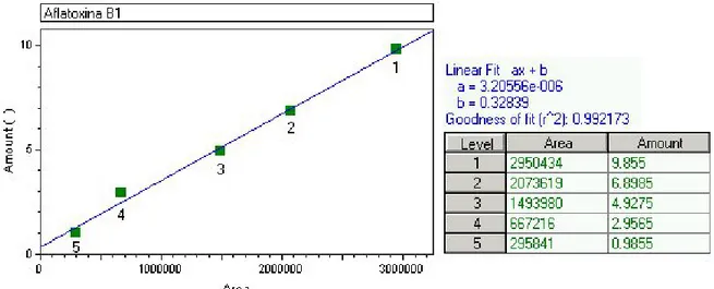
The linearity was evaluated within the range under study
and calculated from the linear regression equation and
determined by the least squares method. The linear correlation
coefficient (r2) was used as the indicator of the straight line as a
mathematical model. The values were always over 0.99 as
recommended by Green (11). Figures 1, 2 and 3 show an
aflatoxin B1 calibration curve, the chromatogram of a mixture
of aflatoxins standards, and the extract of a horse chestnut seed
sample contaminated by 2.96 ng/g of aflatoxin B1, after
post-column derivatization with the electrochemical generation of
bromine.
Figure 1. Standard curve used for the quantification of aflatoxin B1 with the area obtained in the readings, the concentration of
Figure 2. HPLC chromatogram with fluorescence detection post-column derivatization (electrochemically) – Kobra cell of a mixture of
aflatoxin standards; Shim-pack CLC-ODS column and water:metahanol:acetonitrile, 60:20:20,v/v/v as mobile phase. Aflatoxin B1 (4.93
ng/mL).
Figure 3. HPLC chromatogram with fluorescence detection of an extract of horse chestnut seed. Aflatoxin B1-contaminated sample (2.96
ng g-1); immunoaffinity column purification; HPLC using a post-column derivatization (electrochemically) – Kobra cell; Shim-pack CLC-ODS column and water:metahanol:acetonitrile, 60:20:20,v/v/v as mobile phase.
Despite the existence of several methods described
towards the determination of aflatoxins in medicinal plants,
none of such methods can be applied to all types of samples or
all aflatoxins (15). The difficulty stems from the chemical
complexity of the compounds of every different medicinal
plant. Results of recovery experiments and coefficient of
variation for aflatoxin B1 are showed in Table 1. The recovery
values obtained for aflatoxin B1 revealed that the methodology
in use and the analytical conditions developed in the laboratory
are in compliance with the provisions set forth by law no.
401/2006 (02/23/2006) in the European Union (8). All
recovery values are within the range of 70 to 110%, which is
required when the aflatoxin concentration is in the range of
1-10 µg kg-1. In relation to the variation coefficient values, all stayed below 20% as recommended by Horwitz and Albert
(1982), pointing to a good precision of the applied
Table 1. Recovery in medical herbs spiked with AFB1 standard (2.96 ng/g)
As far as the remaining aflatoxins are concerned (data
shown in Table 1), the recovery values for aflatoxin G2 were
below 60% in all tested plants. Recovery values of 56% were
obtained for aflatoxin B2 in green tea and 53% in guarana
powder. The others presented values between 83% and 103%.
Recovery values for aflatoxin G1 were between 95% and 103%,
except for horse chestnut seed, Espinheira-Santa and guarana,
which presented values over 115%. Low recovery, mainly in
relation to aflatoxin G2 might be due to little mycotoxin affinity
towards the antibody, which depends on the extracts used or
the insufficient quantity of antibody bound to the gel of the
immunoaffinity column (1).
The presence of aflatoxin B1 was noticed in just a single
sample of green tea (Camellia sinensis) at a concentration
below the Limit of Quantification (1.0 ng/g). A possible
explanation for the absence of aflatoxins would be the lack of
toxigenic fungi in the samples or the environmental conditions
during harvest and storage. It is well reported in literature that
temperature and water activity are the main factors that
influence fungal invasion and the production of aflatoxins in
stored products (9). Kulshrestha, Gupta, Shukla, Kundu,
Bhatnagar, Katiyar (2008) evaluated that medicinal plants with
water activity below 0.81 under temperatures of 25 ± 2o C, 30 ±
2o C and 40 ± 2o C and water activity over 0.81 and temperature below 10 ± 2o C did not present aflatoxins, even in the presence
of Aspergillus flavus, an aflatoxin producer.
Similar results were found by Romagnoli, Menna,
Gruppioni, Bergamini (2007) in Italy as 48 infusions and
medicinal plants were analyzed. None of the samples
presented detectable levels of aflatoxins for an analytical
methodology with a limit of detection and quantification of 0.5
and 1.5 ng/g of aflatoxin B1,respectively. Ali, Hashim, Saad,
Safan, Nakajima, Yoshizawa (2005) evaluated 23 traditional
medicinal plants from Malaysia and Indonesia. They observed
aflatoxin B1 in 16 samples with an average 0.26 ng/g. In
Thailand, Tassaneeyakul, Razzazi-Fazeli, Porasuphatana,
Bohm (2004) detected aflatoxins in 5 out of the 28 analyzed
samples within a range of 1.7 to 14.3 ng/g. Unlike the quoted
authors, Selim, Popendorf, Ibramim, Sharkawy, Kashory
(1996) observed high contamination of aflatoxin B1 in 9
samples in Egypt, after having analyzed 31 medicinal plants
within a range of 24 to 105 ng/g and an average 49 ng/g.
In Brazil, a small number of papers evaluated the
quantification of aflatoxins in medicinal plants. Aquino,
Sample Amount recovered (ng/g) Recovery mean (%) Coefficient of variation (%)
2.56 Senna
(Cassia angustifolia Vahl) 2.99 94 10.9
3.38 Espinheira-Santa
(Maytenus ilicifolia Martius)
2.89 106 11
2.53 Passion Fruit
(Passiflora sp Sims) 3.13 96 14.9
2.40 Guarana Powder
(Paullinia cupana H.B.K) 2.69 86 8.1
3.04 Valerian Root
(Valeriana officinalis Linné)
3.04 103 0
2.57 Cascara Sagrada
(Rhamnus purshiana D.C.) 2.04 78 16.2
3.10 Horse Chestnut Seed
(Aesculus hippocastanum
L.)
3.02 104 1.2
2.99 Green Tea
Gonçalez, Rossi, Campos Nogueira, Reis, Corrêa (2010)
evaluated boldo (Peumus boldus), green tea (Camellia
sinensis), Espinheira-Santa (Maytenus ilicifolia), and senna
(Cassia angustifolia), when aflatoxins were not detected
through the use of VICAM immunoaffinity columns (AflaTest
kit), a monoclonal antibody-based affinity chromatography
system and posterior confirmation by thin-layer
chromatography. Braga, Medeiros, Oliveira, Macedo (2005)
analyzed different samples of M. illicifolia (undisclosed
figures) sold in stores and drugstores in the city of João Pessoa,
State of Paraíba, when aflatoxins were not detected either. In
this case, the method in use involved immunoaffinity column
purification and high efficiency liquid chromatography,
excluding the derivatization stage.
Owing to the relevance of human exposure to aflatoxin B1
for public health, the lack of information and the Brazilian
climatic conditions, which favor fungal production as well as
mycotoxin production, a constant supervision of the quality of
medicinal plants and phytotherapeutic products is required to
guarantee good health conditions for the users of such
products.
ACKNOWLEDGEMENTS
The authors are grateful to the Research Foundation of the
State of Minas Gerais (Fundação de Amparo à Pesquisa do
Estado de Minas Gerais – FAPEMIG) for the financial support
received.
REFERENCES
1. Ali, N.; Hashim, N.H.; Saad, B.; Safan, K.; Nakajima, M.; Yoshizawa, T. (2005). Evaluation of a method to determine the natural occurrence of aflatoxins in commercial traditional herbal medicines from Malaysia and Indonesia. Food Chem. Toxicol., 43, 1763-1772.
2. Aquino, S.; Gonçalez E.; Rossi, M.H.; Campos Nogueira, J.H.; Reis, T.A.; Corrêa, B. (2010). Evaluation of fungal burden and aflatoxin presence in packed medicinal plants treated by gamma radiation. J. Food Prot., 73, 932-937.
3. Arranz, E.; Egmond, H.V.; Stzoo, E.; Kroegner, K.; Legarda, T.M.; Burdanpal, P.; Reif, K.; Stroka, J. (2006). Determination of aflatoxin B1
in medical herbs: interlaboratory study. J. AOAC Int, 89, 595-605. 4. Association of Official Analytical Chemists (AOAC). OfficialMethods
of the AOAC International. 16. ed. Gaithersburg: AOAC International, 1997.
5. Braga, S.M.L.F.M.; Medeiros, F.D.; Oliveira, E.J.; Macedo, R.O. (2005). Development and validation of a method for the quantitative determination of aflatoxin contaminants in Maytenus ilicifolia by HPLC with fluorescence detection. Phytoch. Anal., 16, 267-271.
6. Brasil. Agência Nacional de Vigilância Sanitária. Resolução no. 10, de 9
de março de 2010. Dispõe sobre limites para contaminantes microbiológicos em drogas vegetais. Diário Oficial [da] República Federativa do Brasil, Brasília, 10 mar. 2010.
7. Bugno, A.; Almodovar, A.A.B.; Pereira, T.C.; Pinto, T.J.A.; Sabino, M. (2006). Occurrence of toxigenic fungi in herbal drugs. Braz. J. Microbiol., 37, 47-51.
8. Commission regulation (EC) No. 401/2006 of 23 february. 2006. Laying down the methods of sampling and analysis for the official control of the levels of mycotoxins in foodstuffs. Official Journal of the European Communitties, L70/12.
9. Council for Agricultural Science ad Technology – CAST. Mycotoxins: economics and health riscks. Ames, Iowa: Council for Agricultural Science and Tecnology, 2003. Task Force Report 139.
10. Efuntoye, M.O. (1996). Fungi associated with herbal drug plants during storage. Mycopath., 136, 115-118.
11. Green, J.M. (1996). A pratical guide to analytical method validation.
Anal. Chem., 68, 305-309.
12. Halt, M. (1998). Moulds and mycotoxins in herb tea and medicinal plants. European J. Epid., 14, 269-274.
13. Horwitz, W.; Albert, R. (1982). The reliability of aflatoxin assays. Assoc. FoodDrug Off. Quart. Bull., 46, 14-24.
14. International Agency for Research on Cancer. (1993). Some naturally occurring substances: food itens and constituents, heterocyclic aromatic amines and mycotoxins. IARC monographs on the evaluation of carcinogenic risks to humans. IARC Sci. Publ., 56, 19-23.
15. Ip, S.P.; Che, C.T. (2006). Determination of aflatoxins in Chinese medicinal herbs by high-performance liquid chromatography using immunoaffinity column cleanup. Improvement of recovery. J Chrom., 1135, 241-244.
16. Junior, V.F.V.; Pinto, A.C.; Maciel, M.A.M. (2005). Plantas medicinais: Cura segura? Quim. Nova, 28, 519-528.
medicinal plants – a review. Planta Med., 68, 5-15.
19. Kosalec, I.; Cvek, J.; Tomic, S. (2009). Contaminants of medicinal herbs and herbal products. Arh Hig Rada Toksikol, 60, 485-501.
20. Kulshrestha, R.; Gupta, C.P.; Shukla, G.; Kundu, M.C.; Bhatnagar, S.P.; Katiyar, C.K. (2008). The effect of water activity and storage temperature on the growth of Aspergillus flavus in Medicinal herbs.
Planta Med, 74, 1308-1315.
21. Martins, H.M.; Martins, M.L.; Dias, M.I.; Bernardo, F. (2001). Evaluation of microbiological quality of medicinal plants used in natural infusions. Int.J. Food Microbiol., 68, 149-153.
22. Prado, G.; Andrade, M.C.; Oliveira, M.S.; Leal, A.S.; Oliveira, B.R.; Batista, L.R. (2009). Efeito da irradiação gama na microbiota fúngica de plantas medicinais. Ciên. Agrotec., 33, 1372-1378.
23. Razak, M.F.A.; Aidon, K.E.; Candlish, A.G.G. (2009). Mixed herbs drugs: Inhibitory effect on growth of the endogenous mycoflora and aflatoxin production. Mycopath., 167, 273-286.
24. Rizzo, I.; Vedoya, G.; Maurutto, S.; Haidukowski, M.; Varsavsky, E. (2004). Assessment of toxigenic fungi on Argentinean medicinal herbs.
Microbiol.Res., 159, 113-120.
25. Romagnoli, B.; Menna, V.; Gruppioni, N.; Bergamini, C. (2007). Aflatoxins in spices, aromatic herbs, herb-teas and medicinal plants
marketed in Italy. (2007). Food Control, 18, 697-701.
26. Roy, A.K.; Sinha, K.K.; Chourasia, H.K. (1988). Aflatoxin contamination of some common drug plants. Appl. Environ. Microbio., 54, 842-843.
27. Selim, M.I.; Popendorf, W.; Ibramim, M.S.; Sharkawy, S.E.; Kashory, S. (1996). Aflatoxin B1 common Egyptian foods.J AOAC Int., 79,
1124-1129.
28. Tassaneeyakul, W.; Razzazi-Fazeli, E.; Porasuphatana, S.; Bohm, J. (2004). Contamination of aflatoxins in herbal medicinal products in Thailand. Mycopath., 158, 239-244.
29. Trucksess, M.W.; Scott, P.M. (2008). Mycotoxins in botanicals and dried fruits: a review. Food Addit. Contam., 25, 181-192.
30. Vieira, I.F.R.; Leal, A.S.; Krambrock, K.; Tambourg, E.B. (2007). Identificação de plantas medicinais irradiadas através da ressonância paramagnética eletrônica. Braz. J. Food Technol., 10, 63-69.
31. Weaver, C.M.; Trucksess, M.W. (2010). Determination of aflatoxins in botanical roots by a modification of AOAC Official MethodSM 991.31:
Single-Laboratpry validation. J AOAC int., 93, 184-189.
32. World Health Organization. 1998. Quality control methods for medicinal plant materials. World Health Organization, Geneva.