Article
, Vol. 29, No. 10, 2137-2143, 2018 Printed in Brazil - ©2018 Sociedade Brasileira de Química*e-mail: [email protected]
#L. Wei and Y. Yan contributed equally to this work.
Determination of Estrogens in Milk Using Polypyrrole Fiber-Mediated Solid-Phase
Extraction Followed by High Performance Liquid Chromatography
Lanlan Wei,a,# Yan Yan,b,# Jianjun Deng,c Yuqin Ma,c Yu Wang,d Xiaoxiao Wue and
Xuejun Kang*,d
aKey Laboratory of Environmental Medicine and Engineering (Ministry of Education),
School of Public Health, Southeast University, 210009 Nanjing, China
bJiangsu Product Quality Testing & Inspection Institute, 210000 Nanjing, China
cSuzhou Dongqi Biological Technology Co., Ltd., 215123 Suzhou, China
dKey Laboratory of Child Development and Learning Science (Ministry of Education),
School of Biological Science and Medical Engineering, Southeast University, 210096 Nanjing, China
eNational Center of Supervision Inspection on Processed Food & Food Additives Quality,
Nanjing Institute of Product Quality Inspection, 210019 Nanjing, China
A solid-phase extraction (SPE) method based on conductive polypyrrole (PPy) nanofibers which were fabricated by electrospinning and in situ polymerization was developed. PPy nanofibers-mediated SPE followed by high performance liquid chromatography (HPLC) was used for the determination of three synthetic estrogens, namely diethylstilbestrol (DES), dienestrol (DIS), or hexestrol (HS) in milk sample. Extraction conditions including extraction nanofibers, donor solution pH and salt concentration were optimized. The target compounds were extracted from a 0.5 mL aqueous sample at pH 5.0 through PPy fibers, and then were eluted with 0.1 mL methanol. After extraction, the eluant was directly injected into an HPLC system for detection. Under the optimized extraction conditions, a large enrichment factor was achieved for three estrogens. The limit of detection (LOD) at a signal to noise ratio (S/N) of 3 ranged from 0.02 to 0.05 µg mL-1 for
the estrogens in milk sample.
Keywords: liquid dairy products, packed-fibers solid-phase extraction, high performance
liquid chromatography, estrogens
Introduction
Estrogens have been considered as one of the groups
of analytes concerned in certain food matrices, especially in milk and other dairy products.1 Synthetic estrogens such
as diethylstilbestrol (DES), dienestrol (DIS) and hexestrol (HS) are potent regulatory factors in physiological response medicine, and have been used widely as growth promoters in livestock and as a treatment for estrogen-deficiency disorders in veterinary medicine.
A variety of techniques have been applied to determine estrogens. The abuse of estrogens is known to be strongly harmful to humans owing to their potential carcinogenic properties. The use of estrogens in food-producing
animals and other products has been prohibited in many countries. Synchronously, more attention is paid to residue analysis of these estrogens that helps us control their illegal use.2-4 Generally, the determination of DES in
wastewater has been carried out using gas
chromatography-mass spectrometry,5,6 liquid chromatography-mass
spectrometry7,8 or high performance liquid chromatography
equipped with diode array detection.9-11 Since the
concentration of DES in complex matrices is usually extremely low, a preconcentration step, such as solid-phase extraction (SPE) and liquid-solid-phase microextraction (LPME), is required. However, the lack of selectivity and low recovery are the main problems in the determination of estrogens by using common samples preconcentration.12-15
widely applied to the extraction of different compounds.16
Recently, a novel solid-phase extraction method based on electrospun polymer nanofibers as adsorbents to enrich
target compounds has been developed.17 Owing to their
large surface area to volume ratio, electrospun nanofibers facilitate the miniaturization of SPE when they are packed into a pipette tip as the sorbent beds. With this packed-fiber solid-phase extraction (PFSPE) platform, the extraction capability could be efficiently improved and the volume of desorption solvent could be reduced to microliter levels. Thus, the evaporation step is not needed any more. PFSPE has been successfully applied in pharmaceutical analysis and pharmacokinetic studies of animals and humans, analysis of functional ingredients in health foods and determination of environmental pollutants in various sample matrices, etc.18 PFSPE using
polystyrene (PS) nanofibers as extracting agent coupled with high performance liquid chromatography (HPLC) tandem mass spectrometry (MS/MS) has been used for the simultaneous determination of three estrogens including
DES, HS and DIS in liquid dairy products,19 but the
target compounds must be extracted by acetonitrile, and an evaporating step was needed to remove the organic solvent before PFSPE.
In recent years, conductive polymers have attracted a great interest for the extraction of polar compounds due to their following advantages. They have a high extraction efficiency for polar compounds because of their inherent and unusual multifunctionality, such as ion-exchange properties, the π-π interactions, hydrogen bonding, acid-base properties, polar functional groups,
and electroactivity.20 Among the conducting polymers,
polypyrrole (PPy) is especially promising for commercial applications because of its better environmental stability and facile synthesis.21 In this paper, we develop an SPE
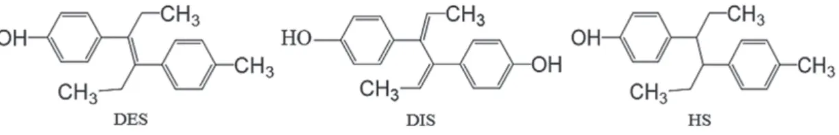
method by combining the advantages of PPy and PFSPE. DES, DIS, and HS in milk are selected as the model analytes to exhibit the potential application of the PPy fibers in the PFSPE of these molecules after a simple precipitation of protein in milk. The structures of DES, DIS and HS are given in Figure 1. Under optimized extraction conditions, the proposed method is calibrated for quantitative analysis of the target compounds in milk samples.
Experimental
Reagents and chemicals
DES, DIS and HS standards were purchased from Sigma-Aldrich (USA). PPy nanofibers were from Dongqi Bio-Technology Co., Ltd. (China). HPLC grade methanol were from Sinopharm Chemical Reagent (China). Each estrogen was dissolved in methanol to obtain a standard solution with a concentration of 0.1 mg mL-1 and stored at 4 oC. The working
solutions with 0.16, 0.47, 1.42, 4.27, and 12.80 µg mL-1 of
DIS; 0.49, 0.99, 1.97, 3.94, and 7.89 µg mL-1 of DES; and
0.17, 0.51, 1.51, 4.62, and 13.85 µg mL-1 of HS were obtained
by diluting the 0.1 mg mL-1 solution with ultrapure water.
Characterization
The morphology images of the PS nanofibers and PPy nanofibers were obtained by using a scanning electron microscope (SEM, Hitachi S-3000N, Japan), at an acceleration voltage of 10 kV and a specific surface and porosity analyzer (Micromeritics ASAP2020, USA).
Instrumentation
Centrifuge was purchased from Ruijiang Corporation (China). XW-80A vortex mixer was from Vimins (China). PFSPE column (styrene copolymer type) and array SPE extraction device were purchased from Suzhou DongQi Biological Technology Co., Ltd. (China). The HPLC system consisted of an LC-20A pump, an SPD-M20A detector, an SIL-20AC autosampler and a CTO-20A oven purchased from Shimadzu Corporation (Japan), equipped with an analytical column (Shimadzu C18, 250 × 4.6 mm2,
5 µm) attached to a photodiode array detector. The mobile
phase consisted of 60% methanol in 20 mmol L-1 sodium
hydrogen phosphate (v/v), pH 6.0. The flow rate was kept constant at 1.0 mL min-1 and the wavelength for UV detector
was set at 230 nm.
Samples preparation
Milk was purchased from a supermarket in Nanjing, China. Typical samples without detectable estrogens
identified by a literature method were used as the blank samples.22
The preparation of milk sample employed the following steps: (i) insertion of 1.8 mL milk into 4 mL centrifuge tube; (ii) addition of 0.2 mL estrogens (DIS, DES and HS) with appropriate concentration; (iii) addition of 1.0 mL 30% acetocaustin; (iv) sonication of the mixture for 10 min and centrifugation at 4000 rpm for 10 min, then separation of the supernatant; (v) transfer of the total supernatant to clean tube and modification to pH 5 by adding 220 µL of 4.0 mol L-1 sodium hydroxide solution before the treatment
of PFSPE.
The PFSPE column, as shown in literature,23 was
preconditioned by 100 µL of methanol and then 200 µL of water. The total supernatant was loaded and pushed through the sorbent by the pressure of air forced by gas tight plastic syringe. Target retained on the PFSPE was eluted with 100 µL of methanol and 20 µL of elution was injected into the HPLC by autosampler.
Results and Discussion
Morphology of PPy fibers
The surface of PPy nanofibers synthesized under ultrasonication appears quite smooth at low magnification (Figure 2a). The nanofibers had an outer diameter of approximately 620 ± 410 nm. PS nanofibers with smooth and uniform morphology and random orientation were generated with a mean diameter of 460 ± 100 nm (Figure 2b). As observed from the images, the presence of the nanostructure could provide a high specific surface area and many interaction sites that could offer increased SPE efficiency.
The textural properties, including the Brunauer-Emmett-Teller (BET) surface area, pore volume and pore size are listed in Table 1. It can be seen that the BET surface area and pore volume of the PPy nanofibers were the lowest.
Solid-phase extraction of estrogens
First, the effect of nanofiber characteristics on the efficiency of extraction was investigated. In this work, two types of nanofibers prepared with different framework were evaluated for their extraction recoveries. As shown in Figure 3, the extraction recoveries of PPy nanofibers for DES, DIS, HS were 27.0, 35.0, and 38.0%, respectively, Figure 2. SEM images of (a) PPy elecrospun nanofibers and (b) PS elecrospun nanofibers.
Table 1. Textural properties of the tested materials
Material BET surface area / (m2 g-1)
Pore volume / (cm3 g-1)
PPy nanofibers 10.50 0.02
PS nanofibers 42.31 0.26
BET: Brunauer-Emmett-Teller; PPy: polypyrrole; PS: polystyrene.
which were higher than those of PS nanofibers. The PPy nanofibers diameter, however, was thicker (Figure 2) and the BET surface area was smaller (Table 1). The result could be explained by the interaction mechanism between PPy and the analytes, because the conjugated π structure was found in PPy backbone, which showed strong interaction with analytes, and hydrogen bonding interaction could also exist between the polymer and analytes, as shown in Figure 4.
Figure 5 described the previously reported conventional extraction method for determining estrogens. Comparing with conventional methods, PFSPE method had advantages like less organic solvent consumption, simplicity, high recovery rate, easy operation and automation.
pH of the donor solution
To investigate the effect of pH, the pH value of the donor solution was adjusted to 3.0, 4.0, 5.0 and 6.0 with
4.0 mol L-1 NaOH. As shown in Figure 6, an inverted
v-shaped outline of elution efficiency vs. the pH of donor solution was observed. The peak area was improved with the increasing pH until its highest point (pH 5), after that, the peak area was decreased. Therefore, pH 5.0 was selected.
Figure 4. The interaction between estrogens and PPy nanofibers.
Figure 5. Flow chart of determination methods: (a) previously reported conventional extraction methods; (b) the method proposed in this paper.
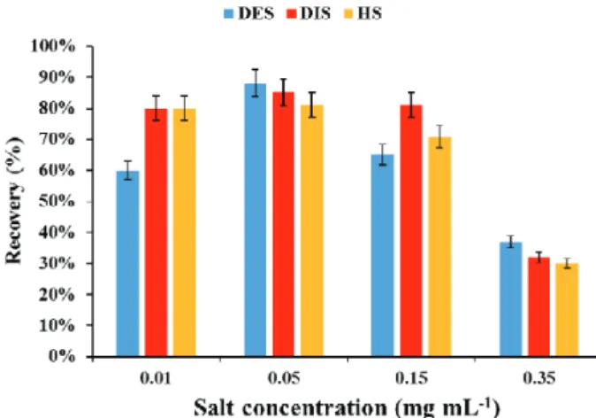
Effect of salt concentration
The effect of salt concentration on the donor phase was also investigated. NaCl is added at concentrations of 0.01, 0.05, 0.15, and 0.35 g mL-1. As shown in Figure 7,
addition of NaCl improved the recovery of the analytes. However, the recovery in 0.15 g mL-1 NaCl is lower than
that in 0.05 g mL-1 NaCl. Two simultaneous processes
could explain this phenomenon. With the addition of salt, hydration spheres are generated around the ionic salt molecules in aqueous solution. These hydration spheres reduce the amount of water which dissolves analyte molecules, resulting in additional analytes moving near the sorbent.24 At the same time, estrogen molecules may
participate in electrostatic interactions with the salt in solution, thereby decreasing their ability to move into the extraction nanofibers. Initially, the predominant process would be the interaction of the salt molecules with water molecules; but as the salt concentration further increased, salt molecules would interact with analyte molecules. It seemed reasonable to add 0.05 g mL-1 NaCl to the donor
phase, since it contributed to the best extraction efficiency. However, in view of the salt introduced in the steps of precipitating protein and pH adjustment, NaCl was not added into the sample in the analysis of the samples.
Validation of the method
The linearity and the limit of detection (LOD) of the method under the optimal extraction conditions were investigated (Table 2) in order to evaluate the practical applicability of the PPy approach. A good linearity was
found within the range 0.49-7.89 µg mL-1 for DES,
0.17-13.85 µg mL-1 for HS and 0.16-12.80 µg mL-1 for
DIS. Correlation coefficients (R2) better than 0.99 were
obtained. The relative standard deviations (RSD) of DES, DIS, and HS were 6.8, 9.5 and 7.9%, respectively. LOD
was 0.02 µg mL-1 for DIS, 0.05 µg mL-1 for DES, and
0.02 µg mL-1 for HS.
Applicability to detection in real sample
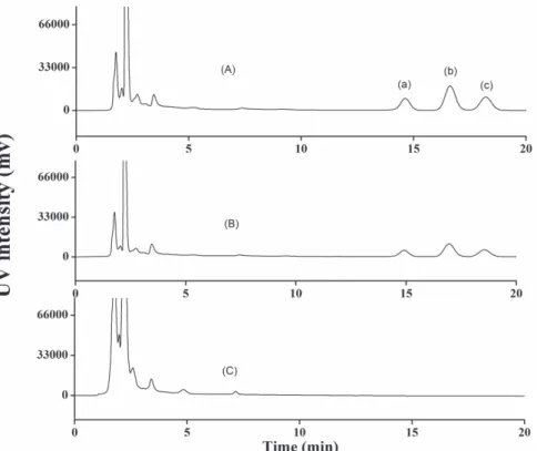
The proposed analytical method was applied to the analysis of milk samples purchased from a supermarket in Nanjing, China, and no target analytes were detected. The samples were then spiked with DES standards at 0.8 and 1.6 µg mL-1 levels, and DIE and HS at 0.3 and 0.9 µg mL-1
levels to assess matrix effects. Estrogens were detected in milk samples (as shown in Figure 8). The recoveries
from the blank milk are presented in Table 3, indicating that the influence of the matrix was not significant for food analysis.
Conclusions
A fast, sensitive HPLC-UV method for the determination of estrogens in milk was presented in this paper. Although PS nanofibers based on PFSPE coupled with HPLC-MS/MS has already been reported by our group, special and expensive equipment, even staff with a high level of operation are required. This manuscript provides a low-cost determination method with a simpler pretreatment for three estrogens in food. The PFSPE pretreatment method offered a simple, fast and low-cost operation. The microliter amount of eluting solvent could improve the conventional enrichment process, which obviously illustrated the environmental friendliness. Moreover, higher extraction recoveries and good reproducibility were especially suitable for trace analysis of the samples.
Acknowledgments
This study was supported by the National Science Foundation of China (No. 81673230, No. 21307086 and No. 81172720); the Social Development Research Program of Jiangsu Province Science and Technology department Figure 7. Effect of salt concentration on the extraction.
Table 2. Performance of the method
Target compound
Linearity range / (µg mL-1)
R2 LOD /
(µg mL-1)
RSD / % (n = 3)
DES 0.49-7.89 0.997 0.05 6.8
DIS 0.16-12.80 0.997 0.02 9.5
HS 0.17-13.85 0.998 0.02 7.9
R2: correlation coefficient; LOD: limit of detection; RSD: relative standard
Table 3. Results from the analysis of DIS, DES, and HS in milk
Sample DIS / (µg mL-1) Recovery / % DES / (µg mL-1) Recovery / % HS / (µg mL-1) Recovery / %
1
–
–
–
–
–
–
2 – – –
3 – – –
1
0.3
97.5
0.8
102.5
0.3
100.0
2 102.5 93.2 97.6
3 100.0 104.3 101.2
1
0.9
98.9
1.6
100.0
0.9
100.0
2 103.4 96.9 97.8
3 97.7 104.7 101.1
DES: diethylstilbestrol; DIS: dienestrol; HS: hexestrol.
Figure 8. HPLC-UV spectra of real samples. (A) Chromatogram of standard solution after extraction; (B) chromatogram of milk sample spiked with each analyte after extraction; and (C) chromatogram of milk sample after extraction. Analytes: (a) DES, (b) DIS, and (c) HS.
(No. BE2016741); Science & Technology Project of China General Administration of Quality Supervision, Inspection and Quarantine (No. 2015QK055); and Postgraduate Research & Practice Innovation Program of Jiangsu Province (No. KYCX17_0189).
References
1. Farlow, D. W.; Xu, X.; Veenstra, T. D.; J. Chromatogr. B: Anal. Technol. Biomed. Life Sci.2009, 877, 1327.
2. Pfaffl, M. W.; Reck, B.; Dreher, R.; Meyer, H. H.; Anal. Chim. Acta2003, 483, 401.
3. Newbold, R. R.; Toxicol. Appl. Pharmacol.2004, 199, 142. 4. Martino, M. A.; Nevadunsky, N. S.; Magliaro, T. J.; Goldberg,
M. I.; Prim. Care Update Ob Gyns2002, 9, 7.
5. Liao, S.; Wu, X.; Xie, Z.; Anal. Chim. Acta2005, 537, 189. 6. Quintan, J. B.; Carpinteiro, I. J.; Rodríguez, R. A.; Lorenzo, A.
M.; Carro-Cela, R.; J. Chromatogr. A 2004, 1024, 177. 7. Chanbasha, B.; Akhila, J.; Meng-Keow, K.; Suresh, V.;
Hian-Kee, L.; J. Chromatogr. A2005, 1100, 137.
8. Rodriguez-Mozaz, S.; López de Alda, M. J.; Barceló, D.; J. Chromatogr. A 2004, 1045, 85.
10. López de Alda, M. J.; Barceló, D.; J. Chromatogr. A2001, 911, 203.
11. Penalver, A.; Pocurull, E.; Borrull, F.; Marcé, R. M.; J. Chromatogr. A2002, 964, 153.
12. Haupt, K.; Analyst2001, 126, 747.
13. López de Alda, M. J.; Barceló, D.; J. Chromatogr. A2001, 938, 145.
14. Thurman, E. M.; Chem. Anal.1998, 147, 5441.
15. Kang, X.-J.; Pan, C.; Xu, Q.; Yao, Y.-F.; Wang, Y.; Qi, D.-J.; Gu, Z.-Z.; Anal. Chim. Acta2007, 587, 75.
16. Kang, X.-J.; Chen, L.-Q.; Zhang, Y.-Y.; Liu, Y.-W.; Gu, Z.-Z.; J. Sep. Sci.2008, 31, 3272.
17. Zhang, Y.-Y.; Kang, X.-J.; Chen, L.-Q.; Pan, C.; Yao, Y.-F.; Gu, Z.-Z.; Anal. Bioanal. Chem. 2008, 391, 2189.
18. Xu, Q.; Wang, M.; Yu, S.; Tao, Q.; Tang, M.; Analyst2011, 136, 5030.
19. Hu, W.-Y.; Kang, X.-J.; Zhang, C.; Yang, J.; Ling, R.; Liu, E.-H.; Li, P.; J. Chromatogr. B: Anal. Technol. Biomed. Life Sci.2014, 957, 7.
20. Li, M.; Wei, Z.-X.; Jiang, L.; J. Mater. Chem.2008, 18, 2276. 21. Mohammadi, A.; Ameli, A.; Alizadeh, N.; Talanta2009, 78,
1107.
22. Boyd-Boland, A. A.; Pawliszyn, J. B.; J. Chromatogr. A1995, 704, 163.
23. Liu, M.; Qiu, B.; Jin, X.; Zhang, L.; Chen, X.; Chen, G.-N.; J. Sep. Sci.2008, 31, 622.
24. Zhao, R.-S.; Chu, L.-L.; Wang, Y.; Song, Y.; Liu, P.; Li, C.; Huang, J.-J.; Kang, X.-J.; Clin. Chim. Acta 2017, 468, 120.
Submitted: December 15, 2017
Published online: April 27, 2018