Presence of enterotoxigenic
Staphylococcus aureus
in artisan fruit salads
in the city of San Luis, Argentina
Cecilia S.M. Lucero Estrada
1, Lucia E. Alcaráz
2, Sara E. Satorres
2,
Eduardo Manfredi
3, Lidia del C. Velázquez
11Microbiología General, Universidad Nacional de San Luis, San Luis, Argentina. 2Bacteriología y Virología, Área Microbiología, Facultad de Química, Bioquímica y Farmacia,
Universidad Nacional de San Luis, San Luis, Argentina.
3Servicio Fisiopatogenia, Departamento de Bacteriología, INEI-ANLIS “Dr. Carlos G. Malbrán”,
Ciudad Autónoma de Buenos Aires, Argentina.
Submitted: May 11, 2011; Approved: April 04, 2013.
Abstract
An increase in the consumption of fruit juices and minimally processed fruits salads has been ob-served in recent years all over the world. In this work, the microbiological quality of artisan fruit sal-ads was analysed. Faecal coliforms, Salmonella spp, Shigella spp, Yersinia enterocolitica and Escherichia coliO157:H7 were not detected; nevertheless, eleven strains ofStaphylococcus aureus were isolated. By multiplex PCR, all isolates showed positive results forS. aureus 16S rRNAgene and 63.6% of them were positive forseagene. Furthermore, PCRseapositive strains were able to produce the corresponding enterotoxin. Finally, the inactivation of these strains in fruit salads by nisin, lysozyme and EDTA, was studied. EDTA produced a totalS. aureusgrowth inhibition after 60 h of incubation at a concentration of 250 mg/L. The presence ofS. aureusmight indicate inade-quate hygiene conditions during salad elaboration; however, the enterotoxigenicity of the strains iso-lated in this study, highlights the risk of consumers’ intoxication. EDTA could be used to inhibit the growth ofS. aureusin artisan fruit salads and extend the shelf life of these products.
Key words:Staphylococcus aureus, enterotoxigenic strains, fruit salads, antimicrobial compounds, multiplex PCR.
Introduction
An increase in the consumption of fruit juices and min-imally processed fruits, in the form of peeled, cut and pack-aged fruit salads, has been observed in recent years. Fruit salads are not heat-processed, contain no preservatives, and therefore, they can be easily spoiled by fungi and bacteria. Many microorganisms, in particular loving or acid-tolerant bacteria and fungi (yeasts and moulds), can use fruits as substrate and cause spoilage, producing flavour and odour alteration, product discoloration, and human illness if the contaminating microorganisms are pathogens (Tournas et al., 2006). Possible contamination sources are soil, faeces, manure, irrigation and washing water, ice, animals (includ-ing insects and birds), handl(includ-ing of the harvest(includ-ing and
pro-cessing equipment, and transport (Johnnessenet al., 2002). Raw fruits and vegetables have been identified as carriers of pathogenic bacteria such asShigellaspp, Salmonellaspp, enterotoxigenic and enterohemorrhagic Escherichia coli, Staphylococcus aureus, Campylobacter spp, Listeria monocytogenes, Yersinia enterocolitica, Bacillus cereus, and parasites asGiardia lamblia, Cyclospora cayetanensis and Cryptosporidium parvum (De Roever et al., 1998; Kumaret al., 2006).
S. aureusis one of the major pathogens that can cause food poisoning. Intoxication is caused by the ingestion of enterotoxins within foods, usually because the food has been left at room temperature (Walls and Scott, 1997). There are five major classical types of staphylococcal enterotoxins (SEs): SEA, SEB, SEC, SED and SEE, as well
Send correspondence to C.S.M.L. Estrada. Microbiología General, Universidad Nacional de San Luis, Ejército de los Andes 950. Bloque I, Piso 1. 5700 San Luis, Argentina. E-mail: [email protected].
as new SEs or SE-like superantigens (Sags) such as SEG to SEU (Chianget al., 2008). Foods requiring considerable handling during preparation and kept without refrigeration are usually involved in staphylococcal poisoning. This bac-terium is able to grow in a wide temperature range (7-48 °C), with an optimal growth at 35-37 °C, a frequent value in warm climates (Baezaet al., 2007).
The increasing demand for high-quality, non-ther-mally processed and microbiologically safe foods requires the development of different treatments for the reduction of microorganisms. Nisin is a bacteriocin that forms pores in cell membranes, it has “generally recognized as safe” (GRAS) status, and has been approved for use as a food preservative (Montville and Chen, 1998). Lysozyme is a protein with hydrolytic activity (Mckenzie and White, 1991), although it has lately been reported to have an anti-bacterial activity that is independent of its enzymatic activ-ity (Ibrain et al., 2001). Sodium ethylenediamine tetra-acetate (EDTA) is a chelating agent used in a wide variety of foods to prevent oxidation and other deteriorating reac-tions catalyzed by metallic ions. It has been claimed that the antimicrobial spectrum and potency of these molecules can be increased when used in combination each another (Chung and Hancock, 2000; Branen and Davidson, 2004) or other antimicrobials (Carneiro De Meloet al., 1998; Gill and Holley, 2003).
The aims of this work were i) to study the microbio-logical quality of artisan fruit salads manufactured in our region, ii) to detect the presence ofsea, seb, sec, sed, see genes encoding staphylococcal enterotoxins by multiplex PCR in ourS. aureusisolates, and iii) to assess the inhibi-tory capacity of nisin, lysozyme and EDTA on this bacte-rium.
Materials and Methods
Sample collection
A total of 71 samples of artisan fruit salads obtained in retail shops in San Luis city, Argentina, were processed. The samples were stored at 4 °C for no longer than 4 h until processing. The pH of the samples was determined with an Orion model 420A pHmeter (Orion Research Inc., Boston, Massachusets, USA).
Microbiological analysis
Twenty-five grams of each sample were homoge-nized in a stomacher (IUL Masticator, Koningswinter, Ger-many) for 1 min. From each homogenate, two serial decimal dilutions, 10-1 and 10-2, were prepared in 0.1% peptone water pH 7 (PW, Britania Laboratories, Buenos Aires, Argentina).
Counts of total mesophilic aerobes
Volumes of 0.1 mL of homogenate and each dilution were spread in duplicate onto plate count agar (PCA, Merck
Laboratories, Darmstadt, Germany) and incubated at 37 °C for 24 h.
Counts of moulds and yeasts
Aliquots of 0.1 mL of homogenate and dilutions were surface spread onto duplicated oxytetracycline-glucose-yeast extract agar (OGY, Merck) and incubated at 22 °C for 5 days.
Counts of total and faecal coliforms
Total (TC) and faecal (FC) coliforms were investi-gated in Mac Conkey broth (Merck) at 37 °C for 48 h by the three-tube Most Probable Number (MPN) method. Pre-sumptive results of TC were confirmed in bile brilliant green lactose broth (BGLB, Merck) at 37 °C for 24 h. FC were confirmed inEscherichia coli(EC) broth (Merck) at 44.5 °C for 24 h (De la Canal, 2004), with subsequent isola-tion on Eosin Methylene Blue agar (EMB; Merck). Suspect colonies from EMB were studied by Gram-staining and biochemical tests.
Investigation of Salmonella spp and Shigella spp.
These bacteria were investigated as described: 1 mL of homogenate were seeded into 9 mL of lactose broth (Merck) and incubated for 24 h at 37 °C. Then, one milli-litre aliquots were transferred to two tubes containing 9 mL of selenite broth (Merck), and two tubes with 9 mL of tetrathionate broth (Merck). One tube of each selective broth was incubated 24 h at 37 °C and the other one was in-cubated 24 h at 42 °C. Isolations were done on Bismuth Sulphite agar (BiSA, Merck) and Salmonella Shigella agar (SS, Merck). Suspect colonies were assayed by Gram stain-ing and classical biochemical tests (Caffer and Terragno, 2001; Tiruneh, 2009).
Counts of E. coli O157:H7
Volumes of 0.1 mL of homogenate and dilutions were seeded on Mac Conkey Sorbitol agar (SMC, Merck) at 37 °C for 24 h and sorbitol non-fermenting colonies were studied by biochemical tests and challenged against O157 antiserum.
Investigation of Y. enterocolitica
One millilitre of homogenate was inoculated in phos-phate buffered saline (PBS, Merck) pH 7.6 added with 1% sorbitol and 0.15% bile salts for 21 days at 4 °C. Isolations were performed on Mac Conkey agar (MC, Merck) for 48 h at 22 °C. Presumptive colonies were subjected to Gram staining and classical biochemical tests according to Ber-covier and Mollaret (1984).
Counts of S. aureus
an opaque zone and with an outer clear zone were selected for counting ofS. aureus.
Characterization ofS. aureus
Phenotypic characterization
The isolates ofS. aureuswere characterized as de-scribed previously (Satorres and Alcaráz, 2007). The inter-pretation of coagulase test was performed according to Sperberg and Tatini (1975). The 4+ reaction corresponded to a very firm opaque clot which remained in place when the tube was tipped on its side. The 2+ and 3+ reactions were not as opaque as 4+ reactions and were surrounded by clear plasma. The 1+ reaction showed a small and disorga-nized clot.
Simultaneously, an automated system (bioMerieux, Marcy I’Etoile, France) and a TECRA immunoassay equipment (International Pty Ltd, Australia) were used to determine the ability ofS. aureus isolates to produce the pool of SEA, SEB, SEC1, SEC2, SEC3, SED, and SEE enterotoxins.
Genotypic characterization
For the detection ofsea, seb, sec, sed, see and16S rRNAgenes ofS. aureus, a multiplex polymerase chain re-action (multiplex PCR) was used (Manfredi, 2007).
DNA extraction
Each strain was conserved in TSB (Britania) with 30% glycerol. From this culture medium each strain was spread on brain heart infusion agar (BHI, Becton Dickinson) and incubated for 24 h at 37 °C. After the incu-bation period, one colony was taken and inoculated in 5 mL of BHI broth, for 24 h at 37 °C. An aliquot of 1.5 mL was taken and centrifuged at 12000 x g for 5 min (Eppendorf 5415C). The supernatant was discarded and the pellet was resuspended in 150mL of Triton X100 (1% in buffer TE 1X, 10 mM Tris-HCl pH 8,1 mM EDTA pH 8). The
sus-pension was boiled for 30 min and then centrifuged at 12,000 x g for 5 min to remove the bacterial debris. A 5-microliter aliquot was used as DNA template.
Multiplex PCR
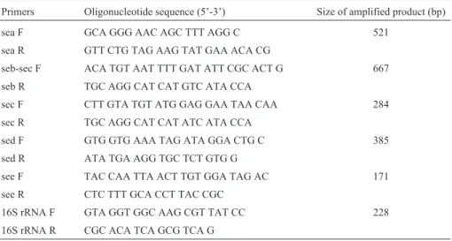
The reaction mix had a final volume of 50mL and contained the following compounds: 300 mM of each pri-mer pair of thesea,seb,sec,sedandseegenes (Table 1); 60 mM of the primer pair of16S rRNAgene (Invitrogen, Buenos Aires, Argentina) (Table 1); 400mM of each dNTP (Promega; Madison, WI, USA); 1X PCR buffer (Fer-mentas, California, USA), 0.4 mM MgCl2 (Fermentas), 0.04 U/mL of Taq polymerase (Fermentas), and 5mL of DNA template. DNAs of enterotoxin producingS. aureus strains [M5 (sea), N14 (seb), MC1 (sec), C1 (sed), N19125 (see)], were used as positive controls, and the S. aureus ATCC 25923 strain was used as negative control. The reac-tion mixture without template was used as system control. The amplification conditions were: 95 °C for 5 min, fol-lowed by 15 cycles of 95 °C for 30 s, 68 °C for 30 s and 72 °C for 30 s; and 20 cycles of 95 °C for 30 s, 60 °C for 30 s, 72 °C for 30 s; with a final extension at 72 °C for 2 min.
A volume of 10mL of a solution of xylene cyanol and glycerol 30% was added to 50mL of amplified DNA, in 10mL agarose gel 2.5% and 1X TAE buffer with 0.5mg of bromide ethidium/mL. A 100 marker ladder (Biodynamics S. R. L., Buenos Aires, Argentina) was used. A model transilluminator 2000 (BioRad, Hercules, California, USA) and a C-5060 Wide Zoom Olympus (Melville, USA) were used for amplicons’ observation. The analysis of the gels was performed with the Doc-It Image Acquisition software (UVP, Inc. Upland CA, USA) (Manfredi, 2007).
Antimicrobial susceptibility testing
TheS. aureusstrains were tested for susceptibility to a panel of antimicrobial agents using the disc diffusion
Table 1- Primers used for the detection of staphylococcal enterotoxin genes.
Primers Oligonucleotide sequence (5’-3’) Size of amplified product (bp)
sea F GCA GGG AAC AGC TTT AGG C 521
sea R GTT CTG TAG AAG TAT GAA ACA CG
seb-sec F ACA TGT AAT TTT GAT ATT CGC ACT G 667 seb R TGC AGG CAT CAT GTC ATA CCA
sec F CTT GTA TGT ATG GAG GAA TAA CAA 284
sec R TGC AGG CAT CAT ATC ATA CCA
sed F GTG GTG AAA TAG ATA GGA CTG C 385
sed R ATA TGA AGG TGC TCT GTG G
see F TAC CAA TTA ACT TGT GGA TAG AC 171
see R CTC TTT GCA CCT TAC CGC
16S rRNA F GTA GGT GGC AAG CGT TAT CC 228
method on Mueller-Hinton agar (MH, Merck) according to the Clinical and Laboratory Standards Institute (CLSI) (2007). The antibiotics tested were: penicillin G (PEN), 10 U; nitrofurantoin (NIT), 300mg; oxacillin (OXA), 1mg; erytromycin (ERY), 15 mg; gentamicin (GEN), 10 mg; rifampin (RIF), 5 mg; trimethoprim-sulfamethoxazole (TMS), 1.25 g/25mg; vancomycin (VAN), 30mg; cipro-floxacin (CIP), 5mg; amikacin (AKN), 30mg; fosfomicin (FOS), 50mg and tetracycline (TET), 30mg. TheS. aureus ATCC 25923 strain was included in each trial as a control. This strain was cultured in BHI broth to 37 °C for 24 h and kept in Luria broth (LB; Merck) at -20 °C.
Inhibition assays
Chemicals
The inhibitory capacity of the following compounds, nisin (2.5% nisin with an activity of 1.020 IU/mg; Sigma-Aldrich), pure lysozyme from egg white (with an activity of 36.000 IU/mg; Fluka Chemie, Buchs, Switzerland) and EDTA (99% purity; Sigma-Aldrich), was assayed on S. aureusin contaminated fruit salads.
Minimum inhibitory concentration (MIC) assessment for S. aureus
The microtechnique of Gill and Holley (2003) was applied for determining the MIC ofS. aureus correspond-ing to each chemical. The MIC values were expressed as mg/L.
Chemical treatments of S. aureus contaminated fruit salad
Taking into account the MIC values obtained as de-scribed, a study was conducted where a concentration equivalent to the MIC of each compound was added to 100 g of a sample of fruit salad. Each compound was tested alone or combining each other. Each sample was inoculated with 105cfu/mL of
S. aureusATCC 25923. The incubation conditions were 24 °C for 0, 4, 6, 24, 48 and 60 h. At each time, 1 mL of sample was taken and serial dilutions (1:10) in 0.1% PW pH 7 were performed. Then, 0.1 mL of each di-lution was spread onto Baird Parker (BP; Britania), plates were incubated for 24 h at 37 °C and finally, counts ofS. aureuscolonies were done.
Samples without addition of any compound were treated under the same time and temperature conditions and used as control.
Statistical analysis
Survival was expressed as log10N – log10No, where Nowas the initialS. aureuscount in fruit salad and N was the bacterial count after antimicrobial treatment. Mean val-ues of three replications were subjected to analysis of vari-ance and Students test by Infostat 1.0 software for deter-mining if significant variations (p < 0.05) in populations of microorganisms existed between treatments. The mean and
the standard deviation were calculated for the log10 reduc-tion of microorganism for each set of experiments.
Results
Microbiological analysis
The mean pH values observed in the fruit salads were 3.85±0.24. The counts of total mesophilic aerobes ranged from 1.60 to 4.70 log10cfu/g (mean 3.25 ± 0.54). Total coliforms varied between 30 and 1.100 MPN/g; faecal coliforms were not detected. The counts of moulds and yeasts varied between 2.30 and 4.33 log10cfu/g (mean 3.17 ±0.54). Enterobacteria such as E. coliO157:H7, Salmo-nellaspp,Shigellaspp, or Y. enterocoliticawere not de-tected in any of the analysed samples.
S. aureuswas isolated from eleven (7.81%) of the in-vestigated samples, with counts varying between 1.30 and 2.47 log10cfu/g (mean 1.75±0.49). All the isolates were thermonuclease positive and coagulase 4+. It was observed no organoleptic or physical difference betweenS. aureus positive and negative fruit salads. All salads were prepared with pieces of oranges, peaches, banana and apples, and had a similar pH. AllS. aureusisolates showed positive re-sults when the presence of16S rRNAgene was studied by multiplex PCR. Furthermore, 7 out of the 11 strains (63.6%) were positive for theseagene (Figure 1) and for the production of the SEA enterotoxin. None of other inves-tigated genes or toxins were detected. All strains were resis-tant to penicillin but susceptible to the remaining anti-microbial agents.
Inhibitory activity of antimicrobial compounds against
S. aureus
The nisin MIC was 500 mg/L and the EDTA MIC was 250 mg/L at 24 h of incubation. Lysozyme did not show inhibitory capacity, either alone or combined with nisin. In fruit salads, with nisin at 60 h of incubation no in-hibition was detected; meanwhile, with EDTA totally
inhi-Figure 1 - Multiplex PCR for S. aureus enterotoxin genes. A) sea
(521 bp),seb(667 bp),sec(284 bp), and16S ARNr(228 bp). Lines 1-7: isolates corresponded to the seven samples obtained from fruit salads; lines 8-10:sea,sebandsecpositive controls respectively; line 11: negative
bition of S. aureus at 60 h of incubation was observed (Figure 2).
Discussion
Over the last years, economic and job changes in Ar-gentina have resulted in the spread of small manufacturers of different types of foods, particularly dairy products, fruit juices and their derivates. Sometimes, these products are elaborated and sold by families or small groups of entrepre-neurs under no optimal sanitary conditions. Fruit salads constitute a common product, usually packed in plastic containers and labelled with expiration date of generally 4 days after elaboration.
The Argentine Alimentary Code (AAC) in Article 969 provides specific microbiological recommendations for fruit salads (De la Canal, 2004). This food must be free of pathogenic microorganisms exhibiting the same micro-biological quality than drinking water (mesophilic£ 500 cfu/mL, total coliforms£3 MPN/100 mL, and absence of faecal coliforms and Pseudomonas aeruginosa). In the present work, although no faecal coliforms were detected in the studied fruit salads, the range of mesophilic aerobic bacteria was between 1.60 and 4.70 log10cfu/g, and the val-ues of total coliforms were between 30 and 1.100 MPN/g. The MPN/g of total coliforms in all the samples was higher than the AAC maximal limit for these microorganisms. The pH was 3.85±0.24, a value that can allow the growth of yeasts and moulds, which were between 2.30 and 4.33 log10cfu/g.
Many authors have studied the microbiological qual-ity of fruit juices (Parish, 1997; Ghendheshet al., 2005), but very little literature is related to mixed fruit salads. Abadiaset al. (2008) studied 21 kinds of fresh-cut fruit sold in streets of Catalonia, Spain, and reported that aerobic mesophilic bacteria ranged from 2.0 to 7.1 log10cfu/g, and yeast and mould counts were between 1.7 and 4.9 log10 cfu/g, but they did not observe any pathogenic microorgan-isms. In a study of 38 fruit salad samples, among which eight were of mixed fruit, Tournaset al.(2006) reported that the yeast and moulds counts in the mixed fruit salads
ranged from 3.41 to 7.11 log10 cfu/g, being higher than those observed in this work.
Many foods depend on a combination of a variety of factors like pH and low temperatures to extend their shelf life. Although the growth of many pathogenic microorgan-isms can be inhibited in such foods, they can survive for long periods at dangerous concentrations. In the present study, neither Salmonella spp, Shigella spp, Y. entero-coliticanorE. coliO157:H7 were detected in the fruit sal-ads, but 11 strains ofS. aureuswere isolated. These results are in agreement with Kumaret al.(2006), who studied the microbiological characteristics of fruit salads sold in streets of Patiala, India, and detected coagulase positiveS. aureus in 66 (44%) samples, 38 of these strains present produced enterotoxins B and C. Although theS. aureusconcentration needed to produce food intoxication is high (105-106cfu/g), this bacterium is able to grow at a wide range of pH (4.5-9.3) and temperature (7-43 °C) (World Health Organi-zation, 1998). This could enable the growth ofS. aureusin fruit salads during storage time and lead to health hazard for consumers. In the present work, the mean count of S. aureuswas 1.75±0.49 log10cfu/g. Among the 11 strains analysed for the presence of enterotoxin genes by multiplex PCR, seven were positive toseagene. In fact, enterotoxin A together with the toxic shock syndrome toxin-1 (TTSS) were the most frequently found toxins inS. aureusisolates from food-poisoning cases in Taiwan (Chianget al., 2008). Theseagene coding for enterotoxin A has also been fre-quently detected inS. aureusstrains isolated from foods (Kerouantonet al., 2007; Lópezet al., 2008; Rallet al., 2008).
Three different antimicrobial components were tested to evaluate their inhibitory activity againstS. aureus. In our work, at the initial 24 h of nisin action,S. aureuswas sensi-tive to 500 mg/mL; however, at 60 h of incubation no con-centration of nisin inhibited the growth of this bacterium. Our results are similar to those obtained by Lima Grisi and Gorlach-Lira (2005), who observed a significant inhibition of S. aureus in pure culture for all nisin concentrations (100-1200mg/mL) used, but only for 8 h of incubation. The inhibition phase was followed by a period of rapid growth of the strain, achieving levels similar to those of the control after 24 h. Piperet al.(2009) studied 55S. aureusstrains and reported that nisin was active against all the assayed strains with a MIC between 0.5 to 8.3 mg/L. Microorgan-isms exhibiting resistance to nisin may act by different ways, inactivating the peptide via enzymatic action, alter-ing membrane susceptibility (Montville and Chen, 1998), or producing the enzyme nisinase that neutralizes the anti-microbial activity of the polypeptide (Hurst and Hoover, 1993). In the present study, lysozyme did not inhibit the growth ofS. aureusneither in pure culture nor in fruit sal-ads. Beraet al.(2007) have shown that modifications in peptidoglycan (PG) by O acetylation, wall teichoic acids,
and a high degree of cross-linking lead to the loss of sensi-tivity to lysozyme byS. aureus.
The obvious implication of the emergence of patho-genic microorganisms resistant to bacteriocins is the poten-tial risk in foods that are preserved exclusively by a single compound. To overcome this hazard, some researchers have suggested using combinations of bacteriocins with an-other antimicrobial. In theory, combinations of bacteriocins could be successfully applied if the mechanisms of action of the bacteriocins were different (Chung and Hancock, 2000). In our case, such interaction was not observed when nisin was combined with lysozyme.
EDTA produced a total inhibition ofS. aureusgrowth at 60 h of incubation in a concentration of 250 mg/L, both in broth and in fruit salads. These results agree with those of other authors who observed a complete inhibitory effect of EDTA, in broth medium, against several microorganisms: E. coli, Enterococcus faecalis, Shewanella putrefaciens andS.Enteritidis, among other bacteria (Boziaris and Ad-ams, 1999; Gill and Holley, 2003).
Although the artisan fruit salads studied in the present work were purchased from different shops, allS. aureus strains were resistant to penicillin and susceptible to the other tested antimicrobial agents. Kérouantonet al.(2007) reported that only two strains of 178 analysed in France were susceptible to all tested agents whereas 63.6% of the strains were resistant to three or more agents.
The presence ofS. aureusin artisan fruit salads could indicate that the hygienic conditions during the elaboration of these products were inadequate. Moreover, the presence of strains carrying theseagene detected in our work in-creases the risk of intoxication for consumers of fruit sal-ads. Although nowadays no additives are added in the artisan products that are commercialized in our city, our re-sults show that EDTA could be useful to extend the shelf life of fruit salads and decrease microorganism number. Furthermore, the obtained results highlight the need of san-itary education led to food handlers for arising awareness and improving the hygienic conditions by preparing foods.
Acknowledgments
This work was supported by National University of San Luis, Argentina (Project 8803). Authors want to thank Dr. María Esther Escudero for kindly read and correct the manuscript.
References
Abadias M, Usall J, Anguera M, Solsona C, Viñas I (2008) Micro-biological quality of fresh, minimally-processed fruit and vegetables, and sprouts from retail establishments. Int J Food Microbiol 123:121-129.
Baeza R, Rossler CE, Mielnicki DM, Zamora MC, Chirife J (2007) Simplified prediction of Staphylococcus aureus growth in a cooked meat product exposed to changing
envi-ronmental temperatures in warm climates. Rev Arg Microbiol 39:237-242.
Bera A, Biswas R, Herbert S, Kulauzovic E, Weidenmaier C, Peschel A, Gotz F (2007) Influence of wall teichoic acid on lysozyme resistance inStaphylococcus aureus. J Bacteriol 189:280-283.
Bercovier H, Morallet HH (1984) Yersinia. Pp: 493-506. En Krieg, N. R. (ed) Bergey`s Manual of Systematic Bacteriol-ogy, vol. 1. The Williams and Wilkins Co., Baltimore. Boziaris IS, Adams MR (1999) Effect of chelators and nisin
pro-duced in situ on inhibition and inactivation of gram-nega-tives. Int J Food Microbiol 53:105-113.
Branen JK, Davidson PM (2004) Enhancement of nisin, lyso-zyme, and monolaurin antimicrobial activities by ethyle-nediaminetetraacetic acid and lactoferrin. Int J Food Mi-crobiol 90:63-74.
Caffer MI, Terragno R (2001) Manual de procedimentos para la caracterización de Salmonella. Ministério de Salud. Subsecretaría de Investigación y Tecnología
Carneiro De Melo MAS, Cassar CL, Miles RJ (1998) Trisodium phosphate increase sensitivity of Gram-negative bacteria to lysozyme and nisin. J Food Prot 61:839-844.
Chiang YC, Liao WW, Fan CM, Pai WY, Chiou CS, Tsen HY (2008) PCR detection of Staphylococcal enterotoxins (Ses) N, O, P, Q, R, U, and survey of SE types inStaphylococcus aureusisolates from food-poisoning cases in Taiwan. Int J Food Microbiol 121:66-73.
Chung W, Hancock RE (2000) Action of lysozyme and nisin mix-tures against lactic acid bacteria. Int J Food Microbiol 60:25-32.
Clinical and Laboratory Standards Institute. 2007. Performance Standards for Antimicrobial Disk Susceptibility Test. Sev-enteenth Informational supplement. CLSI Document. M 100-517. Clinical Laboratory Standards Institute. Wayne, Pennsylvania, USA.
De la Canal JJ (2004) Código Alimentario Argentino. Buenos Ai-res (Argentina), De la Canal & Asociados SRL. In: http://anmat.gov.ar/codigo/caal.htm.
De Roever C (1998) Microbiological safety evaluations and rec-ommendations on fresh produce. Food Control 9:321-347. Ghendhesh KS, Belhaj K, El-Amin WB, El-Nefathi SE, Zalmum
A (2005) Microbiological quality of fruit juices solds in Tri-poli-Libya. Food Control 16:855-858.
Gill AO, Holley RA (2003) Interactive inhibition of meat spoilage and pathogenic bacteria by lysozyme, nisin and EDTA in the presence of nitrite and sodium chloride at 24 °C. Int J Food Microbiol 80:251-259.
Hurst A, Hoover DG (1993) Nisin.In:P.M Davidson and A.L Branen (eds) Antimicrobials in Foods. Marcel Dekker Inc, New York, pp. 369-394.
Ibrahin HR, Matsuzaki T, Aok T (2001) Genetic evidence that an-tibacterial activity of lysozyme is independent of its cata-lytic function. FEBS Letters 506:27-32.
Johnnessen GS, Loncarevic S, Kruse H (2002) Bacteriological analysis of fresh produce in Norway. Int J Food Microbiol 77:199-204.
Kumar M, Agarwal D, Ghosh M, Ganguli A (2006) Microbiologi-cal safety of street vended fruit chats in Patiala city, Indian. J Med Microbiol 24:75-76.
Lima Grisi TC, Gorlach-Lira K (2005) Action of nisin and high pH on growth ofStaphylococcus aureusand Salmo-nellasp. In pure culture and in the meat of land crab (Ucides cordatus) Braz J Microbiol 36:151-156.
López C, Feltri A, Leotta G, González G, Manfredi E, Gottardi G, Elder M, De Las Carreras S, Patri C, Guajardo F, San Martin A, Rivas M (2008) Foodborne disease outbreak in El Huecú community, province of Neuquén. Rev Argent Microbiol 40:198-203.
Manfredi E (2007) Phenotypic and genotypic characterization of strains ofStaphylococcus aureusisolated from food. Buenos Aires, Argentine. Master Thesis in Molecular Biology. ANLIS “Dr. Carlos G. Malbrán” and Nacional University of San Martín. Argentine.
Mckenzie HA, White FH (1991) Lysozyme and alpha-lactal-bumin: structure, function, and interrelationships. Adv Prot Chem 41:173-315.
Montville TJ, Chen Y (1998) Mechanistic action of pediocin and nisin: recent progress and unresolved questions. Appl Microbiol Biotechnol 50:511-519.
Parish ME (1997) Public health and nonpasteurized fruit juices. Crit Rev Microbiol 23:109-119.
Piper C, Draper LA, Cotter PD, Ross RP, Hill C (2009) A compar-ison of the activities of lacticin 3147 and nisin against
drug-resistant Staphylococcus aureus and Enterococcus species. J Antimicrob Chemother 64:546-551.
Rall VLM, Vieira FP, Rall R, Vieitis RL, Fernandes JRA, Can-deias JMG, Cardoso KFG, Araujo JP (2008) PCR detection of staphylococcal enterotoxin genes in Staphylococcus aureusstrains isolated from raw and pasteurized milk. Vet Microbiol 132:408-413.
Satorres SE, Alcaráz LE (2007) Prevalence oficaA and icaD genes in Staphylococcus aureus and Staphylococcus epidermidisstrains isolated from patients and hospital staff. Cent Eur J Public Health 15:87-90.
Sperberg WH, Tatini SR (1975) Interpretation of the tube coa-gulase test for identification ofStaphylococcus aureus. Appl Microbiol 29:502-505.
Tiruneh M (2009) Serodiversity and antimicrobial resistance pat-tern ofShigellaisolates at Gondar University Teaching Hos-pital, Northwest Ethiopia. Jpn J Infect Dis 62:93-97. Tournas VH, Heeres J, Burgess L (2006) Moulds and yeasts in
fruit salads and fruit juices. Food Microbiol 23:684-688. Walls I, Scott VN (1997) Use of predictive microbiology in
mi-crobial food safety risk assessment. Int J Food Microbiol 36:97-102.
World Health Organization (1998) Surface decontamination of fruits and vegetables eaten raw: a review. Food safety unit. WHO, Geneva.