Filipa Nunes
Dissertation presented to obtain the Ph.D degree in Biology
Instituto de Tecnologia Química e Biológica António Xavier | Universidade Nova de LisboaOeiras,
June, 2015
Determinants for the subcellular localization of the
Filipa Nunes
Dissertation presented to obtain the Ph.D degree in Biology
Instituto de Tecnologia Química e Biológica António Xavier | Universidade Nova de LisboaOeiras, June, 2015
iii
Financial support from Fundação para a Ciência e a Tecnologia
ix
To Instituto de Tecnologia Química e Biológica António Xavier ( ITQB-AX) of the Universidade Nova de Lisboa, for receiving me as a PhD student and for providing all the working conditions and the great scientific
environment for the execution of this work.
To Fundação para a Ciência e a Tecnologia (FCT) for awarding me with a PhD fellowship. To Fundação Luso-Americana para o
Desenvolvimento (FLAD), whose financial support allowed me to perform part of my work at Prof. Eichenberger’s lab.
To my supervisor Prof. Adriano O. Henriques and my co-supervisor
Dr. Mónica Serrano, without whom this work would have never been possible. For the opportunity to work with them, for all the knowledge they
shared with me, and for the guidance and encouragement along this journey,
specially is those moments in which the results were not the expected.
To my present and former lab colleagues, for creating a good atmosphere in the lab and for the great times out of it. To Catarina Fernandes and Fátima Pereira, for the direct contributions to this work and the helpful suggestions. To Anabela Isidro and Lia Domingues, that started the studies on the LysM domain of SafA and on the cysteine residues
of SpoVID. To Ana Paiva, Ana Rita Tomé, Cláudia Serra, Fátima Pereira,
Patrícia Amaral and Teresa Costa, who more than lab colleagues, are my friends. Thank you for your constant willingness to help, useful suggestions,
scientific and non-scientific discussions, and specially, for your support
during good and bad moments. To Teresa Silva, who made my life so much easier in the lab.
x
and for the opportunity to give a talk at NYBIG. To all the members of his lab,
specially Tim Chu, Bentley Shuster and Giana D’Aleo, for all the support in the lab and for showing me a little bit of the New York culture. To Marina Raguse, for her friendship and for those wonderful weeks in the lab and exploring New York City.
To Prof. Aida Duarte, my former supervisor, who introduced me into the scientific research world. Thank you for sharing with me your
passion for science, and for all the teaching and support during my first years
in a lab.
To my colleagues of the ITQB PhD Program and the InTeraQB, for the good times at ITQB and out of it. To Leiria Toastmasters and Young Entrepreneur Toastmasters Clubs, for giving me confidence to speak in public.
To my dear friends outside the lab that helped me so much along this
journey. A special thank to Catarina Dourado, Rita Aires, Isabel Guerreiro,
Dina Marques, Célia Rodrigues, Marta Pizzighella, Manuela Gomes, Igor Murta, Sara Focaccia and Marija Vranic for their support and friendship.
To my dear family, for all the love and care during all these years. To my parents, David and Lina Leitão, and to my brother, Diogo Nunes, for your unconditional love, advice and support. You will always be an
inspiration to me, and without you by my side I would never had the strength
to complete this PhD. Finally, to Fabio Silva, for your love, help and constant support. Thank you for pushing me not to give up on my dreams, and for
xi
LIST OF ABBREVIATIONS ……….………..….... xiv
ABSTRACT ……….………... 1
RESUMO ………..…………... 3
CHAPTER 1 – General introduction ………. 7
THE MODEL ORGANISM B. SUBTILIS ………... 9
The discovery of B. subtilis and of bacterial spores ………. 10
THE SPORE ……….… 10
Morphology and properties of the spore ………... 11
Applications of spores ………... 13
AN OVERVIEW OF SPORULATION ……….. 14
The morphological stages of sporulation ……….. 15
Entry into sporulation in B. subtilis……….…... 16
Compartmentalized gene expression in B. subtilis……….. 18
THE SPORE COAT IN B. SUBTILIS ………. 20
Structure of the coat ……….……... 20
Coat composition ……….………. 22
Regulation of coat assembly ……….. 23
The role of morphogenetic proteins in coat assembly ……….. 25
The coat genetic interaction network ……….………. 32
Two steps in coat assembly: targeting and encasement ……….. 34
Successive waves of encasement during coat morphogenesis ………. 35
AIMS OF THIS WORK ……….………….. 37 REFERENCES ……… 38
CHAPTER 2 – Interaction between SpoVID and CotE is necessary for spore encasement ……….. 51
xii
MATERIAL AND METHODS ………. 56
RESULTS ……….. 65
Specific residues in the N-terminal domain of SpoVID are essential for spore encasement ..……….……… 65
SpoVID interacts with CotE in yeast two-hybrid assays ……….. 69
SpoVID and CotE physically interact in vitro ………... 71
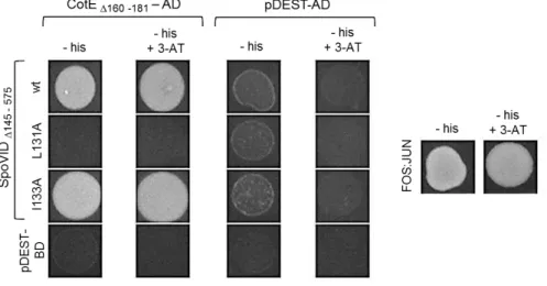
Specific residues in region E of SpoVID are required for binding to CotE ………..… 72
Residue L131 of SpoVID is dispensable for binding to SafA ………….. 73
Probing the function of cysteine residues within the N-terminal domain of SpoVID ………..… 74
The N-terminal domain of SpoVID is structured ……….. 76
DISCUSSION ………..… 80
ACKNOWLEDGEMENTS ……… 85
REFERENCES ………... 86
CHAPTER 3 – Binding of SafA to region E of SpoVID ………...…..….. 91
SUMMARY ……….………….. 93
INTRODUCTION ……….. 94
MATERIAL AND METHODS ……….. 97
RESULTS ………... 107
Specific residues in region E are essential for encasement by SafA ……….……….... 107
Residues involved in encasement by SafA are important for a direct interaction between SafA and SpoVID ………... 113
Residues in region E required for interaction with SafA have a role in anchoring SafA to the spore surface ……….. 114
Region E facilitates binding of SafA and CotE hubs to a second surface in SpoVID ………..………... 117
Residues required for encasement by both SafA and CotE are important for coat integrity ……… 121
xiii
REFERENCES ……….……… 135
CHAPTER 4 – The LysM module of SafA in spore morphogenesis ... 141
SUMMARY ……….…………... 143
INTRODUCTION ……… 144
MATERIAL AND METHODS ………...… 147
RESULTS ……… 155
Conserved features of the LysM domain of SafA ……….... 155 The LysM domain is required for SafA subcellular localization …... 157 The LysM domain is required for SafA association with
the cortex ……… 161
The LysM domain of SafA binds to spore peptidoglycan in vitro .… 163 The localization of SafA is dependent on cortex biogenesis ……...… 164 The LysM domain of SpoVID in peptidoglycan recognition
in vitro………... 167
DISCUSSION ………... 169
ACKNOWLEDGEMENTS ……….… 173
REFERENCES ………. 174
CHAPTER 5 – General discussion ………. 183 The major role of the region E of SpoVID in spore encasement …... 186 SpoVID organization as the driving force for encasement……… 188 SafA and CotE as hubs for inner and outer coat proteins ……….…… 188 SafA and CotE belong to the same kinetic class of coat proteins …. 190 The LysM modules of SpoVID and SafA as localization
determinants ……… 191
The interdependency on assembly of the spore protective
layers ……….……… 194
An updated model for spore coat assembly ……..……… 194
REFERENCES ……….……….… 196
xiv
DSM: Difco sporulation medium
DTT: dithiothreitol
EDTA: ethylenediamine tetraacetic acid
g: gram
GlcNAc: N-Acetylglucosamine
H: hour
IPTG: isopropyl β-D-1-thiogalactopyranoside
L: liter
LB media: Luria-Bertani medium
M: molar
MALDI-TOF: matrix assisted laser desorption ionization – time of flight
MCD pole: mother cell distal pole
MCP pole: mother cell proximal pole
MS: mass spectrometry
MurNAc: N-Acetylmuramic acid
PBS(-T): phosphate buffered saline(-tween)
PMSF: phenylmethanesulfonylfluoride
PVDF: polyvinylidene difluoride
SDS: sodium dodecyl sulfate
SDS-PAGE: sodium dodecyl sulfate polyacrylamide gel electrophoresis
SM: sporulation medium
TBS-T: tris-buffered saline- tween
TSDS-PAGE: tricine-sodium dodecyl sulfate polyacrylamide gel
1 Endospores, or spores for simplicity, are a highly resistant cell type
produced by some bacterial species under adverse conditions. Two main
protective layers contribute to the resilience of spores: the cortex, composed
of peptidoglycan, and the outermost proteinaceous coat. In Bacillus subtilis,
the coat comprises up to 80 different proteins, organized into four sublayers:
the basement layer, the inner coat, the outer coat and the crust. These
proteins are synthesized at different times during sporulation and deposited
at the spore surface in multiple coordinated waves. Central to coat formation
is a group of morphogenetic proteins that guide the assembly of the coat
components. Targeting of the coat proteins to the surface of the developing
spore is mainly controlled by the SpoIVA morphogenetic ATPase. In a second
stage, the coat proteins fully encircle the spore, a process termed encasement
that requires the morphogenetic protein SpoVID. Assembly of the inner coat
requires SafA, whereas formation of the outer coat and the crust requires
CotE. SafA interacts directly with the N terminus of SpoVID.
In Chapter 2, we started by demonstrating that a stretch of 12
residues at the N-terminal domain of SpoVID, that we named region E, is
required for spore encasement by all the coat sublayers. We identified
specific residues within this region that are important for binding and
encasement by CotE, linking encasement by the outer coat to a specific
protein-protein interaction. Encasement is presumably facilitated by the
oligomerization of SpoVID at the spore surface, and we showed that SpoVID
oligomers contain disulfide bonds. Finally, we addressed some structural
features of SpoVID, and we proposed an updated model for coat assembly
that incorporates the interactions between the major coat morphogenetic
proteins.
In Chapter 3, we focus on the interaction between SpoVID and SafA.
2
encasement by both the inner and the outer coat modules require specific
interactions of their hubs (SafA and CotE) with the same region of SpoVID.
Our results also suggest that region E facilitates binding of SafA and CotE to a
second surface in SpoVID. Furthermore, two residues within region E are
required for encasement by both SafA and CotE hubs, and consequently, by
inner and outer coat/crust components, having a major role in coat integrity.
As the substitution of one of them do not preclude binding of CotE to SpoVID
and mislocalization of CotE is suppressed by the deletion of safA, we conclude that a mislocalized inner coat acts as an attractor for CotE. This
reveals a tight connection between assembly of the inner and the outer coat
modules.
In Chapter 4, we analysed the role of the peptidoglycan-binding LysM
domain of SafA on the subcellular localization and function of the protein.
Within this domain, we identified five conserved, surface-exposed residues
that are required for SafA-YFP localization and peptidoglycan recognition in vitro. Of those, two are important for earlier SafA-YFP deposition, whereas the other three are involved in late localization, at a time where the cortex is
already present in the spore. Late localization of SafA-YFP also requires the
cortex, reinforcing that cortex and coat morphogenesis are linked. Finally, we
provide evidence that the LysM module of SpoVID does not recognize
peptidoglycan in our experimental conditions. Altogether, we propose a
model for SafA localization that includes the interactions with SpoIVA and
SpoVID (via region A), as well as peptidoglycan recognition (via LysM).
Overall, our work expands the current model for coat assembly, and
highlights the role of protein-protein interactions during coat
3 Endósporos, ou simplesmente esporos, são um tipo celular
extremamente resistente produzido por algumas espécies bacterianas em
resposta a condições adversas. Duas camadas protetoras contribuem para a
resiliência destas estruturas: o cortex, composto por peptidoglicano, e mais
externamente o manto, de natureza proteica. Em Bacillus subtilis, o manto é composto por mais de 80 proteínas diferentes, organizadas em quatro
subcamadas: a camada basal, o manto interno, o manto externo e a crosta.
Estas proteínas são sintetizadas em diferentes etapas da esporulação e
depositam-se à superfície do esporo de uma forma coordenada, através de
múltiplas ondas. As proteínas morfogenéticas tem um papel central na
formação do manto, guiando a deposição dos seus componentes. A
localização das proteínas à superfície do esporo em desenvolvimento é
essencialmente controlada pela ATPase morfogenética SpoIVA. Numa
segunda fase, as proteínas do manto rodeiam completamente o esporo, num
processo designado por envolvimento que requer a proteína morfogenética
SpoVID. A montagem do manto interno depende de SafA, enquanto que as do
manto externo e da crosta necessitam de CotE. SafA interage diretamente
com o N-terminal de SpoVID.
No Capítulo 2, começámos por demonstrar que uma região de 12
resíduos no domínio N-terminal de SpoVID, a que chamámos região E, é
essencial para o envolvimento do esporo pelas quatro subcamadas do manto.
Nesta região, identificámos resíduos específicos importantes para a interação
com CotE e para a sua migração em redor do esporo, estabelecendo uma
relação entre o envolvimento pelo manto externo a uma interação
proteína-proteína específica. Presume-se que o envolvimento seja facilitado pela
oligomerização de SpoVID à superfície do esporo, e nós demostrámos que os
oligomeros de SpoVID contêm pontes dissulfídicas. Finalmente,
4
interações entre as proteínas morfogenéticas mais importantes.
No Capítulo 3, focámo-nos na interação entre SpoVID e SafA.
Identificámos substituições de aminoácidos na região E de SpoVID que levam
à deslocalização de SafA e previnem a sua ligação a SpoVID, demostrando que
o envolvimento do esporo pelos mantos interno e externo requer interações
específicas das suas proteínas hub (SafA e CotE) com a mesma região de SpoVID. Os nossos resultados sugerem ainda que a região E facilita a ligação
de SafA e CotE a uma segunda superfície em SpoVID. Além disso, dois dos
resíduos da região E são importantes para o envolvimento do esporo tanto
por SafA como por CotE, e consequentemente, pelos componentes dos
mantos interno e externo e da crosta, tendo um papel determinante na
integridade do manto. Tendo em conta que a substituição de um desses
resíduos não compromete a ligação de CotE a SpoVID e que a deslocalização
de CotE é suprimida pela deleção de safA, concluímos que a deslocalização do manto interno leva à deslocalização de CotE. Assim, concluímos que existe
uma estreita ligação entre a montagem destas subcamadas do manto.
No Capítulo 4, analisámos o papel do domínio de ligação a
peptidoglicano de SafA, LysM, na localização e função desta proteína.
Identificámos cinco resíduos conservados expostos à superfície do domínio
que são importantes para a localização de SafA-YFP e para o reconhecimento
de peptidoglicano in vitro. Destes, dois são necessários para a deposição inicial de SafA-YFP, enquanto os outros três estão envolvidos na sua
localização tardia, quando o cortex já está presente no esporo. A localização
tardia de SafA-YFP também requer a presença do cortex, reforçando que as
morfogéneses do cortex e do manto estão intimamente ligadas. Finalmente,
mostrámos que o domínio LysM de SpoVID não reconhece peptidoglicano
nas nossas condições experimentais. Deste modo, propusemos um modelo
5 esporo (via LysM).
Em suma, o nosso trabalho aprofunda o modelo atual da morfogénese
do manto, enfatizando a importância das interações proteína-proteína
Chapter
1
THE MODEL ORGANISM BACILLUS SUBTILIS
Bacillus subtilis is a Gram-positive, rod-shaped bacterium that is both
a soil organism and a gut commensal of several animals, including humans
(Nicholson, 2002; Tam et al., 2006; Hong et al., 2009). This facultative aerobe
(Hoffmann et al., 1995; Nakano and Zuber, 1998) is able to differentiate
highly resistant dormant cells named endospores, or spores for simplicity,
that contribute for its persistenceunder unfavorable conditions. B. subtilis is
an important model for studies of cell differentiation and one of the
best-characterized organisms. The main advantages of its use are the natural
competence, easy manipulation and maintenance, high growth rates and
non-pathogenicity, as well as the availability of its whole genome for almost
20 years (Kunst et al., 1997). These features, together with the spore
properties and the capacity to secrete high concentrations of several
products directly into the culture medium, makes B. subtilis well suited for
applications in industry, biotechnology and biomedicine (see below in
“Applications of spores”).
Spore-forming bacteria are diverse and ubiquitous. They include
several Bacillus and Clostridium species that form spores through an
evolutionary conserved mechanism, such as the pathogens B. cereus, B.
anthracis and C. difficile (de Hoon et al., 2010; Galperin et al., 2012; Abecasis
et al., 2013). Sporeformers have been isolated, for example, from fresh and
saline water, hot springs, arctic sediments, gastro-intestinal tracts of
mammals and insects and soil (Nazina et al., 2004; Yukimura et al., 2009;
Maughan and Van der Auwera, 2011), Their capacity to colonize such a
The discovery of B. subtilis and of bacterial spores
Although infections by sporeformers were reported in ancient
literature thousands of years ago (Torred et al., 2012), the first known
descriptions of spores are only dated from the 19th century. In 1852, Maximilian Perty observed light-refractile bodies in bacteria. Almost 20
years later (1870), Luis Pasteur confirmed these observations and correlated
the presence of spores to a greater resistance to injurious agents (as cited in
Morrison and Rettger, 1930). At that time, Henry Charlton Bastian showed
that some bacteria could regrow in organic fluids after boiling them in sealed
flasks (Bastian, 1872; in Torred et al., 2012), and Ferdinand Cohn proposed
that it may be due to a special developmental stage of some bacteria that
survive high temperatures (as cited in Torred et al., 2012). Cohn found that
the bacteria growing in boiled cheese infusions was not the typical
putrefactive Bacterium termo, but rather bacillus rods he renamed Bacillus
subtilis (Cohn, 1872; in Torred et al., 2012). This organism was described 40
years earlier and baptized Vibrio subtilis (Ehrenberg, 1833–1835; in Rasmussen et al., 2009). Cohn also observed the formation of spores in B.
subtilis and proved that they survive strong heat and germinate to form new
bacilli, justifying the microbial growth in boiled organics infusions (Cohn,
1876; in Torred et al., 2012 and Rasmussen et al., 2009). In the same year,
Koch relied on Cohn's observations in his work on B. anthracis (Koch, 1876;
in Torred et al., 2012). Their independent experiments marked the beginning
of the field of spore research.
(Morrison and Rettger, 1930; Rasmussen et al., 2009)
THE SPORE
Spores are a highly resistant dormant cell-type produced via a
differentiation process known as sporulation. They are one of the most
stresses that kill other cells. These include extreme heat, freeze, high
pressure, ionizing radiation, noxious chemicals — oxidizing agents, acids, bases and organic solvents — exogenous lytic enzymes and digestion by predators (Nicholson et al., 2000; Nicholson et al., 2002; Klobutcher et al.,
2006; Setlow, 2006).
In laboratory cultures, most of the strains initiate sporulation in
response to adverse conditions. First, the bacterial cell divides
asymmetrically and the larger compartment, named mother cell, encases the
smaller one. Then, the smaller compartment differentiates into a mature
spore, assisted by the mother cell. Finally, the mother cell lyses and the
mature spore is released into the environment (Errington, 2003), where it
can persist for long periods of time, perhaps even for millions of years
(Nicholson et al., 2000; Vreeland et al., 2000).
Morphology and properties of the spore
Spores exhibit a conserved concentric, multilayered architecture that
largely contributes to their resistance (de Hoon et al., 2010). In those
produced by B. subtilis, thin-section transmission electron microscopy
revealed three main concentric compartments: the core, the cortex and the
coat (Fig. 1.1). Species such as B. cereus and B. anthracis also have an
additional outer layer, named exosporium (Driks, 1999; Henriques and
Moran, 2007).
The core is the innermost compartment of the spore, and houses the
chromosome. It has a reduced water content, which is important for spore
dormancy and resistance to wet heat. The presence of dipicolinic acid,
presumably chelated with divalent cations (predominantly Ca2+), contributes to dehydration, protection of the DNA and spore resistance. This
compartment also contains small acid-soluble proteins of the α/β-type that saturate the DNA, protecting it from a wide range of damaging agents, such
1989; Setlow, 2006). The core is delimitated by the inner forespore
membrane, with similar composition to the plasma membrane. This
membrane is a strong permeability barrier to noxious chemicals and
contributes for the reduced water content in the core. Additionally, it
becomes the membrane of the new vegetative cell that results from spore
germination (Nicholson et al., 2000; Cortezzo et al., 2004; Setlow, 2006).
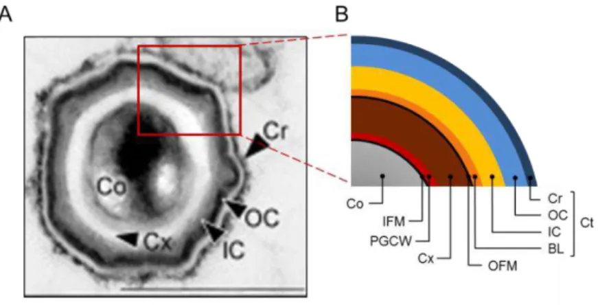
Figure 1.1 – Structure of the B. subtilis spore. A) Thin-section transmission electron micrograph of a B. subtilis spore fixed and stained with ruthenium red. Scale bar: 500nm (in McKenney et al., 2010); B) Schematic representation of the structure of B. subtilis
spores. Co: core, IFM: inner forespore membrane, PGCW: primordial germ cell wall, Cx: cortex, OFM: outer forespore membrane, Ct: coat, BL: basement layer, IC: inner coat, OC: outer coat, Cr: crust.
Two layers of peptidoglycan surround the inner forespore
membrane: the primordial germ cell wall, in immediate contact with this
membrane, and the cortex. The primordial germ cell wall has an identical
composition to the vegetative cell peptidoglycan and, upon germination,
gives rise to the cell wall of the vegetative cell. The cortex is composed of
peptidoglycan with several spore-specific modifications, namely the
presence of muramic δ-lactam residues, the reduced number of peptide side chains and the absence of teichoic acids (see more details on Chapter 4). As a
result, the cortex peptidoglycan shows reduced cross-linking of the glycan
of the core, spore mineralization, dormancy, and also for protection against
organic solvents and heat (Popham, 2002; Setlow, 2006; Higgins and
Dworkin, 2012). At least during the initial steps of spore development, the
cortex is surrounded by the outer forespore membrane. This membrane is
essential during spore formation, but may be absent from mature spores
(Piggot and Hilbert, 2004; Setlow, 2006).
The next protective structure is the proteinaceous coat, arranged into
several layers. From the inside to the outside, the B. subtilis spore coat
comprises the basement layer, the inner coat, the outer coat and the crust
(Fig. 1.1). The coat prevents access of peptidoglycan lytic enzymes, such as
lysozyme, to the cortex and confers resistance to noxious chemicals, ionizing
radiation and predation. Moreover, it has a major role in spore adhesion,
infection by pathogenic sporeformers and in germination. In some species,
including B. subtilis, the coat is the outermost layer of the spore, mediating
the interaction with the surrounding environment. Thus, as an adaptation to
distinct ecological niches, the coat composition differs among species
(Nicholson et al., 2000; Riesenman and Nicholson, 2000; Klobutcher et al.,
2006; Setlow, 2006). In organisms such as B. cereus, B. anthracis and some
Clostridia, the spore coat is encased by an exosporium. This additional
protective layer is loose-fitting, with a paracrystalline basal layer and an
external hair-like nap, and is separated from the outer coat by an interspace
(Henriques and Moran, 2007; Higgins and Dworkin, 2012).
The spore protective surface layers — the cortex, the coat and the
exosporium — are flexible and elastic. This allows spore expansion or retraction according to environmental conditions, such as relative humidity
(Westphal et al., 2003).
Applications of spores
Bacterial spores, especially those produced by B. subtilis, have been
commercialized as probiotic preparations for animal and human
consumption (Barbosa et al., 2004; Cartman and La Ragione, 2004), or as
biocontrol agents in agriculture, promoting plant growth or preventing plant
diseases (Nicholson, 2002). Some of their applications exploit the capacity to
display bioactive molecules at the surface by genetic manipulation. This way,
they can be vehicles for vaccine delivery, biosensors for specific compounds,
or display systems for proteins or peptides of biotechnological interest
(Isticato et al., 2001; Ferreira and Schumann, 2012). In addition to their
resistance properties, they are easy and cheap to produce in larger scales and
can remain stored at room temperature for long periods without losing
viability. Understanding the mechanism of sporulation in B. subtilis is not
only important in fundamental research, but also in the development of
products of interest in industry and healthcare.
AN OVERVIEW OF SPORULATION
Endospore formation is an ancient, conserved process. The basic
sequence of steps along this developmental program is essentially
maintained in Bacilli and Clostridia species studied so far (de Hoon et al.,
2010). Sporulation is regulated by a program of gene expression, directed by
specific RNA polymerase sigma factors and additional transcription
regulators (Piggot, 2002; Errington, 2003; Hilbert and Piggot, 2004; Higgins
and Dworkin, 2012; Robleto et al., 2012). Cell-cell signaling pathways that
operate between the developing spore and the mother cell is also a hallmark
of the process. The genetic program dictates a series of morphological
changes that culminate in the formation of a mature spore. Spore
development represents a considerable investment of energy and time.
Laboratory cultures of B. subtilis take about 8-10 hours to complete the
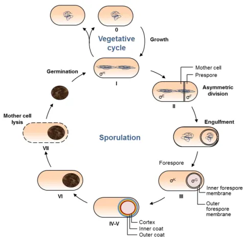
The morphological stages of sporulation
The basic sequence of morphologic changes along sporulation was
elucidated by electron microscopy. Although this is a continuous process,
several stages were considered for convenience (Ryter, 1965; Piggot and
Coote, 1976) (Fig. 1.2). Vegetative growth is known as stage 0. Stage I
corresponds to entry in sporulation, after a round of DNA replication. At this
stage, the two sister chromosomes rearrange into an axial filament that
stretches along the longitudinal plan of the cell. Then, an asymmetric septum
is formed by membrane invagination, resulting in two compartments with
different sizes and cell fates (stage II). The smaller compartment is the
prespore that differentiates into a mature spore. The larger one is the mother
cell, which participates in spore formation and ultimately lyse (Ryter, 1965;
Piggot and Coote, 1976).
After asymmetrical division, the septum bulges into the cytoplasm
and the membranes migrate around the prespore in a process similar to
phagocytosis, named engulfment. This results in a double-membrane free
protoplast, termed forespore, within the mother cell (stage III). During stage
IV, the primordial germ cell wall and the cortex deposit between the two
forespore membranes. Almost in parallel, the coat becomes visible around
the surface of the forespore, corresponding to stage V. Forespores, previously
phase-dark, become gradually phase-bright. Stage VI designates the
maturation of the spore and acquisition of resistant properties (Ryter, 1965;
Piggot and Coote, 1976).
In stage VII, the mother cell lyses through programed cell death and
the mature spore is released. The spore has the ability to remain dormant for
long periods of time in the absence of nutrients, and can be transported to
different, more favorable, locations. Even in a dormant state, it constantly
monitors the surrounding environment conditions through receptors in the
inner forespore membrane. As soon as the environmental conditions become
acids and sugars, the spore exits the dormant state and germinates (Ryter,
1965; Piggot and Coote, 1976).
Figure 1.2 – The vegetative and sporulation cycles in B. subtilis. The key morphological stages during sporulation (I-VII) are represented. 0: vegetative growth; I: chromosomal replication and formation of the axial filament; II: asymmetric division, followed by engulfment; III: engulfment completion; IV-V: cortex and coat deposition; VI: spores maturation; VII: mother cell lysis, releasing the spore that then germinates and returns vegetative growth or sporulation cycles. The RNA polymerase sigma factors are indicated in the stages were they become active.
Entry into sporulation in B. subtilis
The decision-making apparatus that regulates entry into sporulation
is precisely regulated and responds to a series of internal and external
signals (Errington, 2003; de Hoon et al., 2010). In domesticated strains of B.
nitrogen or phosphate source, although high population density is also
important (Piggot and Coote, 1976; Sonenshein, 2000; Piggot and Hilbert,
2004). These factors led to activation of Spo0A, the key transcription factor
for triggering sporulation. Spo0A is activated by phosphorylation through an
expanded two-component signal transduction system known as
phosphorelay (Burbulys et al., 1991; Jiang et al., 2000; Sonenshein, 2000;
Fujita and Losick, 2005; Chastanet et al., 2010). The levels of phosphorylated
Spo0A (Spo0A~P) are negatively regulated by specific phosphatases that
dephosphorylate Spo0A~P or other components of the phosphorelay system
(Perego et al., 1996; Bongiorni et al., 2007; Smits et al., 2007). Spo0A~P
controls the expression of more than 120 genes directly and 520 indirectly,
including several genes coding for transcription factors (Fawcett et al., 2000;
Molle et al., 2003). The concentration of Spo0A~P defines the physiological
outcome in the cell. High cellular levels of Spo0A~P led to sporulation by
activation of the transcription of genes coding for the first
sporulation-specific regulators (σF and σE), as well as of genes required for the formation of the axial filament and asymmetric division of the cell (Fujita et al., 2005;
Chastanet et al., 2010; Robleto et al., 2012). However, in moderate levels,
Spo0A~P triggers other less energy consuming stress responses, such as
biofilm formation, sessile behavior, cannibalism, antibiotic production, or
competence (Fujita et al., 2005; Robleto et al., 2012).
Another important positive regulator for entry into sporulation is the
RNA polymerase sigma factor σH, whose regulatory pathways are interconnected with those leading to the production of Spo0A~P. σH regulates transcription in the predivisional cell, controls the expression of
several genes involved in the phosphorelay, including spo0A, and is essential
for asymmetric division (Britton et al., 2002; Molle et al., 2003; Hilbert and
Piggot, 2004).
Asymmetric septum formation is initiated by redirecting the division
finishing the asymmetrical division, only 1/3 of the chromosome is trapped
in the prespore (Fig 1.2, stage II). Thus, a DNA translocase is required for
translocation of the remaining chromosome (Wu and Errington, 1997).
Compartmentalized gene expression in B. subtilis
Immediately after asymmetric division, two parallel programs of
gene expression start in each daughter cell, directed by different,
sporulation-specific, RNA polymerase sigma factors. The first sigma factor to
become active is σF in the prespore, followed by σE in the mother cell; after engulfment, they are replaced by σG and σK, respectively (Piggot and Hilbert, 2004).
σF is synthesized before asymmetric division and held inactive by association with an anti-σ factor. This association is untied by interaction with an anti-anti-σ factor, activating σF. The anti-anti-σ factor is regulated by phosphorylation and dephosphorylation cycles that involve a phosphatase
present in higher levels in the prespore. This contributes to restrict σF activity to this compartment (Min et al., 1993; Arigoni et al., 1996; Decatur
and Losick, 1996; Duncan et al., 1996). Another factor that regulates σF activity is the transient chromosomal asymmetry generated during the
asymmetrical division. Since the gene encoding anti-σ is one of the latest being translocated into the prespore, there is a time gap where it is not
expressed in that compartment, allowing the presence of non-associated,
active σF (Errington, 2003). The products of genes controlled by σF mediate activation of σE in the mother cell, DNA repair and communication with the mother cell compartment. Also, σF directs the transcription of genes encoding σG and germination receptors (Robleto et al., 2012).
whose products are involved in engulfment, mother cell metabolism and
cortex and coat formation. It also directs activation of σG and synthesis and activation of σK and the transcription factors SpoIIID and GerR. These two regulators act together with σE to activate or repress the expression of several genes involved in sporulation and germination (Robleto et al., 2012).
About 1 h after asymmetric division, the prespore is engulfed by the
mother cell. Membrane migration around the prespore only occurs if the
septal peptidoglycan is hydrolyzed by several σE-dependent enzymes (Guinand et al., 1974; Abanes-De Mello et al., 2002; Morlot et al., 2010).
Engulfment also requires a channel composed by proteins synthesized in
both compartments that may function as a feeding tube to nurture the
forespore or may drive the membrane movements around it (de Hoon et al.,
2010; Higgins and Dworkin, 2012; Crawshaw et al., 2014). After engulfment
completion, σG is activated in the forespore to direct the final stages of spore development, together with σK in the mother cell. The gene coding for an inactive form of σG is transcribed only in the forespore under σF control, and its synthesis and activation require signals from the two compartments (Sun
et al., 1989). σG activation also involves at least one component of the channel mentioned above. Thus, either the channel is required to import specific
regulatory factors to activate σG, or its assembly is an engulfment checkpoint (Errington, 2003; Blaylock et al., 2004; Meisner et al., 2008; de Hoon et al.,
2010). A third factor that regulates activity of σG is an anti-σ factor that prevents pre-engulfment activation and avoids a rapid increase of its levels
in the forespore (Serrano et al., 2011). σG-dependent genes encode important factors for σK activation, cortex morphogenesis, acquisition of the spore resistance properties and germination (Robleto et al., 2012).
σK is the last sporulation-specific sigma factor being activated, and its activity is regulated both at transcriptional and post-translational levels. The
SpoIIID-dependent recombinase (Stragier et al., 1989; Sato et al., 1990). Also, σK is transcribed as an inactive precursor, and is proteolytically activated by a
protease that requires σE and σG-dependent elements (Dong and Cutting, 2003). σK directs the expression of factors involved in coat formation, germination and mother cell autolysis. It is also required for activation of the
transcription factor GerE, which activates and represses the expression of
several genes under σK control (Robleto et al., 2012).
THE SPORE COAT IN
B. SUBTILIS
Morphogenesis of the spore coat in B. subtilis is a model system to
study the formation of supramolecular structures at specific subcellular sites,
in coordination with gene expression cascades, during a cell differentiation
pathway. Understanding these mechanisms is a major goal in developmental
biology, since all organisms share the challenge of directing the assembly of a
vast number of proteins into functional molecular machines. Viral capsids
(Dokland et al., 1997), bacterial flagellum (McCarter, 2006), the divisome
(Goehring and Beckwith, 2005) or the clathrin-coated vesicles in eukaryotic
cells (Kaksonen et al., 2005) are a few examples. Despite the diversification
of these supramolecular machines in terms of structure and function, their
assembly follows the same basic rules.
Structure of the coat
In mature spores of B. subtilis, the coat commonly varies between 60
and 250 nm width, appearing thicker at the spore poles (Driks, 1999;
Henriques and Moran, 2007). This structure is composed of more than 80
different proteins, whose main features are referred in Table A1 of the
Appendices. Transmission electron microscopy revealed that the B. subtilis
the basement layer or undercoat, the inner coat, the outer coat and the crust
(Fig. 1.1). The basement layer, with an amorphous appearance, is the
innermost one. It contains proteins involved in initial steps of coat assembly
and is presumably important to bridge the cortex and the coat (Driks, 1999;
Henriques and Moran, 2007; McKenney et al., 2013). Surrounding the
basement layer, there is the lightly staining, lamellar inner coat, delimited by
the electron-dense, striated outer coat. Together, these two substructures fill
most of the coat volume (Driks, 1999; Henriques and Moran, 2007;
McKenney et al., 2013). The outermost layer is the crust, which was
identified by staining the spores with ruthenium red before electron
microscopy (Waller et al., 2004). This compound marks acid polysaccharides,
indicating that the crust comprises glycosylated components. As this is a
characteristic of the exosporium of other sporeformers, the crust may
represent a rudimentary exosporium in B. subtilis (Waller et al., 2004;
McKenney et al., 2010; McKenney et al., 2013). The existence of these four
spatially distinct coat sublayers is supported by fluorescence microscopy
coupled with high-resolution image analysis. By mapping the subcellular
localization of several coat proteins fused to GFP or its derivatives and
determining their genetic dependencies, coat components were grouped in
four substructures that correspond to the layers described before (Imamura
et al., 2010; McKenney et al., 2010).
Recently, B. subtilis mutant spores missing some important proteins
for coat assembly were examined by high-resolution atomic force
microscopy. This study supports the existence of the four coat layers
previously reported and add others not identified before. According to the
authors, the coat is organized into the basement layer or undercoat, a
multilayer structure that possibly corresponds to the inner coat, a layer of ‘‘nanodot’’ particles that may be CotE molecules, a fibrous/granulous layer that likely corresponds to the outer coat, a honeycomb layer, a rodlet layer
Coat composition
About 70% of the B. subtilis coat proteins can be solubilized and
extracted from purified spores by alkali treatment or using detergents and
reducing agents. Proteins within this soluble fraction can then be resolved by
SDS-PAGE, individually extracted from the gel and partially sequenced
(Henriques and Moran, 2007; Aronson, 2012). This approach, coupled with
reverse genetics, led to the identification of the first coat proteins. Nowadays,
mass spectrometry, fluorescence microscopy and global transcriptional
profiling of the mother cell compartment have been used to find additional
coat components (van Ooij et al., 2004; Kim et al., 2006). Coupling
fluorescence microscopy with high-resolution image analysis, it is possible
not only to identify proteins that compose the coat, but also to assign them to
a particular sublayer (Imamura et al., 2010; McKenney et al., 2010).
Most of the identified coat proteins are structural components,
although there are also enzymes with important roles, such as
post-translational modifications, spore resistance and germination (Table A1 of
Appendices). For example, Tgl is a transglutaminase that cross-links several
coat proteins (Zilhao et al., 2005), SodA is a superoxide dismutase that
provides protection against toxic oxidative compounds (Henriques et al.,
1998), and CwlJ is a cortex lytic enzyme essential for germination (Ragkousi
et al., 2003).
As mentioned before, the composition of the coat varies between
species, reflecting the physical and nutritional conditions where the spores
developed and persist (Nicholson et al., 2000; Riesenman and Nicholson,
2000; Klobutcher et al., 2006; Setlow, 2006; Aronson, 2012). Thus, most coat
proteins are not conserved among sporeformers. Genome sequence
comparison suggests that only about half of the known B. subtilis coat
proteins have orthologous in other Bacillus, and the number is even lower if
comparing to Clostridium species (Henriques and Moran, 2007;
McKenney et al., 2013) (Table A1 of Appendices). However, some
species-specific coat proteins have functional analogues in other sporeformers. For
example, SpoVID is a Bacillus protein essential for migration of the coat
components around thespore (Beall et al., 1993; Wang et al., 2009), and SipL
is its functional analogous in Clostridium difficile (Putnam et al., 2013).
SpoVID and SipL have a high degree of divergence, but both interact with
SpoIVA (that directly recruits SpoVID to the surface of the spore) and their
absence result in similar phenotypes in sporulating cells (Beall et al., 1993;
Wang et al., 2009; Putnam et al., 2013). This is a case where two organisms
developed different structural solutions for the same biological challenge. In
opposition, SpoIVA is one of the most conserved coat proteins in Bacillus and
Clostridium species, but while in B. subtilis it has a role in both cortex and
coat assembly, it is not required for cortex formation in C. difficile (Roels et
al., 1992; Henriques and Moran, 2007; Putnam et al., 2013).
Regulation of coat assembly
Coat assembly begins as soon as the asymmetric septum starts to
curve and proceeds even after spore release into the environment. Along the
time, coat proteins are synthesized in the mother cell and deposit at the
surface of the developing spore. Initially, they accumulate at the mother
cell-proximal (MCP) pole and then they encircle the spore in successive waves
(Henriques and Moran, 2000; Errington, 2003; Henriques and Moran, 2007;
McKenney and Eichenberger, 2012; McKenney et al., 2013). Coat
morphogenesis is regulated at three levels: the time and localization of
synthesis of coat components, the guiding role of a small group of coat
proteins named morphogenetic proteins (see below) and the
post-translational modifications of the coat components (Henriques and Moran,
2007).
The genes coding for coat proteins are organized in four regulons
expressed in a hierarchical cascade, forming feed forward loops in which one
of the regulators is required for synthesis of a second one, and together they
control the expression of several target genes. This assures that coat proteins
are synthesized in a specific order and in the right compartment (Driks,
1999; Henriques and Moran, 2000; Eichenberger et al., 2004; Steil et al.,
2005). Genes within the first regulon are expressed under control of σE just before the asymmetric septum begins to curve. They include spoVM, spoIVA,
spoVID, safA and cotE, which encode essential proteins for initiation of coat
assembly. This regulon also comprises the genes coding for GerR, SpoIIID
and σK. GerR represses the transcription of some genes under control of σE, while SpoIIID acts as a positive or negative regulator of the expression of
several σE-dependent genes. σK, whose activation requires the concerted action of σE and SpoIIID, promotes the transcription of a large group of genes coding for coat proteins, as well as of gerE. In turn, GerE represses the
expression of several σE-dependent genes, and together with σK, activates transcription of the genes that compose the last regulon (Driks, 1999;
Henriques and Moran, 2000; Eichenberger et al., 2004; Steil et al., 2005;
Henriques and Moran, 2007).
As the coat components are synthesized, they are guided to their
specific subcellular sites by morphogenetic proteins (see below) and the coat
is gradually formed. Protein-protein interactions, cross-linking, proteolytic
processing, glycosylation and other post-translational modifications
contribute to the assembly process (Henriques and Moran, 2007; Aronson,
2012). For instance, in the final stages of coat morphogenesis, several
proteins are cross-linked by enzymes, such as Tgl and probably SodA, or via
disulfide bridges. Tgl-dependent cross-linking in fact occurs mainly following
spore release through lysis of the mother cell; hence, the released spore has
an unfinished coat whose maturation path will depend on the environmental
The role of morphogenetic proteins in coat assembly
Morphogenetic proteins are a small subset of coat proteins whose
function is to guide coat assembly. They localize at the surface of the
prespore at earlier stages and direct the other coat components to their
specific cellular sites. The role of these proteins is exclusively in protein
localization, not interfering with their synthesis. In B. subtilis, the most
important morphogenetic proteins are SpoVM, SpoIVA, SpoVID, SafA and
CotE. The absence of each of these proteins results in spores missing the coat
or exhibiting a coat with severe defects (Driks et al., 1994; Henriques and
Moran, 2000, 2007; McKenney et al., 2013).
SpoVM:
SpoVM is among the first proteins assembling at the surface of the
prespore. The transcription of its gene initiates 2 hours after the onset of
sporulation, and the protein starts localizing at the asymmetric division
septum shortly after it begins to curve. Then, SpoVM tracks along the
engulfing membrane, surrounding the forespore (Levin et al., 1993; van Ooij
and Losick, 2003; Le and Schumann, 2008).
This morphogenetic protein is important for assembly of both the
cortex and the coat. In a spoVM null mutant, spores exhibit an incipient cortex
and the coat is thin, disorganized and loosely attached. Fluorescence
microscopy experiments using 40 coat proteins-GFP fusions showed that in
the absence of SpoVM, most coat proteins are recruited to the MCP pole of
the spore, but fail to encircle it. As a result, mutant spores are sensitive to
heat, organic solvents and lysozyme (Levin et al., 1993; Wang et al., 2009).
SpoVM is a 3 kDa, 26 residues peptide that folds into an amphipathic
α-helix in the presence of lipids. This helix recognizes the positive curvature of the outer forespore membrane and localizes at its surface according to a
specific orientation. The cytosolic side of the helix contains six positive
opposite face, buried in the phospholipid bilayer (Levin et al., 1993; Prajapati
et al., 2000; van Ooij and Losick, 2003; Ramamurthi et al., 2006; Le and
Schumann, 2008; Ramamurthi et al., 2009) (Fig. 1.3 A). The three
hydrophobic residues on the cytosolic side of the helix (F3, I6 and P9)
localize at the N terminus and are essential for SpoVM localization and
function. Localization of SpoVM also requires SpoIVA to restrict it to the
spore surface, and residue I6 is involved in a direct interaction between these
proteins (Prajapati et al., 2000; van Ooij and Losick, 2003; Ramamurthi et al.,
2006). Moreover, residue I15 is essential for cortex formation, but not for
SpoVM localization or for initiation of coat assembly (Ebmeier et al., 2012).
SpoIVA:
SpoIVA is present in all sporeformers examined to date, which is a
strong indication of its importance. It starts localizing earlier at the MCP pole
of the prespore and then encircles it (Roels et al., 1992; Driks et al., 1994;
Price and Losick, 1999; Henriques and Moran, 2007). As SpoVM, SpoIVA is
involved in both cortex and coat formation. spoIVA null mutant spores miss
the cortex, and coat proteins fail to assemble at the spore surface, forming
swirls dispersed at the mother cell cytoplasm (Roels et al., 1992; Stevens et
al., 1992). These swirls are composed by partially structured material,
supporting that coat components are able to self-assemble (Aronson and
Fitz-James, 1971; Aronson et al., 1992).
SpoIVA has 492 residues and comprises an N and a C-terminal
conserved domains (Price and Losick, 1999) (Fig 1.3 B). The N-terminal
domain, corresponding to the first 240 residues, has the characteristic fold of
GTPases. It includes a highly conserved Walker A motif between residues 24
and 31 that is responsible for ATP binding and hydrolysis. This motif is
essential for SpoIVA self-assembly into cable-like structures in an
ATP-dependent manner, a mechanism presumably required for the formation of a
Figure 1.3 – Schematic representation of the main morphogenetic proteins in B. subtilis
spore coat. A) The amphipathic α-helix SpoVM, with its hydrophobic and positive charged faces; B) SpoIVA, comprising the GTPase N-terminal domain (G), with the Walker A motif, and the C-terminal localization region (C); C) SpoVID consists of an N-terminal domain (N), a middle extended region (M) and a C-terminal targeting region composed by region A and the LysM domain; D) SafA has a LysM domain at the N terminus, followed by region A, essential for interaction with SpoVID. The region that composes the SafAC30 form is
assembly. Furthermore, the Walker A motif, in particular residue K30, is
involved in SpoIVA localization, indicating that the ATPase activity may be
important for its targeting at the spore surface (Ramamurthi and Losick,
2008; Mullerova et al., 2009; Castaing et al., 2013). The SpoIVA C-terminal
domain comprises the last 82 residues and interacts with SpoVM. These two
morphogenetic proteins are co-dependent for localization: SpoIVA requires
SpoVM to encircle the spore, and in turn SpoIVA helps restricting SpoVM to
the spore surface (Price and Losick, 1999; Ramamurthi et al., 2006). SpoIVA
is also essential for subcellular localization of three others morphogenetic
proteins: SpoVID, SafA and CotE (Driks et al., 1994; Ozin et al., 2001b; Wang
et al., 2009). Direct interactions of SpoIVA with SpoVID and SafA were
already demonstrated in vitro (Mullerova et al., 2009; Wang et al., 2009; Qiao
et al., 2012).
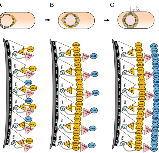
SpoVID:
SpoVID is synthesized 2 hours after asymmetrical division and
encircles the spore in a SpoVM-dependent manner. In the absence of this
protein, sporulating cells exhibit a phenotype reminiscent of the spoIVA null
mutant, in the sense that coat proteins also accumulate in the mother cell
cytoplasm in partially structured swirls (Beall et al., 1993; Driks et al., 1994).
Fusions of coat proteins to GFP localize at the MCP pole in spoVID null mutant
spores, but fail to encircle them, similarly to what happens in cell missing
SpoVM (Wang et al., 2009). However, contrarily to spoVM and spoIVA null
mutants, the cortex is not affected. As a result, spoVID mutant spores are
susceptible to lysozyme, but not to high temperatures, and are deficient in
germination (Beall et al., 1993; Driks et al., 1994).
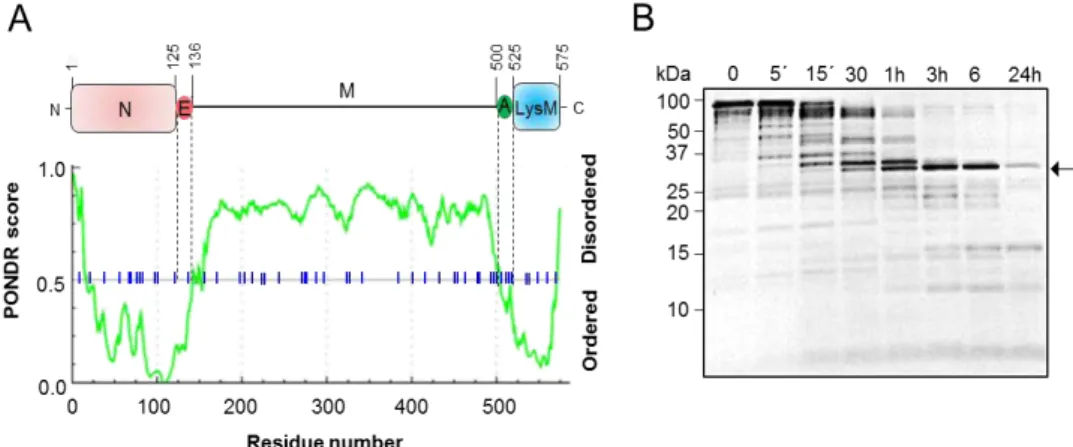
This 575 residues long, highly acidic protein is composed by a
N-terminal domain, a middle domain with an extended coiled-coil conformation
and a C-terminal region comprising the 24 residues region A and a LysM
2009) (Fig. 1.3C). The N-terminal domain has 137 residues, resembles the
phage-capsid protein PP7, and is essential for the morphogenetic function of
SpoVID. In particular, a small region at the end of this domain (residues
86-136) is required for migration of coat components around the developing
spore (Wang et al., 2009) (see Chapters 2 and 3). The N-terminus of SpoVID
interacts directly with the morphogenetic protein for the inner coat
assembly, SafA (Costa et al., 2006).
The C-terminal region of SpoVID is required for the assembly at the
spore surface. Both region A, via interaction with SpoIVA, and the LysM
domain are essential for SpoVID localization, although it seems that the
major contribution comes from region A. (Mullerova et al., 2009; Wang et al.,
2009). Since LysM motifs are commonly glycan-binding modules, it has been
suggested that its role in SpoVID localization might implicate spore
peptidoglycan recognition (see Chapter 4). However, it remains to be
explained how the LysM domain binds to the spore peptidoglycan in the
presence of the outer forespore membrane.
SpoVID oligomerizes in vitro (Ozin et al., 2000; Mullerova et al.,
2009), and oligomerization may be important for the encasement process.
Disulfide bridges may be involved, as SpoVID has five cysteine residues, two
of them in the N-terminal domain. The homologous phage-capsid protein PP7
also has two cysteines that are responsible for oligomerization during
assembly of the viral capsid (Tars et al., 2000; Caldeira and Peabody, 2007).
SafA:
SafA, also known as YrbA, localizes at the cortex/coat interface and is
essential for inner coat morphogenesis. safA null mutant spores exhibit a
reduced inner coat and a loosely attached outer coat that tends to peel off.
These spores are sensitive to lysozyme and deficient in germination
(Takamatsu et al., 1999; Ozin et al., 2000). Moreover, at least CotG is absent
inner coat assembly, it also makes important contributions for outer coat
formation (Ozin et al., 2000).
SafA is a proline-rich protein that has at least three forms in
sporulating cells: the 45 kDa full-length form, comprising 387 residues, a 30
kDa form named SafAC30 that results from alternative internal translation starting from M164 (Fig. 1.3 D), and a 21 kDa N-terminal form termed
SafAN21 (Ozin et al., 2000; Takamatsu et al., 2000b; Ozin et al., 2001a). The full-length form starts accumulating earlier, as the asymmetric septum
begins to curve (Ozin et al., 2000; Takamatsu et al., 2000a), and has the
morphogenetic role in coat assembly (Ozin et al., 2001a). In contrast, neither
SafAN21 nor SafAC30 are essential for coat formation. SafAC30 requires the full-length form of SafA for localization, and interacts with the full full-length protein
in vitro (Ozin et al., 2001b).
SafA has a LysM domain at its N-terminal end, followed by a
conserved region A (residues 51-63) that interacts directly with SpoVID (Fig.
1.3 D). This interaction is essential for SafA localization and function, and
consequently for proper coat assembly. At the C-terminal half of SafA there is
another region of interaction with SpoVID, named region B or PYYH motif
(residues 203 and 206). Region B is important for formation of the
SpoVID-SafA complex and its deletion causes deficient formation of the coat (Ozin et
al., 2000; Ozin et al., 2001b; Costa et al., 2006).
SafA requires SpoVID to encircle the spore, yet its initial localization
at the MCP pole depends on SpoIVA (Ozin et al., 2001b; Wang et al., 2009).
SafA establishes a direct interaction with this protein, but it appears weaker
than the interaction with SpoVID (Mullerova et al., 2009; Qiao et al., 2012). It
has been suggested that the LysM domain may also have a role in SafA
localization via spore peptidoglycan binding, but again it is not clear how the
LysM crosses the outer forespore membrane to reach it (Ozin et al., 2000;
Ozin et al., 2001b) (see Chapter 4).
exception of the N-terminal region, it is possible that it extends radially from
the cortex until reaching the outer coat. The LysM domain would be buried
into the peptidoglycan layer, while the C-terminal region interacts with other
coat proteins. The SafAC30 form may contribute to increase the surface available for interactions with coat components (Ozin et al., 2000; Ozin et al.,
2001b).
CotE:
CotE is essential for assembly of the outer coat and the crust. Proteins
that constitute these layers fail to localize properly in cotE null mutants,
resulting in germination-deficient spores with susceptibility to lysozyme
(Zheng et al., 1988; Kim et al., 2006). However, likewise the major
morphogenetic protein for inner coat formation, SafA, has a role in outer coat
integrity, CotE also contributes to inner coat morphogenesis. It is important
for the retention of at least some inner coat proteins, such as OxdD, at the
spore surface and may be involved in a direct interaction with some of them
(Zheng et al., 1988; Kim et al., 2006; Henriques and Moran, 2007).
Production of CotE is regulated at the transcription level by two
tandem promotors. At first, cotE is expressed from promotor P1, controlled
by σE, while in a second stage it requires promotor P2, initially regulated under jointed control of σE and SpoIIID and that remains active under control of σK (Costa et al., 2007). CotE starts localizing at a 75 nm distance from the surface of the forespore, forming a ring that may be the basal layer for
assembly of the outer coat. The gap between the basement layer and this
ring, whose composition is unknown, is called matrix or precoat and defines
the place for inner coat assembly after engulfment (Driks et al., 1994;
Henriques and Moran, 2007).
CotE, with 181 residues, has a modular design. The N-terminal
domain and the C-terminal region (residues 1-106 and 153-181,
107-141 are involved in CotE localization and, together with a second region
in the N-terminal half of the protein (residues 58-75), in oligomerization (Fig.
1.3 E). The ability to form oligomers may facilitate the formation of the CotE
ring around the spore (Bauer et al., 1999; Little and Driks, 2001; Krajcikova
et al., 2009).
CotE depends on SpoIVA to localize at the surface of the prespore and
on SpoVID to encircle it (Driks et al., 1994; Wang et al., 2009). In turn, CotE is
essential for the localization of CotH and CotO, two other morphogenetic
proteins with roles in assembly of small subsets of coat proteins, in
germination and in lysozyme resistance (Naclerio et al., 1996; Zilhao et al.,
1999; Eichenberger et al., 2003; McPherson et al., 2005).
Moreover, CotE is required for assembly of CotW, CotX and CotZ,
which are important in crust morphogenesis. This outermost layer is mainly
composed by proteins synthesized at later stages. Most of them are
cysteine-rich and highly cross-linked, being part of the coat insoluble fraction. In the
absence of the crust, spores show altered surface properties, specially
hydrophobicity, and increased accessibility to germinants (Zhang et al., 1994;
Kim et al., 2006; Henriques and Moran, 2007; Krajcikova et al., 2009;
McKenney et al., 2010; Imamura et al., 2011).
The coat genetic interaction network
Subcellular localization, genetic dependencies and direct interactions
between coat proteins provide us clues on the mechanisms governing their
recruitment and assembly at specific subcellular sites. SpoVM, SpoIVA and
SpoVID are required for formation of all the coat sublayers (Roels et al.,
1992; Stevens et al., 1992; Beall et al., 1993; Levin et al., 1993; Wang et al.,
2009). SpoVM and SpoIVA interact directly and are co-dependent for
localization, while SpoVID depends on both to localize and binds at least
SpoIVA (Price and Losick, 1999; Ramamurthi et al., 2006; Mullerova et al.,