Characterization of the
oml
A gene from different serotypes
of
Actinobacillus pleuropneumoniae
: A new insight into an old approach
Ciro César Rossi, Elza Fernandes de Araújo, Marisa Vieira de Queiroz and Denise Mara Soares Bazzolli
Laboratório de Genética Molecular de Micro-organismos, Departamento de Microbiologia,
Universidade Federal de Viçosa, Viçosa, MG, Brazil.
Abstract
The OmlA protein is a virulence factor ofActinobacillus pleuropneumoniae, an important pathogen in pigs. The polymorphisms present in the omlA gene sequence of 15 reference serotypes of A. pleuropneumoniae and non-serotypable isolates were assessed to determine the possible evolutionary relationship among them and to vali-date the importance of this gene as a molecular marker for the characterization of this bacterium. Divergence among the 15 serotypes ofA. pleuropneumoniae probably resulted initially from two major evolutionary events that led to subsequent differentiation into nine groups. This differentiation makes it possible to characterize most of the serotypes by using bionformatics, thereby avoiding problems with immunological cross-reactivity. A conserveda -he-lix common to all the serotypes was most likely involved in connecting the protein to the outer membrane and acting as a signal peptide. A previously unknown gene duplication was also identified and could contribute to the genetic variability that makes it difficult to serotype some isolates. Our data support the importance of theomlA gene in the bi-ology ofA. pleuropneumoniae and provide a new area of research into the OmlA protein.
Keywords:Actinobacillus pleuropneumoniae, omlAgene, phylogenetic reconstruction, porcine pleuropneumonia.
Received: November 30, 2012; Accepted: January 29, 2013.
Introduction
Swine pleuropneumonia (SPP) is a significant respi-ratory disease and has been reported in all countries where pig farming is intensively practiced. The etiological agent of SPP is the Gram-negative coccobacillusActinobacillus pleuropneumoniae, currently divided in 15 serotypes that are defined based on the antigenic properties of capsule polysaccharides. Although all of the serotypes are capable of causing SPP, differences in virulence make the sero-typing of field isolates ofA. pleuropneumoniaea key factor in the epidemiological study and control of this disease (Schuchertet al., 2004).
Numerous assays have been developed for the sero-logical characterization ofA. pleuropneumoniae isolates. Although immunological assays are relatively fast, their main limitation is that they commonly show cross-reactivity (Jessinget al., 2003). Various molecular tech-niques have also been used to study the molecular epidemi-ology of bacterial pathogens and are extremely important in monitoring the characteristics of a given population (Ashis
et al., 2012). Current approaches seek to develop molecular markers that can complement the sometimes inconclusive
information obtained using serological techniques. Molec-ular phylogenies based on gene polymorphisms have been used to characterize and distinguish serotypes or isolates of microorganisms (Nightingale et al., 2005; Gonzalez-Es-calonaet al., 2008).
The objective of this study was to analyze poly-morphisms of the omlA gene, which codes for an outer membrane protein, in isolates from different serotypes ofA. pleuropneumoniae. This is the first in-depth study of the polymorphisms and phylogeny of the omlA gene in A.
pleuropneumoniaeand provides new insights on the struc-ture and organization of this gene. This work provides additional molecular tools for genotypingA. pleuropneu-moniae.
Materials and Methods
Microorganisms, culture conditions, DNA extraction and PCR
Clinical isolates of A. pleuropneumoniae and the reference strains used in this study were kindly provided by Microbiologia Veterinária Especial Ltda (MICROVET - Viçosa, MG, Brazil). The isolates were obtained from the lungs and tonsils of animals with clinical signs of pleuropneumonia from different areas of southeastern Brazil, most of them in the state of Minas Gerais (MG)
Send correspondence to Denise Mara Soares Bazzolli. Avenida Peter Henri Rolfs s/n, Departamento de Microbiologia/BIOAGRO, Campus Universitário, Universidade Federal de Viçosa, 36570-000, Viçosa, MG, Brazil. E-mail: [email protected].
(Table 1). The isolates ofA. pleuropneumoniaewere iden-tified by biochemical tests (Gottschalket al., 2003), sero-typed by immunodiffusion according to Williams et al.
(2000) and genotyped by multiplex PCR (Gram et al., 2000a). All isolates were grown at 37 °C for 24 h in a 5%
CO2 atmosphere in brain-heart infusion (Oxoid,
Hamp-shire, UK) supplemented with NAD (10mg/mL; Sigma-Aldrich, Poole, UK).
Genomic DNA from A. pleuropneumoniae strains was obtained using the Wizard Genome DNA purification
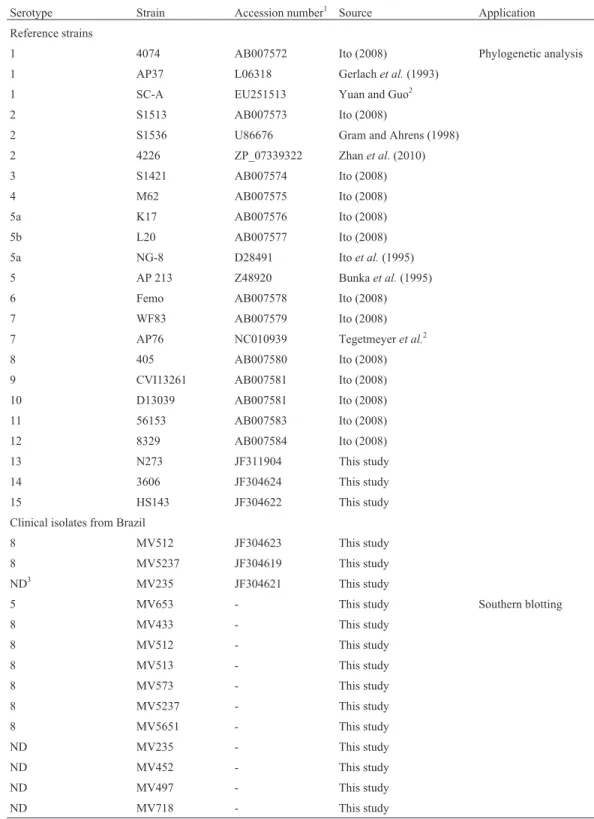
Table 1- Strains, clinical isolates and nucleotide sequences used in this study.
Serotype Strain Accession number1 Source Application Reference strains
1 4074 AB007572 Ito (2008) Phylogenetic analysis 1 AP37 L06318 Gerlachet al.(1993)
1 SC-A EU251513 Yuan and Guo2
2 S1513 AB007573 Ito (2008)
2 S1536 U86676 Gram and Ahrens (1998) 2 4226 ZP_07339322 Zhanet al.(2010) 3 S1421 AB007574 Ito (2008) 4 M62 AB007575 Ito (2008) 5a K17 AB007576 Ito (2008) 5b L20 AB007577 Ito (2008) 5a NG-8 D28491 Itoet al.(1995) 5 AP 213 Z48920 Bunkaet al.(1995) 6 Femo AB007578 Ito (2008) 7 WF83 AB007579 Ito (2008) 7 AP76 NC010939 Tegetmeyeret al.2
8 405 AB007580 Ito (2008) 9 CVI13261 AB007581 Ito (2008) 10 D13039 AB007581 Ito (2008) 11 56153 AB007583 Ito (2008) 12 8329 AB007584 Ito (2008) 13 N273 JF311904 This study 14 3606 JF304624 This study 15 HS143 JF304622 This study Clinical isolates from Brazil
8 MV512 JF304623 This study 8 MV5237 JF304619 This study ND3 MV235 JF304621 This study
5 MV653 - This study Southern blotting
8 MV433 - This study
8 MV512 - This study
8 MV513 - This study
8 MV573 - This study
8 MV5237 - This study
8 MV5651 - This study
ND MV235 - This study
ND MV452 - This study
ND MV497 - This study
ND MV718 - This study
kitTM(Promega, Madison, WI, USA) according to the man-ufacturer’s instructions. A pair of oligonucleotides, LPF1
(5’-ATTGTAAACTTTAGAGCTTTATATT-3’) and
LPR1 (5’-ATTAAAAAGTAAAAAAGCTATCCC-3’)
(Gram and Ahrens, 1998), was used to amplify theomlA
gene (the amplicon had an expected size of approximately 1270 bp). The PCR was done using 1.25 U of GoTaqDNA polymerase (Promega) in a final volume of 50mL of en-zyme buffer containing 1.5 mM MgCl2-, 0.2 mM of each
dNTP, 0.2mM of each oligodeoxynucleotide and 50 ng of DNA in a C1000TM thermal cycler (BioRad, Richmond, CA, USA). The DNA was initially denatured at 94 °C for 3 min, followed by 35 cycles of denaturation at 94 °C for 1 min, annealing at 50 °C for 45 s, and an extension step at 72 °C for 1.5 min, followed by a final extension step at 72 °C for 10 min. The reaction products were analyzed by electrophoresis in 1.0% agarose gels, purified using a Wiz-ard SV gel and PCR clean-up system (Promega) and se-quenced using the Sanger sequencing method.
Nucleotide sequences
The omlA gene nucleotide sequences used in this study were fromA. pleuropneumoniaeisolates from differ-ent serotypes and origins. The NCBI GenBank database ac-cession numbers and the serotypes of their respective isolates are listed in Table 1. The nucleotide sequences of isolates from serotypes 13, 14 and 15 that were previously unavailable in the databases were obtained in the present study and deposited under accession numbers JF311904, JF304624 and JF304622, respectively. In addition,omlA
genes from isolates with serotypes that could not be defined by immunological methods because of cross-reactivity were sequenced (accession numbers JF304619, JF304621 and JF304623).
Structural analysis of theomlAgene
The +1 point of translation and the termination codon of the omlA gene were predicted using the analysis tool ORFfinder(Rombelet al., 2002). Sequences correspond-ing to the promoter region (-10 and -35) of theomlAgene were predicted using the Bacterial Promoter Prediction Program BPROM, which was also used to predict possible
ciselements for the recognition of transcription factors.
Organization of theomlAgene inA.
pleuropneumoniaeisolates
The copy number and organization of theomlAgene in the genomes of the different isolates was studied using Southern blotting (Sambrooket al., 1989). We selected 11
A.pleuropneumoniaeclinical isolates (Table 1) that were obtained between 2003 and 2010 from six farms located in southeastern Brazil and included four non-serotypable
iso-lates. The primers omlAhF
(5’-CGGTTTAGTCGCAGGTTTAGT-3’) and omlAhR (5’-TCCTTAACCCCTAATTCCTTAAGA-3’) were used
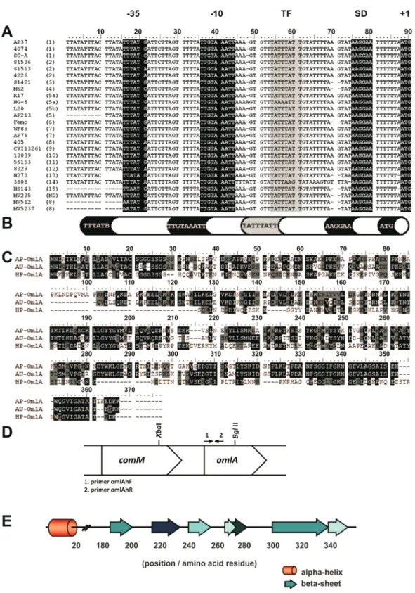
to synthesize a 372-bp probe for hybridization (Figure 1D). The probe was labeled using a PCR DIG Probe Synthesis kit (Roche, Mannheim, Germany), according to the manu-facturer’s instructions. Five micrograms of total DNA from the isolates was digested for 16 h with the restriction en-zymesXbaI andBglII to generate an expected fragment of 943 bp. The fragments were separated by electrophoresis in a 0.8% agarose gel and transferred to a nylon membrane (Amersham Hybond N+; GE Healthcare, Chalfont St. Gi-les, UK). The hybridization was done under high strin-gency conditions using the DIG High Prime DNA Labeling and Detection Starter kit IITM(Roche) to ensure the high specificity of hybridization. The results were visualized and documented using L-Pix Chemi photodocumentation system (Loccus, São Paulo, SP, Brazil).
Structural analysis of the OmlA protein
The amino acid sequences coded by theomlAgenes were predicted using the bacterial genetic code in the pro-gram Mega 5.03 (Tamuraet al., 2007). The putative sec-ondary structure was predicted using the Phyre algorithm (Kelley and Sternberg, 2009) and the Jnet algorithm of Jpred3 (Coleet al., 2008). The resulting structures were compared and only motifs with probability scores > 80% were used to construct the consensus structure model of the OmlA protein. The membrane protein topology prediction method TMHMM, based on the Markov model, was used to predict transmembrane helixes (Krogh et al., 2001). Conserved domains were located using the PROSITE data-base (Sigristet al., 2010) and the Conserved Domain Data-base (NCBI).
Phylogenetic molecular analysis
Results
Sequence analyses
The sequences used in this study contained 1092-1125 base pairs and 506 variable sites (~43%) in the aligned positions. There were 450 parsimony-informative sites (39%) and the high variability in the nucleotide se-quences resulted in 216 base substitutions, 77 (36%) in the first base of the codon, 84 (39%) in the second and 55 (25%) in the third (data not shown). Since most of the sub-stitutions occurred in the first and second bases, there was a large number of variable sites in the amino acidic se-quences deduced by Mega 5.03. Among the 383 aligned amino acids, 260 sites were variable (~68% of the total). The extensive number of alterations in the primary se-quence of the OmlA protein resulted from transitions and transversions. Typically, transition rates are approximately two times higher than transversion rates (Zhang and Gerstein, 2003) since the latter are usually rapidly detected by DNA repair mechanisms. However, for theomlAgene, the transition/transversion ratio was ~0.99 (data not shown). Thus, the high transversion rate reflected the high genetic variability acquired during the evolution of differ-entA. pleuropneumoniaeserotypes.
Structural analysis of theomlAgene
The open reading frame (ORF) ofomlAwas flanked by the codons ATG and TAA as the initiation and termina-tion codons, respectively, and the alignment of these se-quences showed that the initial region of the gene was very conserved, as also pointed by Gram and Ahrens (1998). Be-tween the +1 point of translation and point +160, 87.5% of the nucleotides were identical in all the serotypes and dif-ferences were observed in only a few isolates. The BPROM program identified the probable positions of the -10 and -35 regions of the promoters in theomlAgenes (Figure 1A). The sequence TATTTATT was prevalent in the promoter region of the gene and the BPROM program suggested that this might be a binding site for the regulatory protein Lrp, a global transcription regulator and member of a widely dis-tributed family among Bacteria and Archaea (Brinkmanet al., 2003). The putative Shine-Dalgarno region (ribosome binding site) was inferred based on the composition and po-sition of the consensus sequence. Figure 1B shows the schematic organization of the promoter region of theomlA
gene based on analyses of all available sequences.
Structural analysis of the OmlA protein
The deduced sequence of the OmlA protein was 362-375 amino acid residues long and 80% of the first 58 resi-dues were identical in all of the sequences analyzed. The primary sequence of theA. pleuropneumoniaeOmlA pro-tein was similar to OmlA in Actinobacillus ureae and
Haemophilus parasuis, both belonging to the family Pasteurellaceae. Alignment of the primary sequences of
this protein from these three species (Figure 1C) revealed conservation of the OmlA structure, including the N-ter-minal region, which reinforces the importance of this por-tion of the molecule. The secondary structure model of OmlA based on the profiles generated by Jpred3 and Phyre revealed that a large part of the protein is organized inb
sheets (Figure 1E), the positions of which vary only slightly according to the serotype; there was only onea-helix and this was located between the 5thand 20thamino acids and was conserved in all serotypes. In most serotypes, this
re-gion contained the amino acid sequence
KLIAGLVAGLVLTAC, with variations only in serotypes 1, 8, 9 and 11. In these cases, the third (isoleucine), fifth (glycine), ninth (glycine) and tenth (leucine) amino acids were replaced by methionine, serine (two) and valine, re-spectively, to yield the sequence KLMASLVASVVLTAC. This sequence variation probably does not significantly af-fect the formation of thea-helix since the substituted amino acids are from the same charge groups as the original resi-dues. The TMHMM model showed that thea-helix did not have a transmembrane insertion, which suggested involve-ment in another function. In the PROSITE database, the first 20 amino acids of the sequence matched a lipid-bin-ding site (profile PS51257) and a signal peptide. Hence, the
a-helix of the OmlA protein may serve to anchor the pro-tein to the external membrane, in addition to its function in directing the transport of the protein to its extracellular lo-cation. Analysis using the Conserved Domain Database in-dicated that the OmlA protein belonged to the lipoprotein 5 superfamily, which contains proteins that bind to trans-ferrin and is distinctly related to other protein families that bind to solutes.
Phylogenetic analysis
The phylogenetic trees obtained with the different methods used had very similar topologies. Figure 2A shows a Bayesian tree and the nine distinct groups identified based on the microorganism serotype: group 1 – serotypes 1, 9 and 11, group 2 – serotypes 2 and 8, group 3 – serotypes 3, 4 and 6, group 4 – serotype 13, group 5 – serotype 15 and our clinical isolates, group 6 – serotype 7, group 7 – serotypes 5 and 10, group 8 – serotype 14 and group 9 – serotype 12. The bootstrap values were equally high for all of the meth-ods used to construct the phylogenetic trees, as were the posteriori probability values obtained in Bayesian analysis (close to or equal to 100%).
if the group was analyzed separately from the others, given their almost identicalomlAsequences. For group 2, the cor-responding values for variable sites were 1.5% and 0.9%, and for groups 3 and 7, these values were 2.5% and 3.6%, respectively. Serotypes 13, 14 and 15 were distantly related and the clinical isolates grouped with serotype 15. The ra-dial topology (Figure 2B) of the tree shown in Figure 2A strongly suggested that the differentiation of the 15 A. pleuropneumoniaeserotypes may have involved two dis-tinct evolutionary events (highlighted by arrows in Figu-re 2B). This radial analysis separated the isolates into two major clusters: the first comprising serotypes 1, 2, 8, 9, 11, 12 and 14 and the second, serotypes 3, 4, 5, 6, 7, 10, 13 and 15. These cluster profiles were very similar to those ob-tained when the reconstruction was done using the pre-dicted amino acid sequences (data not shown), the only difference being that in the latter case, the serotype 4 organ-isms had distanced themselves from serotypes 3 and 6. This divergence most likely resulted from the duplication of a sequence present between positions +144 and +176
(GenBank accession number AB007575) and added 11 amino acids to the OmlA protein.
Organization of theomlAgene in clinical isolates of
A. pleuropneumoniae
Since the nucleotide sequence of theomlAgene varies significantly among serotypes, the primer pair omlAhF/omlAhR was synthesized based on the conserved region located in the initial extremity of theomlAgene; the resulting probe contained 372 bp. The organization of the
since the fragment size was > 10 kb the genome of this iso-late most likely did not have the same recognition se-quences for cleavage byXbaI andBglII present in the other isolates.
Discussion
The OmlA protein belongs to a family of small, poorly characterized bacterial lipoproteins widely distrib-uted inbandgproteobacteria (Vaniniet al., 2008). OmlA protein is a virulence factor ofA. pleuropneumoniaeand has an important role in binding to transferrin to facilitate the acquisition of iron from the host (Balteset al., 2002).
The genetic diversity of theomlAgene was first ob-served when a 970-bp amplicon was digested with restric-tion enzymes and the resulting fragments then used to classify 12 serotypes into five groups (Osakiet al., 1997). Subsequent analyses took advantage of the variability of the internal region of theomlAgene and used different tech-niques to try to distinguish the then known 12 serotypes, but could still only separate them into four or five groups (Gram and Ahrens, 1998; Gramet al., 2000a). In the pres-ent study, part of the work by Gram and Ahrens (1998) was reassessed and the polymorphisms of theomlAgene in the 15 currently known serotypes were identified and used to build a phylogenetic tree based on more recent methods and their respective best fit models. This approach allowed us to separate the A. pleuropneumoniae serotypes into nine (when using the nucleotide sequences) or ten (when using the amino acid sequences) groups and also to infer possible evolutionary relationships between them. Additionally, the
omlAgenes from clinical isolates ofA. pleuropneumoniae
were sequenced and analyzed.
The existence of various serotypes and the range of antigenic differences among them has made effective vac-cination againstA. pleuropneumoniaedifficult since only serotype-specific immunity is generally observed (Nielsen, 1984). This situation reinforces the importance of the
pre-cise characterization of isolates present in a given area. In addition, many A. pleuropneumoniae isolates are non-typable by currently used techniques but are nevertheless capable of causing disease (Fenwick, 2002). As shown here, the polymorphisms present in the nucleotide sequence of theomlAgene can be used to characterize isolates con-sidered nontypable by conventional methods. In addition, the phylogenetic reconstruction described here for theomlA
gene reinforced the characteristics noted elsewhere and confirmed the usefulness of this gene in distinguishing amongA. pleuropneumoniaeserotypes. For example, the clustering of serotypes 1, 9 and 11 agreed with the cross-reactivity observed amongst these serotypes (Paradiset al., 1999). A similar conclusion is applicable to serotypes 3, 6 and 8 which also show similar tube agglutination, coagglutination and indirect hemagglutination (Mittal et al., 1988). In this case, however, the molecular phylogeny separated serotype 8 from the other two. Cross-reactivity between isolates of serotypes 4 and 7 has also been ob-served (Mittal and Bourdon, 1991) and suggests evolution-ary proximity between them. This conclusion was validated by their close clustering in the phylogenetic tree, although they were placed in monophyletic branches.
Together, these results indicate that the poly-morphisms present in the nucleotide sequence of theomlA
gene and in the amino acid sequence of the OmlA protein can be used as markers to distinguish among the serotypes of many isolates. This is a useful approach for understand-ing the characteristics and origin of isolates in a delimited region.
All of the clinical isolates examined here were grouped with serotype 15, even those designated by other molecular techniques as serotype 8,i.e., some serotype 8 isolates ofA. pleuropneumoniaein Brazil can show varia-tion in theomlAgene that is actually closer to serotype 15. This finding agrees with Gramet al.(2000b) who identified some serotype 8 isolates in which theomlAgene was simi-lar to serotypes 3, 4, 6 and 7. We also believe that although the isolate MV235 was not serotypable by commonly used methods, it was almost certainly a variation of serotype 8 because of its cluster position in the phylogenetic tree.
Although the expression of theomlAgene is constitu-tive in other organisms (Ochsneret al., 1999), there is not much information on the expression of this gene in A. pleuropneumoniae.The structural model of the promoter region of the omlAgene suggests that there are possible binding sites for a transcription factor, the leucine-res-ponsive regulatory protein (Lrp), that may control gene ex-pression, particularly under stress (Wagner and Mulks, 2007), as has been observed in iron-restricted conditions (Deslandeset al., 2007). Additionally, the predicted sec-ondary structure of the OmlA protein indicates the exis-tence of a conserved region that may be involved in the binding of this protein to the lipid region of the outer mem-brane ofA. pleuropneumoniaeand have a role as a signal Figure 3 - Organization of the omlA gene in Actinobacillus
peptide. The tertiary structure of this protein cannot be pre-dicted because of a lack of homologous proteins in the data-bases.
In contrast to the conserved organization of theomlA gene previously reported for other genomes (Gerlachet al., 1993), we have shown that there are important variations in the organization of this gene in different A. pleuropneumoniaeisolates obtained in Brazil, including a surprising duplication. Such variability has not been ob-served before and the duplication event may confer some advantage to this microorganism,e.g., in adapting to new environments or even making it more virulent.
Although some aspects of the structural characteriza-tion of theomlAgene and the corresponding protein de-scribed here require additional experiments to confirm their functional relevance, we nevertheless believe that further detailed analysis of the genetic variability of this gene can yield important information on its role in the 15 serotypes ofA. pleuropneumoniaeidentified in this work. The result-ing information will improve our understandresult-ing of infec-tion byA. pleuropneumoniae.
Acknowledgments
The authors thank the Fundação de Amparo à Pes-quisa do Estado de Minas Gerais (FAPEMIG - APQ-00886-11), Coordenação de Aperfeiçoamento de Pessoal de Nível Superior (CAPES) and Conselho Nacional de Desenvolvimento Científico e Tecnológico (CNPq) for fi-nancial support.
References
Ashis SK, Torok VA, Percy NJ, Abimosleh SM and Howarth GS (2012) Microbial fingerprinting detects unique bacterial communities in the faecal microbiota of rats with experi-mentally-induced colitis. J Microbiol 50:218-225.
Baltes N, Hennig-Pauka I and Gerlach GF (2002) Both transferrin binding proteins are virulence factors in Actinobacillus pleuropneumoniae serotype 7 infection. FEMS Microbiol Lett 209:283-287.
Brinkman AB, Ettema TJ, de Vos WM and van der Oost J (2003) The Lrp family of transcriptional regulators. Mol Microbiol 48:287-294.
Bunka S, Christensen C, Potter AA, Willson PJ and Gerlach GF (1995) Cloning and characterization of a protective outer membrane lipoprotein ofActinobacillus pleuropneumoniae serotype 5. Infect Immun 63:2797-2800.
Cole C, Barber JD and Barton GJ (2008) The Jpred 3 secondary structure prediction server. Nucleic Acids Res 36:197-201. Deslandes V, Nash JH, Harel J, Coulton JW and Jacques M (2007)
Transcriptional profiling of Actinobacillus pleuropneu-moniae under iron-restricted conditions. BMC Genomics 8:e72.
Fenwick B (2002) Porcine infectious pleuropneumonia. In: Bethel MA (ed) Pork Industry Handbook. Michigan State Univer-sity Extension, East Lansing, 1178 pp.
Gerlach GF, Anderson C, Klashinsky S, Rossi-Campos A, Potter AA and Willson PJ (1993) Molecular characterization of a
protective outer membrane lipoprotein (OmlA) from Actinobacillus pleuropneumoniaeserotype 1. Infect Immun 61:565-572.
Gonzalez-Escalona N, Martinez-Urtaza J, Romero J, Espejo RT, Jaykus LA and DePaola A (2008) Determination of molecu-lar phylogenetics of Vibrio parahaemolyticus strains by multilocus sequence typing. J Bacteriol 190:2831-2840. Gottschalk M, Broes A, Mittal KR, Kobisch M, Kuhnert P,
Lebrun A and Frey J (2003) Non-pathogenicActinobacillus
isolates antigenically and biochemically similar to
Actinobacillus pleuropneumoniae: A novel species? Vet Microbiol 92:87-101.
Gram T and Ahrens P (1998) Improved diagnostic PCR assay for Actinobacillus pleuropneumoniae based on the nucleotide sequence of an outer membrane lipoprotein. J Clin Micro-biol 36:443-448.
Gram T, Ahrens P, Andreasen M and Nielsen JP (2000a) An Actinobacillus pleuropneumoniaePCR typing system based on the apx and omlA genes-evaluation of isolates from lungs and tonsils of pigs. Vet Microbiol 75:43-57.
Gram T, Ahrens P and Angen O (2000b) TwoActinobacillus
pleurpneumoniaeserotype 8 reference strains in circulation. J Clin Microbiol 38:468.
Huelsenbeck JP and Ronquist F (2001) MRBAYES: Bayesian in-ference of phylogenetic trees. Bioinformatics 17:754-755. Ito H (2008) Development of typing methods ofActinobacillus
pleuropenumoniaebased on the antigenic and genetic diver-sity of the protective outer membrane lipoprotein.Jpn Agr Res Q 42:261-266.
Ito H, Uchida I, Sekizaki T, Ooishi E, Kawai T, Okabe T, Taneno A and Terakado N (1995) Molecular cloning of an Actinobacillus pleuropneumoniaeouter membrane lipopro-tein (OmlA) from serotype 5a. Microb Pathog 18:29-36. Jessing SG, Angen O and Inzana TJ (2003) Evaluation of a
multi-plex PCR test for simultaneous identification and serotyping ofActinobacillus pleuropneumoniaeserotypes 2, 5, and 6. J Clin Microbiol 41:4095-4100.
Kelley LA and Sternberg MJ (2009) Protein structure prediction on the Web: A case study using the Phyre server. Nat Protoc 4:363-371.
Krogh A, Larsson B, von Heijne G and Sonnhammer EL (2001) Predicting transmembrane protein topology with a hidden Markov model: Application to complete genomes. J Mol Biol 305:567-580.
Larkin MA, Blackshields G, Brown NP, Chenna R, McGettigan PA, McWilliam H, Valentin F, Wallace IM, Wilm A, Lopez R, et al.(2007) Clustal W and Clustal X ver. 2.0. Bioin-formatics 23:2947-2948.
Mittal KR and Bourdon S (1991) Cross-reactivity and antigenic
heterogeneity among Actinobacillus pleuropneumoniae
strains of Serotype-4 and Serotype-7. J Clin Microbiol 29:1344-1347.
Mittal KR, Higgins R and Lariviere S (1988) Quantitation of serotype-specific and cross-reacting group-specific antigens by coagglutination and immunodiffusion tests for differenti-ating Actinobacillus (Haemophilus) pleuropneumoniae strains belonging to cross-reacting serotypes 3, 6, and 8. J Clin Microbiol 26:985-989.
Nielsen R (1984) Haemophilus pleuropneumoniae serotypes
Nightingale KK, Windham K and Wiedmann M (2005) Evolution and molecular phylogeny of Listeria monocytogenes iso-lated from human and animal listeriosis cases and foods. J Bacteriol 187:5537-5551.
Nylander JAA (2004) MrModeltest ver. 2. Program distributed by the author. Evolutionary Biology Centre, Uppsala Univer-sity .
Ochsner UA, Vasil AI, Johnson Z and Vasil ML (1999) Pseudo-monas aeruginosafur overlaps with a gene encoding a novel outer membrane lipoprotein, OmlA. J Bacteriol 181:1099-1109.
Osaki M, Sato Y, Tomura H, Ito H and Sekizaki T (1997) Genetic diversity of the genes encoding the outer membrane lipopro-tein (omlA) ofActinobacillus pleuropneumoniae. J Vet Med Sci 59:213-215.
Paradis SE, Dubreuil JD, Gottschalk M, Archambault M and Jacques M (1999) Inhibition of adherence ofActinobacillus pleuropneumoniae to porcine respiratory tract cells by monoclonal antibodies directed against LPS and partial characterization of the LPS receptors. Curr Microbiol 39:313-0320.
Posada D and Crandall KA (1998) Modeltest: Testing the model of DNA substitution. Bioinformatics 14:817-818.
Rombel IT, Sykes KF, Rayner S and Johnston SA (2002) ORF-FINDER: A vector for high-throughput gene identification. Gene 282:33-41.
Sambrook J, Fritsch EF and Maniatis T (1989) Analysis of ge-nomic DNA by Southern hybridization. In: Nolan C (ed) Molecular Cloning - A Laboratory Manual. Cold Spring Harbor Laboratory Press, New York, pp 9.32-9.58. Schuchert JA, Inzana TJ, Angen O and Jessing S (2004) Detection
and identification ofActinobacillus pleuropneumoniae sero-types 1, 2, and 8 by multiplex PCR. J Clin Microbiol 42:4344-4348.
Sigrist CJ, Cerutti L, de Castro E, Langendijk-Genevaux PS, Bulliard V, Bairoch A and Hulo N (2010) PROSITE, a
pro-tein domain database for functional characterization and an-notation. Nucleic Acids Res 38:D161-166.
Swofford DL (2003) Paup*: Phylogenetics analysis using parsi-mony (*and other methods) ver. 4.0b10. Sinauer Associates, Sunderland.
Tamura K, Dudley J, Nei M and Kumar S (2007) MEGA4: Molec-ular Evolutionary Genetics Analysis (MEGA) software ver. 4.0. Mol Biol Evol 24:1596-1599.
Vanini MM, Spisni A, Sforca ML, Pertinhez TA and Benedetti CE (2008) The solution structure of the outer membrane li-poprotein OmlA fromXanthomonas axonopodispv. citri re-veals a protein fold implicated in protein-protein interaction. Proteins 71:2051-2064.
Wagner TK and Mulks MH (2007) Identification of the Actino-bacillus pleuropneumoniae leucine-responsive regulatory protein and its involvement in the regulation of in vivo-induced genes. Infect Immun 75:91-103.
Williams JJ, Torres-León MA, Escheverria-Coelho P and Matos-Medina MC (2000) Aislamiento e identificación de Actinobacillus pleuropneumoniae en pulmones de cerdos con pleuroneumonía crónica sacrificados en el rastro munic-ipal de Mérida, Yucatán, México. Rev Biomed 11:175-185. Zhan B, Angen O, Hedegaard J, Bendixen C and Panitz F (2010)
Draft genome sequences of Actinobacillus pleuropneu-moniaeserotypes 2 and 6. J Bacteriol 192:5846-5847. Zhang Z and Gerstein M (2003) Patterns of nucleotide
substitu-tion, insertion and deletion in the human genome inferred from pseudogenes. Nucleic Acids Res 31:5338-5348.
Internet Resources
Bacterial Promoter Prediction Program BPROM,
http://www.softberry.com (March 16, 2013).
Associate Editor: Célia Maria de Almeida Soares