Document heading doi:10.12980/JCLM.2.201414J2 襃 2014 by the Journal of Coastal Life Medicine. All rights reserved.
B
iodegradation of carcinogenic textile azo dyes using bacterial isolates of
mangrove sediment
Guru Prasad Srinivasan1*, Asnar Sikkanthar1, Anandajothi Elamaran1, Caroline R Delma1, Kumaran Subramaniyan2,
Somasundaram Thirugnanasambandan Somasundaram1
1Faculty of Marine Sciences, Center of Advanced Study in Marine Biology, Annamalai University, Parangipettai, TN, India
2Department of Microbiology, Sri Sankara Arts and Science College, Enathur, Kanchipuram, India
Journal of Coastal Life Medicine
*Corresponding author: Guru Prasad Srinivasan, Faculty of Marine Sciences, Center of Advanced Study in Marine Biology, Annamalai University, Parangipettai 608502, Tamil Nadu, India.
Tel: +919976355585, +914144243223; 243533
Fax: 04144-243555
E-mail: [email protected]
Foundation Project: Supported by Department of Biotechnology, Government of India (BT/AAQ/Indo-Norway/183196/2007).
1. Introduction
Until the end of the 19th century, all colors used for
dyeing were obtained from natural sources, but today the number of synthetic colorants exceeds 7000. Synthetic dyes are cost effective, offer a wide range of new colors, and they impart better properties upon the dyed materials. During the dyeing process around 30% of the dye quantity remains in
the aqueous phase, mainly in the hydrolyzed form, leading to the colorization of the resulting effluent system[1]. Even though azo dyes are in general less harmful than older types, many are found to be toxic to fish, mammals, as well as to different kind of microorganisms[2-4]. Azo dyes and their metabolites are reported to be mutagenic or carcinogenic[5,6]. Azo dye compounds have been reported to cause bladder cancer in humans and hepatocarcinoma, PEER REVIEW ABSTRACT
KEYWORDS
Biodegradation, Azo dye, Mangrove, 16S rDNA, Paenibacillus sp.
Objective:To evaluate the biodegrading property against carcinogenic azo dyes using bacterial
isolates of mangrove sediment.
Methods:The bacterial isolates were subjected to submerged fermentation and their growth
kinetics were studied. The potential strain was characterized using 16S rDNA sequencing.
Results:In the present study, dye degrading bacterial colonies were isolated from the mangrove
sediment samples of Parangipettai estuarine area, Tamil Nadu. Of the 30 morphologically different strains isolated, 5 showed antagonistic property. The growth kinetics of the two strains, P1 and G1, which showed potent activity were calculated. One particular isolate (P1) showing promising dye degrading potential in the submerged fermentation was further characterized. The strain was identified as Paenibacillus sp. by 16S rDNA sequencing.
Conclusions: This study reveals the less explored microflora of mangrove sediments. The novel
strain may further be analyzed and used in the treatment of effluent from dye industry so as to reduce the impact of carcinogenic contaminants.
Peer reviewer
Dr. NB Dhayanithi, Postdoctoral Researcher, Centre For Biotechnology, Anna University, Chennai–600 025,
India.
Tel: 044-22358365, Fax: 044-22350299
E-mail: [email protected]
Comments
This is a valuable research work in which authors have demonstrated the degradation/decolouration pattern by novel bacteria, Paenibacillus sp. The decolouration effects, growth kinetics were assessed. In addition, potent dye degrading property of the bacteria can be used by the scientific community for the welfare of our society.
Details on Page 161
Article history:
Received 12Jan 2014
Received in revised form 16Jan, 2nd revised form 22Jan, 3rd revised form 27Jan 2014 Accepted 22Feb 2014
nuclear anomalies in intestinal epithelial cells in mouse models[7-10]. Aromatic amines, which are known human carcinogens, have been found in the urine of dyestuff workers and also in test animals following the administration of azo dyes[8,11,12].
Synthetic dyes are common contaminants of water, it is estimated that the production of these compounds is around of 10000 tonnes per year and it is assumed that the quantity
of dye discharged in the environment is about 1%-10%[13].
The discharge of this wastewater to the environment causes aesthetic problems due to the remaining color and also damages the quality of receiving water. The color impedes sunlight penetration disturbing the ecology, and the dyes and/or their degradation derivatives can prove toxic to aquatic life [2,4,14-19].
In the last few years, environmental legislation, about the appearance of colors in discharges, combined with the increasing cost of water for the industrial sector, has made the treatment and reuse of dyeing effluents increasingly attractive to the industry. The conventional treatment produces a lot of sludge, but does not remove all dyes, thus preventing recycling of the treated wastewater.
Scores of physico-chemical methods have been used for the treatment of dye with effluent[20,21]. Physico-chemical methods such as coagulation/flocculation, activated carbon adsorption and reverse osmosis technique have been developed in order to remove the color[20,22]. However the latter methods are not economically feasible can only transfer the contaminants (dyes) from one phase to other
leaving the problem essentially unsolved. Microorganisms have now been preferred to other sources because of their unique ability to degrade the toxic chemicals using their cytoplasmic enzymes. Bacterial enzymes like peroxidases, nitro reductases and azo reductases were found to decolorize dyes. These enzymes possess significant potential for the treatment of waste waters, which are colored by azo dyes.
Therefore microbial dye degradation has become a very promising approach for effluent treatment.
Various bacterial strains reduce azo dyes under anaerobic conditions. The most generally accepted hypothesis for this phenomenon is that many bacterial strains possess rather unspecific cytoplasmic enzymes, which act as azo reductases and under anaerobic conditions transfer electrons via soluble flavins to the azo dyes[23]. Microbial metabolites are preferred over plant and animal source due to their economic production, consistency, ease of process modification and optimization. They are relatively more stable than corresponding metabolites from plant or animals. Diverse categories of microorganisms, such as the actinomycetes, fungi and anaerobic and aerobic bacteria,
bring about the degradation of dyestuff[24,25]. Various lignolytic fungi were shown to decolorize azo dyes using ligninases, manganese peroxidases, and laccases. Wood rotting fungi have been found to effectively degrade a variety of azo dyes under aerobic conditions[26]. Dye degrading fungi have been frequently used in bioreactors for the decolorization and degradation of azo dyes[27].
Immobilized enzymes could have potential effect for dye decolorization and recycling of effluents[28] without the need of growth substrates. Aerobic bacteria have been described to oxidatively decolorize several classes of dyes, among which azo dyes always turned out to be the most recalcitrant compounds. In contrast, under anaerobic conditions, the decolorization of many azo dyes via reduction of the azo bond has been shown by anaerobic (Bacteroides sp., Eubacterium
sp., and Clostridium sp.) and facultative anaerobic bacteria (Proteus vulgaris and Streptococcus faecalis)[29-33].
The marine biosphere is one of the richest of the earth’s innumerable habitats, one of the least studied and characterized fauna[34]. Marine bacteria require sodium, potassium, magnesium ions and some require chloride and ferric ions[35]. Yet one another fact which is leading an increasing interest for exploring and exploiting the marine microfauna for industrial application is their high salt tolerance ability[34]. Most biological degradation of azo dye is carried out by anaerobic bacteria[32,36,37]. Mangrove ecosystems are rich in bacterial flora. The mangrove bacteria exist as symbionts with the plants and animals, saprophytes on dead organic matter, and as parasites on living organisms. Fertility of the mangrove waters results from the decomposition of organic matter and recycling of nutrients[38].
They perform varied activities like photosynthesis, nitrogen fixation, methanogenesis, production of antibiotics and enzymes, etc. Mangrove microbes have been reported to degrade a varied range of pollutants such asPAH, crude oil,
etc[39,40]. Although reports on the dye degrading ability of mangrove are available[41], works on the mangrove associated microbes are very scanty. With this view, the present study was targeted to identify novel dye degrading aerobic bacteria from mangrove environment.
2. Materials and methods
2.1. Sample collection
Mangrove sediment samples were collected from
Parangipettai estuarine area (Latitude 11°29’ N; Longitude
crimson, nito green B and methyl red were collected from the retail vendors (Kumbakonam).
2.2. Screening for dye degrading bacteria
One gram of sediment sample was added to a flask containing 1% peptone in distilled water/sea water (1:1) with
25 mg of azo dye. The flask was incubated at room for 72 h
in an orbital shaker. The decolorization of the sample was checked at different time points. All the studies were carried out with the distilled water /sea water in the ratio of 1:1.
2.3. Isolation of dye degrading bacteria
The sediment which showed decolorization was serially diluted using sterile distilled water up to 10-5 dilutions. An
aliquot of 0.1 mL from 10
-² to 10-5 dilutions was taken and
spread on sets of nutrient agar plates containing 25 mg of
Nitomill Brill crimson, Nito green B and methyl red. The uninoculated plate was kept as control. The plates were incubated at room temperature for 24 h and observed for
growth. Decolorization in plates was observed by fading of dye surrounding the bacterial colonies, compared with the original dye medium as control. Colonies which were morphologically different were counted and streaked on nutrient agar slants. All the strains were preserved at 4 °C until further investigation.
2.4. Evaluation of decolorization
Based on the above observation, the dye degrading bacterial strains were selected and further studied by adopting shake flask technique. The strains were inoculated in flasks containing the azo dyes and incubated in a orbital shaker at 200 r/min for 3 d at 28 °C. The biomass, decoloration and pH were observed every 6 h. The biomass and decoloration were calculated by measuring the OD at specific wavelengths
(520 nm, 640 nm and 560 nm for Nitomill brill crimson, Nito green B and methyl red respectively). Decoloration OD was
measured by filtering the biomass using 0.2 µm syringe
filter[42].
2.5. Growth kinetics
The growth kinetics of the 2 strains which showed
promising results in the screening were calculated.
2.5.1. Specific growth rate
The rate of biomass growth per unit biomass concentration is called specific growth rate. This can be formulated based
on the following equation
µ=(1/X)*(dX/dt)
On integration the above equation results ln X/X0=µt
Where, µ=specific growth rate of biomass (h-1), X=Biomass concentration (g/L), X0=Initial biomass concentration, (dX/
dt)=Biomass growth rate (g/L/h).
2.5.2. Doubling time
The time required to double the biomass concentration is the doubling time. This can be calculated using the following equation
td=ln 2/µ
Where, td=doubling time (h), µ=specific growth rate of biomass (h-1).
2.6. Characterization of dye degrading bacterial strains
2.6.1. Phenotypic characterization
The colony morphology, gram’s staining and the motility of two strains showing antagonistic dye degrading activity were demonstarted.
2.6.2. Biochemical characterization
Cultural and biochemical characters like indole, methyl red, voges prousaker, citrate utilization and oxidase, of the potential strains were studied by adapting standard procedure recommended by Bergey’s Manual of Systematic
Bacteriology.
2.6.3. Molecular characterization
The potential bacterial isolate (P1), was characterized
by 16S rDNA analysis for phylogenetic classification. The exponential phase culture of the isolate was centrifuged at 9000×g for 5 min. Supernatant was discarded and the pellet was dissolved in 567 µL TE buffer, 30 µL of 10%
sodium dodecyl sulfate, and 3 µL proteinase K (60 µg).
After 1 h incubation at 37°C, 100µL of 5 mol/LNaCl was added and mixed thoroughly[43]. The DNA was then purified as previously described[44]. The 16s rDNA sequences were amplified by polymerase chain reaction (PCR) using
universal primers of 27f (AGAGTTTGATCCTGGCTCAG) and
1490r (AAGGAGGTGATCCAGCC)[45]. All the primer sequences are presented in 5’ to 3’ orientation. The reaction mixture consisted of 5 µL of 10x buffer (Mg2+ free), 5 µL of MgCl2,
10µL of dNTP mix, 1 µL of each primer 2 µL of template
DNA and 0.5 µL of Taq DNA polymerase (5U/µL) (Genei,
Bangalore), in a final volume of 50 µL. The amplification
°C for 1 min, 72°C for 90 seconds and final extension at
72°C for 7 min. PCR products were electrophoresed on 1% agarose gel and documented under UV- transillumination.
The sequence was generated using an automated DNA
sequencer (Applied Biosystems, USA). The work was carried out at Bioserve Biotechnologies, Hyderabad. The partial
16S rRNA gene sequence of the potent bacterial isolate was
submitted in NCBI and an accession number was assigned.
3. Results
3.1. Preliminary screening for dye degrading bacteria
Dye decoloration was observed in the flask from the second day which indicated the presence of dye degrading aerobic bacteria in the mangrove sediment samples.
3.2. Isolation of dye degrading bacteria
Morphologically different bacterial colonies which were found to decolorize the specific dyes were observed on the plates. Colony forming units were 34×10-4CFU/g, 45×10-4CFU/ g and 58×10-4 CFU/g for plates incorporated with Nitomill
Brill crimson, methyl red and nito green B respectively.
Thirty colonies were found to be effective in decolorising the dye as evidenced by the zone clearance. Among them, five strains (P1, P3, G1, M1 and M2) which were observed
in all the three plates were selected for further studies.
3.3. Evaluation of decolorization
Decolorization of dye in the flasks was observed (Figure
1-4). The pH variation during the study was noted given
in Table 1. The rate of decoloration of the three dyes by all the five strains with respect to their biomass is graphically represented in Figure 5 to 11. Two strains P1 and G1
exhibited maximum degrading potential.
Figure 1.Nito Green B decolorization by P1.
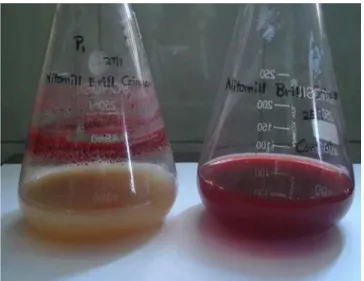
Figure 2. Nitomill Brill crimson decolorization by P1.
Figure 3.Methyl red decolorization by P1.
Figure 4.Dye decolorization by P1 after shake flask fermentation. Table 1
pH variation during decoloration.
Stains 1st Day pH (initial) 2nd Day pH 3rd Day pH
P1 8.0 9.3 7.6
P3 8.0 9.1 7.8
G1 8.0 9.7 7.9
M1 8.0 9.5 7.3
Figure 5. P1 against Nitomill Brill crimson. 0.8
0.7 0.6 0.5
0.4
0.3 0.2 0.1 0.0
OD
at
520
nm
0 6 12 18 24 30 36 42 48 54 Time (h)
Biomass Decolorization
Figure 6.P1 against nito green B. 0.8
0.7
0.6
0.5
0.4
0.3
0.2
0.1
0.0
OD
at
640
nm
0 6 12 18 24 30 36 42 48 54 Time (h)
Biomass Decolorization
Figure 7. P1 against methyl red. 0.8
0.7 0.6 0.5 0.4 0.3 0.2 0.1 0.0
OD
at
560
nm
0 6 12 18 24 30 36 42 48 54 Time (h)
Biomass Decolorization
Figure 8.G1 against Nitomill Brill crimson. 0.8
0.7 0.6 0.5 0.4 0.3 0.2 0.1 0.0
OD
at
520
nm
0 6 12 18 24 30 36 42 48 54 Time (h)
Biomass Decolorization
Figure 9.G1 against nito green B. 0.8
0.7
0.6
0.5
0.4
0.3
0.2
0.1
0.0
OD
at
640
nm
0 6 12 18 24 30 36 42 48 54 Time (h)
Biomass Decolorization
Figure 10.G1 against methyl red. 0.8
0.7
0.6
0.5
0.4
0.3
0.2
0.1
0.0
OD
at
560
nm
0 6 12 18 24 30 36 42 48 54 Time (h)
Biomass Decolorization
Figure 11.Specific growth rate of the isolates. 2.5
2.0
1.5
1.0
0.5
0.0
ln
OD
/
OD
0 10 20 30 40 50 Time (h)
P1 crim, µ=0.04329 h-1
P1 crim, µ=0.04592 h-1
P1MR, µ=0.04502 h-1
G1 crim, µ=0.04551 h-1
G1 green, µ=0.04373 h-1
G1MR, µ=0.07373 h-1
3.4. Growth kinetics
The specific growth rate and the doubling time of the two strains P1 and G1 was estimated and plotted graphically
(Figure 7). Plotting ln X/X0 against ‘t’ results in a straight line
at the exponential growth phase. µ value was found to be
0.04329, 0.04592, and 0.04502(h-1) for P1 and 0.04551, 0.04373
3.5. Characterization of potential strain
Table 2 shows the phenotypic characteristics such as
microscopic appearance, Gram’s staining and motility of
P1. Cultural and biochemical characteristics of P1 were
tabulated (Table 3) and found to be G+ve rod. The amplified
16S rDNA sequence was found to exhibit 96% homology
with Paenibacillus sp. (Table 3). The new subspecies was named as Paenibacillus DDAU. The GenBank accession number for the 16S rDNA sequence of Paenibacillus DDAU is GQ327970. Phylogenetic tree was constructed for the same.
Table 2
Screening of isolated potential stains.
Phenotypic Characters P1
Shape Rod
Gram’s staining G+ve
Motility Motile
4. Discussion
Treating waste water from textile industries has become a real challenge in recent years. The main problem occurring is that the color that remains due to the dyestuff used may cause disturbance to the ecological system of the receiving water[15-17]. The removal of dyes from aqueous effluent has received considerable attention within environmental research[19]; furthermore a recent review reveals that biodegradation is a promising method for dye removal[46].
Different combinations of treatment methods have been proposed in order to effectively manage the above waste water.
Anaerobic digestion of the azo dyes decomposes due to the cleavage of the azo bond consequently eliminating the color of waste water[47]. The reduction products (aromatic amines)
should then be further treated using aerobic biological treatment methods[18,48-52]. However works on aerobic degradation are scanty. In that view, the present study was attempted to identify bacterial strains that effectively
degrade azo dyes.
Mangrove sediments from Parangipettai estuarine area were used for isolation of dye degrading bacteria. Results showed that the microbes of mangrove sediment were potent in degrading both the industrial and laboratory dyes used. Sediment samples showed relatively less growth and the task of recovering the isolates was thriving. Results demonstrated that it is essential to provide the dye for the survival of these bacterial isolates and also the dye alone can supplement these organisms for their further endurance.
Results revealed the existence of the dye degrading bacteria in aerobic conditions. Categorization of the colony was done by naked eye observation.
Nutrient source plays a vital role in the isolation of microbes as evident from the present study. Many reports on aerobic degradation of dye were noted. Components like peptone, beef extract, yeast extract, nutrient broth medium[53,54], supplemented with mineral sources like Na2HPO4, KH2PO4, (NH4)2SO4, MgSO4, CaCO3 and H3BO3 have been
used for microbial growth[53]. Other enrichment sources include P2O5, K2O, MgO with sulfate salts (Fe, Cu, Mn, B,
Zn, Mo)[47]. In the present study 1% peptone along with the
dye exhibit bacterial colonies of different size, color and texture. This indicates that the trace amount of peptone with dye has accentuated the microbes to take up the dye.
Once the peptone in the media gets depleted, the microbes start utilizing the azo dyes as the sole source of carbon and nitrogen[55]. The dyes used in this study are powdery in nature, grouped under the aryl azo groups in which the azo dyes have a double bonded (N=N) structure for its
stability and binding. Possibly these bacterial strains could have cleaved the double bonding thereby using the carbon from the pyrrole rings of the chromophore (dye)[18,48,52]. In the study, the media were prepared using sea water to substitute the low nutrient source. Carbon, oxygen, hydrogen, nitrogen, sulfur and phosphorus are the major elements in the cell’s most important macromolecules namely nucleic acids, proteins, carbohydrates and lipids. A number of cations (K, Ca, Mg and Fe) are also required in significant
Table 3
16S rDNA sequence homology.
amounts to maintain the structure and function of various cellular components such as enzymes and co-enzymes.
In addition to these major nutrients, most bacteria require small quantities of trace elements, such as manganese, cobalt, zinc, molybdenum, copper and nickel. Some heterotrophs require low concentration of preformed growth factors or micronutrients such as aminoacids, pyrimidines, purines, and vitamins because they lack the biochemical pathways for the synthesis of these key intermediates.
Results established that it is essential to provide the naturally occurring microelements as added nutritive source so as to fulfill the eco-physiological parameter that the particular population requires. A particular feature of the marine Bacteria and the Archaea is their requirement for sodium. True marine prokaryotes have an absolute requirement for Na+ ions (usually in the range of 0.5% to
5.0%) and fail to grow in the culture medium substituted with
K+. This distinguishes the marine microbes from the closely
related terrestrial and freshwater species. As expected, most of the marine microbes grow at a concentration of NaCl similar to that of sea water (3.0%-3.5%NaCl). The majority of the marine prokaryotes are moderate halophiles and grow in the media supplemented with up to 15%NaCl[56].
Another important criterion is pH. After 24 h, there was
arise in pH and this may be due to the liberation of ammonia which increased the ionic concentration of hydrogen. This shows the idiophase of the culture. On the second day, there was a fall in pH due to the depletion of ammonia utilized by the culture. This indicated the production of metabolites of the bacterial isolates which require high nutrients. After
3rd day the increase in pH can be attributed to the excess ammonia liberated. This shows that when working with sediment from an extreme environment like mangrove it has to be seen that the initial pH should be taken with the native pH at which the population survives. Furthermore, the strains after the idiophase might have utilized the ammonia liberated in the first phase as the nitrogen source.
The biomass and decoloration data showed that they were positively correlated in the present study. This is concordant with the previous reports[14,47,57,58]. However, those studies dealt with the effluents rather than dye as a whole.
As per 16S rDNA sequence analysis, the dye degrading
strain reported in this study shared a 96% similarity with
Paenibacillus sp. Phylogenetic tree construct reveals that the strain coincides only with the Genus Paenibacillus sp. and not any specified species. Reports on dye degrading Paenibacillus are[59] very few and that too from the mangrove ecosystem reports are diminutive.
Hence, this strain Paenibacillus sp.DDAU can be used
as a candidate strain for dye degradation and can be taken
for scale up processes. Therefore, the bacterial strain P1
with potent dye degrading property can be exploited in a manner so as to stamp out the chore of scientific community and for the welfare of human and marine biosphere.
Conflict of interest statement
We declare that we have no conflict of interest.
Acknowledgements
The authors thank the former Dean Prof. T.
Balasubramanian, CAS in Marine Biology, Annamalai
University, for providing infrastructural facilities and the Department of Biotechnology, Government of India for financial support (BT/AAQ/Indo-Norway/183196/2007).
The authors declare that they have no conflict of interest
Comments
Background
Synthetic dyes are common contaminants of water especially, azo dyes and their metabolites are carcinogenic nature. So, there is need to detect the biological agents to degrade/convert the azo dye.
Research frontiers
The present research work was targeted to identify potential and novel dye degrading bacteria from mangrove environment.
Related reports
Carliell et al. (1995), Georgiou et al. (2004), Pandey et al.
(2007) and Murty et al. (2012) have conducted a research on
the degradation of various dyes using microbes and have also reported different ratio of dye decolouration/degrading capabilities.
Innovations and breakthroughs
Various reports are available on azo dye degrading bacteria from industrial and terrestrial environment with different range of results. But, the present study reported high decolouring/degrading effects expressed by bacteria.
Further, the strain was isolated and identified from the marine region which is novel.
Applications
problems on the waste water treatments of textile industries.
Further, the ecological systems of the receiving water bodies
(River, canal, etc) might be protected from the discharge of
dye pollution.
Peer review
This is a valuable research work in which authors have demonstrated the degradation/decolouration pattern by novel bacteria, Paenibacillus sp. The decolouration effects, growth kinetics were assessed. In addition, potent dye degrading property of the bacteria can be used by the scientific community for the welfare of our society.
References
[1] Dyes, general survey. In: Ulmann’s encyclopedia of industrial chemistry, Vol. A9. 5th ed. New York: VCH; 1987, p. 73.
[2] Ivanov VB, Tatarchenko VE. Reactive dyes in biology, Soviet scientific reviews supplement series, physicochemical biology. Switzerland: Harwood academic publisher; 1987.
[3] Zhang MM, Chen WM, Chen BY, Chang CT, Hsueh CC, Ding Y, et al. Comparative study on characteristics of azo dye decolorization by indigenous decolorizers. Bioresour Technol 2010; 101(8): 2651-2656.
[4] Bhattacharya S, Das A, Mangai G, Vignesh K, Sangeetha J. Mycoremediation of congo red dye by filamentous fungi. Braz J Microbiol 2011; 42(4): 1526-1536.
[5] Franciscon E, Grossman MJ, Paschoal JA, Reyes FG, Durrant LR. Decolorization and biodegradation of reactive sulfonated azo dyes by a newly isolated Brevibacterium sp. strain VN-15. Springerplus
2012; 1: 37.
[6] Stingley RL, Zou W, Heinze TM, Chen H, Cerniglia CE. Metabolism of azo dyes by human skin microbiota. J Med Microbiol 2010; 59: 108-114.
[7] Hartman CP, Fulk GE, Andrews AW. Azo reduction of trypan blue to a known carcinogen by a cell-free extract of a human intestinal anaerobe. Mutat Res 1978; 58: 125-132.
[8] Pan H, Feng J, He GX, Cerniglia CE, Chen H. Evaluation of impact of exposure of Sudan azo dyes and their metabolites on human intestinal bacteria. Anaerobe 2012; 18(4): 445-453.
[9] Johnson GE, Quick EL, Parry EM, Parry JM. Metabolic influences for mutation induction curves after exposure to Sudan-1 and para red. Mutagenesis 2010; 25(4): 327-333
[10] Chequer FM, Angeli JP, Ferraz ER, Tsuboy MS, Marcarini JC, Mantovani MS, et al. The azo dyes disperse red 1 and disperse orange 1 increase the micronuclei frequencies in human lymphocytes and in HepG2 cells. Mutat Res 2009; 676(1-2): 83-86. [11] Sayed HM, Fouad D, Ataya FS, Hassan NH, Fahmy MA. The
modifying effect of selenium and vitamins A, C, and E on the
genotoxicity induced by sunset yellow in male mice. Mutat Res
2012; 744(2): 145-153.
[12] Chequer FMD, Lizier TM, de Felicio R, Zanoni MV, Debonsi HM, Lopes NP, et al. Analyses of the genotoxic and mutagenic potential of the products formed after the biotransformation of the azo dye Disperse Red 1. Toxicol In Vitro 2011; 25: 2054-2063. [13] Nduka JK. Application of chemically modified and unmodified
waste biological sorbents in treatment of wastewater. Int J Chem Eng 2012; 2012: doi:10.1155/2012/751240.
[14] Georgiou D, Melidis P, Aivasidis A. Use of a microbial sensor; inhibition effect of azo-reactive dyes on activated sludge.
Bioprocess Biosyst Eng2002; 25(2): 79-83.
[15] Ong S, Toorisaka E, Hirata M, Hano T. Combination of adsorption and biodegradation processes for textile effluent treatment using a granular activated carbon-biofilm configured packed column system. J Environ Sci (China) 2008; 20: 952-956.
[16] Junkin R. Textile wastes, case history: pretreatment of textile wastewater. In: Proceedings of 37th Industrial Waste Conference, Purdue University. USA: Ann Arbor science; 1982, p. 139-146. [17] Kertell RC, Hill FG. Textile dye house wastewater treatment: a
case history. In: Proceedings of 37th Industrial Waste Conference, Purdue University. USA: Ann Arbor science; 1982, p. 147-156. [18] Balamurugan B, Kannadasan T. Photocatalytic oxidation of
anaerobically degraded reactive red 2 dye bath effluent. J Environ Res Develop 2012; 7: 827-837.
[19] Meyer U. Biodegradation of synthetic organic colorants. In: Leisinger T, editor. Microbial degradation of xenobiotics and recalcitrant compounds: FEMS symposium No. 12. Salt Lake City: Academic Press; 1981, p. 371-378.
[20] Eilbeck WJ, Mattock G. Chemical process in wastewater treatment. UK: Ellis Horwood Ltd; 1987.
[21] Holt PK, Barton GW, Wark M, Mitchell CA. A quantitative comparison between chemical dosing and electrocoagulation.
Colloids Surf A Physicochem Eng Asp 2002; 211(2-3): 233-248. [22] Tchobanoglous G, Burton FL, Stensel HD. Wastewater engineering:
treatment and reuse. New York: McGraw-Hill Inc.; 2003.
[23] Russ R, Rau J, Stolz A. The function of cytoplasmic flavin reductases in the reduction of azo dyes by bacteria. Appl Environ Microbiol 2000; 66(4): 1429-1434.
[24] Pandey A, Singh P, Iyengar L. Bacterial decolorization and degradation of azo dyes. Int Biodeterioration Biodegradation 2007; 59(2): 73-84.
[25] Ali N, Hameed A, Siddiqui MF, Ghumro PB, Ahmed S. Application of Aspergillus nigerSA1 for the enhanced bioremoval of azo dyes in simulated textile effluent. Afr J Biotechnol 2009; 8(16): 3839-3845.
2005, p. 269-288.
[27] Singh AK, Singh R, Soam A, Shahi SK. Degradation of textile dye orange 3R by Aspergillus strain (MMF3) and their culture optimization. Curr Disc 2012; 1(1): 7-12.
[28] Yesilada O, Birhanli E, Ercan S, Ozmen N. Reactive dye decolorization activity of crude laccase enzyme from repeated-batch culture of Funalia trogii. Turk J Biol 2014; 38: 103-110. [29] Murty Srinivas D, Patel Suhagi D, Rakesh S, Nikhil B. Isolation and
identification of bacterial culture for azo dye degrading capability.
Int J Res Chem Environ 2012; 2(4): 69-79.
[30] Hong YG, Gu JD. Physiology and biochemistry of reduction of azo compounds by Shewanella strains relevant to electron transport chain. Appl Microbiol Biotechnol 2010; 88(3): 637-643.
[31] Gingell R, Walker R. Mechanisms of azo reduction by Streptococcus faecalis. II. The role of soluble flavins. Xenobiotica 1971; 1: 231-239. [32] Nakamura J, Kubota Y, Miyaoka M, Saitoh T, Mizuno F, Benno
Y. Comparison of four microbial enzymes in Clostridia and
Bacteroides isolated from human feces. Microbiol Immunol 2002; 46: 487-490.
[33] Pearce CI, Christie R, Boothman C, von Canstein H, Guthrie JT, Lloyd JR. Reactive azo dye reduction by Shewanella strain J18 143.
Biotechnol Bioeng 2006; 95: 692-703.
[34] Kiruthika J, Saraswathy N. Production of L-glutaminase and its optimization from a novel marine isolate Vibrio azureusJK-79. Afr J Biotechnol 2013; 12(50): 6944-6953
[35] Kathiresan K, Ajmal Khan S, editors. Coastal biodiversity in mangrove ecosystems: UNU-INWEH-UNESCO international training course, June, 1-15 2006, course manual. India: Annamalai Univ.; 2006, p. 179-196.
[36] Alalewi A, Jiang CL. Bacterial influence on textile wastewater decolorization. J Environ Protect 2012; 3(8A): 889-903.
[37] Zissi U, Lyberatos G. Azo dye biodegradation under anoxic conditions. Wat Sci Tech 1996; 34: 495-500.
[38] Kathiresan K. Why are mangroves degrading? Curr Sci 2002; 83(10): 1246-1249.
[39] Guo CL, Zhou HW, Wong YS, Tam NFY. Isolation of PAH -degrading bacteria from mangrove sediments and their biodegradation potential. Mar Pollut Bull 2005; 51: 1054-1061. [40] do Carmo Maciel-Souza M, Macrae A, Volpon AGT, Ferreira
PS, Mendonça-Hagler LC. Chemical and microbiological
characterization of mangrove sediments after a large oil-spill in Guanabara Bay-RJ-Brazil. Braz J Microbiol 2006; 37(3): 262-266. [41] Gomathi V, Saravanakumar K, Kathiresan K. Bio removal of
malachite green by mangrove-derived Aplanochytrium sp., KGA2512. Afr J Microbiol Res 2013; 7(24): 3056-3065.
[42] Walker GM, Weatherley LR. Biodegradation and biosorption of acid anthraquinone dye. Environ Pollut 2000; 108: 219-223. [43] Ausubel FM, Brent R, Kingston RE, Moore DD, Seidman JG, Smith
JA, et al. editors. Current protocols in molecular biology. New York: Wiley and Sons; 1993.
[44] Onuk EE, Ciftci A, Findik A, Durmaz Y. Development and evaluation of a multiplex PCR assay for simultaneous detection of
Flavobacterium psychrophilum, Yersinia ruckeri and Aeromonas salmonicida subsp. salmonicida in culture fisheries. J Vet Sci 2010; 11(3): 235-241.
[45] Weisburg WG, Barns SM, Pelletier DA, Lane DJ. 16S ribosomal DNA amplification for phylogenetic study. J Bacteriol 1991; 173(2): 697-703.
[46] Li T, Guthrie JT. Colour removal from aqueous solutions of the reactive azo dye remazol black B using the immobilised cells (Shewanella strain J18 143) -cellulose-g.co-monomer system. J Water Resource Prot 2010; 2(1): 77-84.
[47] Zhang Y, Liu Y, Jing Y, Zhao Z, Quan X. Steady performance of a zero valent iron packed anaerobic reactor for azo dye wastewater treatment under variable influent quality. J Environ Sci (China)
2012; 24: 720-727.
[48] Growther L, Meenakshi M. Biotechnological approaches to combat textile effluents. Int J Microbiol 2008; 7(1): 384-470.
[49] Hong Y, Xu M, Guo J, Xu Z, Chen X, Sun G. Respiration and growth of Shewanella decolorationis S12 with an azo compound as the sole electron acceptor. Appl Environ Microbiol 2007; 73(1): 64-72. [50] Bromley-Challenor KCA, Knapp JS, Zhang Z, Gray NCC,
Hetheridge MJ, Evans MR. Decolorization of an azo dye by unacclimated activated sludge under anaerobic condition. Water Res 2000; 34(18): 4410-4418.
[51] Glasser A, Liebelt U, Hempel DC. Design of a two stage process for total degradation of azo dyes. In: Kreysa G. DECHEMA Biotechnology Conferences; Germany: Vch Verlagsgesellschaft Mbh; 1992, p. 1085-1088.
[52] Wu JY, Chen KC, Chen CT, Hwang SCJ. Hydrodynamic characteristics of immobilized cell beads in a liquid-solid fluidized-bed bioreactor. Biotechnol Bioeng 2003; 83: 583-594. [53] Kodam KM, Gawai KR. Decolorisation of reactive red 11 and 152
azo dyes under aerobic conditions. Indian J Biotechnol 2006; 5: 422-424.
[54] Jirasripongpun K, Nasanit R, Niruntasook J, Chotikasatian B. Decolorisation and degradation of C.I reactive red 195 by
Enterobacter sp. Thammasat Int J Sci Technol 2007; 12(4): 6-11. [55] Shah MP, Patel KA, Nair SS, Darji AM. An innovative approach to
biodegradation of textile dye (Remazol Black B) by Bacillus spp.
Int J Environ Bioremediation Biodegradation 2013; 1(2): 43-48. [56] Munn CB. Marine microbiology: ecology and applications. London
and New York: Taylor & Francis Publishers; 2004.
[57] Olukanni OD, Osuntoki AA, Gbenle GO. Textile effluent biodegradation potentials of textile effluent-adapted and non-adapted bacteria. Afr J Biotechnol 2006; 5(20): 1980-1984.
[58] Papinutti L, Mouso N, Forchiassin F. Removal and degradation of the fungicide dye malachite green from aqueous solution using the system wheat bran-Fomes sclerodermeus. Enzyme Microb Technol